Pheochromocytoma and pregnancy: A case report and review
Authors
Davi Rettori Pardo dos Santos 1
Cinthia Callegari Barbisan 1 Claudio Marcellini 1 Rubia Marina Vieira Rettori dos Santos 1,2
1 UNILUS. 2 UNIFESP.
Submitted on: 01/30/2015. Approved on: 06/24/2015.
Correspondence to:
Rubia Marina Vieira Rettori dos Santos.
Centro Universitário Lusíada, UNILUS, Fundação Lusíada. Rua Oswaldo Cruz , nº 179, Santos, SP, Brasil. CEP: 11.045-101
E-mail: [email protected]
I
NTRODUCTIONPheochromocytomas originate in the chromaffin cells in the adrenal medulla. Although this is the preferential site of involvement - accounting for 85%-95% of the cases - this tumor type may also affect the sympathetic paraganglia, in which
DOI: 10.5935/0101-2800.20150078
Pheochromocytoma is a catecholamine-producing adrenal tumor, being a rare cause of hypertension in pregnancy. It's prevalence in hypertensive patients is 0.2%, and 0.002% of pregnancies. We follow hypertensive pregnant 24 year old on her third pregnancy, admitted to 33 weeks with hypertensive emergency cesarean section indicated by fetal distress evolving with acute pulmonary edema in the post-partum period. Indicated laparoscopy after 13 days for acute abdominal pain, with no significant finding. In the postoperative, due a severe and resistant hypertension, suspected of pheochromocytoma and confirmed by biochemical tests and imaging. Performed unilateral adrenelectomia with cure of hypertension. The pathology and immunohistochemistry confirmed the diagnosis. We conclude that atypical cases of hypertension in pregnancy should be investigated early and differentiated pre-eclampsia. Despite the low prevalence, pheochromocytoma in pregnancy increases fetal maternal morbidity and mortality and the early recognition and treatment drastically change their outcome.
A
BSTRACTKeywords: hypertension; pheochromocy-toma; pregnancy.
case they are called paragangliomas.1 The rule of the 10% (10% of the tumors are malignant, 10% are extra-adrenal, 10% bilateral, and 10% familial) has been recently challenged.2 Approximately 24% of these tumors are inherited or stem from genetic mutations, associations with neurofibromatosis type 1 and Von Hippel-Lindau syndrome, or occur in cases of multiple endocrine neoplasia type 2.2,3 They are mostly benign single tumors cured through surgical resection.
abdomen. Pheochromocytomas are present in 0.002% of pregnancies and cause maternal/fetal death in up to 50% of the cases. Early diagnosis and treatment decrease maternal and fetal death rates to less than 5% and 15%, respectively. In 20% of the cases, diagnosis is not made during pregnancy.4
C
ASE DESCRIPTIONThe patient was a 24-year-old Caucasian female in her third pregnancy. She had been pregnant for 33 weeks with a single fetus when she was hospitalized and diagnosed with severe preeclampsia, with a blood pressure level of 230/170 mmHg. She was started on intravenous hydralazine 10 mg and magnesium sulfate 40 mEq, and was later given oral methyldopa 2g/ day and nifedipine retard 40 mg/day. On day three of hospitalization the fetus was in distress due to centralization of the blood flow and the patient was referred for a Cesarean section. She gave birth to a baby weighing 1,389 grams with APGAR scores of 4 and 8; the baby died 14 days later. The patient was transferred to the ICU with a hypertensive emergency and acute pulmonary edema controlled with intravenous sodium nitroprusside; she was also given oral hydrochlorothiazide 25 mg/day, captopril 100 mg/day, and amlodipine besylate 5 mg/day. She was discharged on a prescription for these three drugs and with a blood pressure reading of 90/70 mmHg. Thirteen days after discharge the patient was hospitalized with a hypertensive crisis and acute diffuse abdominal pain; she underwent laparoscopic examination, but no significant findings were reported. The review of her clinical history showed she had episodes of hypertension in her first pregnancy when she was 17; she had preeclampsia and a stillbirth then. She was pregnant again three years later and although her blood pressure was not monitored, her pregnancy was apparently uneventful. She was diagnosed with pulsating holocranial headaches, profuse sweating, palpitations, and hypertension when she was 23. At the age of 24 the patient was pregnant for the third time. She was
diagnosed with hypertension and preeclampsia, and her baby died a few days after birth. She had acute pulmonary edema and was suspected for acute abdomen in immediate postpartum care. Her family had a history of hypertension. The patient stated she did not smoke, drink, or take drugs. Patient clinical examination revealed profuse diaphoresis and severe hypertension, with systolic and diastolic pressure levels in a supine position ranging between 160 and 230 mmHg and 120 and 130 mmHg, respectively, and standing systolic and diastolic pressure levels ranging between 140 and 110 mmHg and 90 to 60 mmHg, respectively, a finding characteristic of postural hypotension. Her heart rate ranged from 94 to 120 beats per minute. Her face was not swollen and her radial, brachial, pedal, and posterior tibial artery pulses could be felt through palpation and were symmetrical. Ophthalmoscopy revealed diffuse arteriolar spasm and diseased arteriovenous crossings. The results of her workup were as follows: fasting glucose of 124 mg/dl; serum creatinine of 0.78 mg/dl; potassium level of 3.6 mEq/L; creatinine clearance of 83 ml/min; and proteinuria of 170 mg/24hours; her complete blood count, uric acid, total cholesterol and cholesterol fractions, triglycerides, and urinalysis tests were normal. Her electrocardiogram showed she had left ventricular hypertrophy (LVH); her echocardiogram showed an interventricular septum and a left ventricular posterior wall thickness of 16 mm indicative of LVH. Serum levels of aldosterone, calcitonin, cortisol, PTH, dopamine, and adrenaline, and urine levels of dopamine and adrenaline were normal. Urine noradrenaline was elevated and plasma renin activity was about 10 times higher than normal levels. Plasma renin activity was 55.9 ng/ml/h (reference value of 1.9 to 6 ng/ml/h), urine noradrenaline 339 mcg/24hs (reference value < 97 mcg/24hs), and serum noradrenaline 4,102.6 pg/ml (reference value 114 to 352 pg/ml).
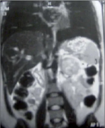
the left adrenal region (Figure 1). Abdominal nuclear magnetic resonance imaging revealed an expansive formation characterized by mildly heterogeneous T2 hyperintensity measuring 5.6 x 4.9 cm located in the retroperitoneal space in the left adrenal area, suggestive of pheochromocytoma (Figure 2). The patient’s blood pressure was managed with carvedilol 50 mg/day, prazosin 8 mg/day, amlodipine 10 mg/ day, and captopril 75 mg/day. She underwent an open left adrenalectomy (Figure 3). Pathology tests and immunohistochemistry (positive for chromogranin A and synaptophysin) confirmed the diagnosis of pheochromocytoma. Her hypertension ceased after surgery. She was discharged with a blood pressure of 130/80 mmHg and a heart rate of 84 beats per minute. The patient got pregnant again and had a normal delivery after her hypertension was resolved.
D
ISCUSSIONAlthough 90% of the pregnant individuals with pheochromocytoma present a series of classical symptoms (hypertension, headache, diaphoresis, and palpitations), not all patients are diagnosed during pregnancy.1 The patient described in this case report started having severe hypertension before the age of 30, accompanied by the triad of symptoms indicative of pheochromocytoma.
If she had been diagnosed earlier, her pregnancy could have been avoided or delayed until her hypertension was managed or the diagnostic study completed. Symptoms tend to manifest more often as patients get closer to the end of pregnancy.5 Other described symptoms are pallor, dizziness, dyspnea, polyuria after hypertensive crisis, seizures, nuchal pain, nausea, vomiting, and abdominal pain.5,6 Patients often report weight loss and bowel obstipation. The postpartum acute abdominal pain the patient
Figure 1. Computed tomography scan of the abdomen with contrast uptake showing a large heterogeneous tumor in the left adrenal territory.
Figure 2. Nuclear magnetic resonance image of the abdomen showing an expansive formation measuring 5.6 x 4.9 cm in the area of the medulla of the adrenal with a hypersensitive signal in T2.
had, which led to her being offered surgery with a negative outcome, may have been caused by an adrenergic crisis due to pheochromocytoma. Counselman et al.7 described a case of pheochromocytoma in a patient whose main complaint was abdominal pain.
In the series published by Oliva et al.8 the six patients were initially treated for preeclampsia. Hypertension in individuals with pheochromocytoma is paroxysmal and does not have a pattern of onset; conversely, preeclampsia starts usually after the first 20 weeks of pregnancy and is not paroxysmal. Proteinuria, edema, and elevated uric acid are not suggestive of pheochromocytoma, while postural hypotension is a classical sign of this tumor type. Café-au-lait spots, freckles, and fibromas - signs of the association of pheochromocytoma and neurofibromatosis - are present in 2% of these patients.4,8 Cardiovascular involvement - characterized by angina and acute myocardial infarction caused by coronary artery spasm secondary to increased catecholamine levels, increased platelet aggregation, and increased oxygen consumption - was one of the main reported complications.4,9 Cardiac arrhythmias such as atrial and ventricular fibrillation cause the sudden death of individuals with pheochromocytomas, often when patients are under anesthesia or during tumor resection procedures.. Acute pulmonary edema may also occur, as described in this case report, in addition to heart failure and ischemic or hemorrhagic stroke.10 Obstetric risks revolve around vasoconstriction and the possible premature detachment of the placenta and intrauterine hypoxia. The fetus is protected against exposure to catecholamines by noradrenaline transporters expressed in placenta cells.4
Clinical diagnosis, biochemical tests, and imaging are equally important. Most pheochromocytomas and paragangliomas produce, store, metabolize, and secrete catecholamines and their metabolites.2 In this process, noradrenaline is metabolized into normetanephrine and adrenaline into
metanephrine. Blood and urine metanephrines are highly sensitive and yield a significant negative predictive value. The continuous metabolization and secretion of metanephrines into the bloodstream, and not the episodic release of catecholamines by the tumor, explain the high levels of sensitivity mentioned above.4 The clonidine suppression test is used when metanephrines are slightly increased and diagnosis is dubious. However, it cannot be used in pregnant individuals due to possible adverse side effects.4 In the case presented in this paper, urine and serum noradrenaline were significantly elevated, the latter at levels ten times greater than reference values. Normal levels of calcitonin and PTH ruled primary hyperparathyroidism and medullary thyroid carcinoma out. Computed tomography and scintigraphy with metaiodobenzylguanidine are highly sensitive, but contraindicated for pregnant patients. Therefore, the imaging test of choice is nuclear magnetic resonance for its high sensitivity, the bright T2 images, and for not emitting ionizing radiation.10
The preoperative care offered to patients with pheochromocytoma is designed to treat hypertension, prevent paroxysms, and correct hypovolemia, once the removal of the tumor leads to an abrupt decrease in catecholamines and places patients at risk for hypovolemic shock.11 The definition of blood pressure targets for pregnant patients is controversial. According to the 6th Brazilian Hypertension Guideline, antihypertensive drugs should not be prescribed to individuals with a BP < 150/100 mmHg associated to preeclampsia or chronic hypertension.12 Very low BP levels may compromise uterine-placental circulation and impair fetal growth.12
less tachycardia and their shorter action allows for quicker dosage adjustments and fewer occurrences of postoperative hypotension.4,9 Beta-adrenergic antagonists exacerbate alpha-adrenergic response and consequently worsen hypertension. These drugs are indicated for the treatment of tachycardia and arrhythmia and should be introduced after alpha-adrenergic antagonists. Carvedilol, the drug prescribed to our patient, is a non-cardioselective beta-blocker and an alpha-1 antagonist that further reduces blood pressure.13 Carvedilol is not recommended during pregnancy. Calcium channel blockers are also an option for hypertensive pregnant patients, but pheochromocytomas may accentuate the tachycardia already established in this condition. Many authors have reported on the beneficial effects of intravenous magnesium sulfate and listed it as an attractive option for severely hypertensive pregnant subjects.14 Patients should also be offered a sodium-rich diet and hydration in order to prevent postoperative hypotension. The patient described in this paper was administered vigorous hydration for 24 hours before surgery, but suffered from severe hypotension immediately after the removal of the tumor.
The best time to remove the tumor surgically is within the first 24 weeks of gestation or after delivery, the second trimester being the safest moment due to the risk of miscarriage present in the first trimester.5,10 Patients diagnosed with pheochromocytoma after 24 weeks of gestation may be treated with the same drugs used in preoperative preparation for prolonged periods until the fetus is viable; in these cases, the tumor may be removed during of after a C-section has been performed.
Although not very prevalent, pheochro-mocytomas are a curable cause of hypertension. The recognition and surgical removal of the-se tumors significantly alters the outcomes for pregnant women and their fetuses, in addition to
mimicking specific pregnancy hypertensive con-ditions. Therefore, atypical and resistant cases of hypertension affecting pregnant and non-preg-nant individuals must be carefully analyzed and diagnosed to rule out causes of secondary hyper-tension and provide patients with effective and potentially curable therapies for hypertension.
R
EFERENCES1. Blake AM, Sweeney AT. Pheochromocytoma. Oncology CME& Education Collection [serial online] [Acessed Sep 7 2015]. Avai-lable from: http://emedicine.medscape.com/article/124059 2. Chen H, Sippel RS, O'Dorisio MS, Vinik AI, Lloyd RV, Pacak
K; North American Neuroendocrine Tumor Society (NANETS). The North American Neuroendocrine Tumor Society consensus guideline for the diagnosis and management of neuroendocrine tumors: pheochromocytoma, paraganglioma, and medullary thyroid cancer. Pancreas 2010;39:775-83. PMID: 20664475 DOI:http://dx.doi.org/10.1097/MPA.0b013e3181ebb4f0 3. Sarathi V, Bandgar TR, Menon PS, Shah NS.
Pheochromo-cytoma and medullary thyroid carcinoma in a pregnant mul-tiple endocrine neoplasia-2A patient. Gynecol Endocrinol 2011;27:533-5. DOI: http://dx.doi.org/10.3109/09513590. 2010.507285
4. Lenders JW. Pheochromocytoma and pregnancy: a deceptive connection. Eur J Endocrinol 2012;166:143-50. DOI:http:// dx.doi.org/10.1530/EJE-11-0528
5. Juszczak K, Drewa T. Adrenergic crisis due to pheochromo-cytoma - practical aspects. A short review. Cent European J Urol 2014;67:153-5.
6. Dugas G, Fuller J, Singh S, Watson J. Pheochromocytoma and pregnancy: a case report and review of anesthetic mana-gement. Can J Anaesth 2004;51:134-8. DOI: http://dx.doi. org/10.1007/BF03018772
7. Counselman FL, Brenner CJ, Brenner DW. Adrenal pheo-chromocytoma presenting with persistent abdominal and flank pain. J Emerg Med 1991;9:241-6. DOI: http://dx.doi. org/10.1016/0736-4679(91)90420-K
8. Oliva R, Angelos P, Kaplan E, Bakris G. Pheochromo-cytoma in pregnancy: a case series and review. Hyperten-sion 2010;55:600-6. PMID: 20083723 DOI: http://dx.doi. org/10.1161/HYPERTENSIONAHA.109.147579
9. Sarathi V, Lila AR, Bandgar TR, Menon PS, Shah NS. Pheo-chromocytoma and pregnancy: a rare but dangerous com-bination. Endocr Pract 2010;16:300-9. DOI: http://dx.doi. org/10.4158/EP09191.RA
10. Biggar MA, Lennard TW. Systematic review of phaeo-chromocytoma in pregnancy. Br J Surg 2013;100:182-90. DOI:http://dx.doi.org/10.1002/bjs.8976
11. Pacak K. Preoperative management of the pheochromo-cytoma patient. J Clin Endocrinol Metab 2007;92:4069-79. DOI:http://dx.doi.org/10.1210/jc.2007-1720
12. Sociedade Brasileira de Cardiologia/Sociedade Brasileira de Hipertensão/Sociedade Brasileira de Nefrologia. VI Diretri-zes Brasileiras de Hipertensão. Arq Bras Cardiol 2010;95:1-51.
13. Leonetti G, Egan CG. Use of carvedilol in hypertension: an update. Vasc Health Risk Manag 2012;8:307-22.