ABSTRACT
Objective: To describe the trends in tumor histology, gender and age among patients with non-small cell lung cancer (NSCLC) treated with lung resection. The histology of lung cancer has changed in developed countries, and there is still little information available on the topic for developing countries. Methods: This was a retrospective study of 1,030 patients with NSCLC treated with lung resection between 1986 and 2015 at a university hospital in southern Brazil. Differences in histology, stage, and type of surgery were analyzed by gender and for three periods (1986-1995, 1996-2005, and 2006-2015). Results: Most (64.5%) of the patients were males, and the main histological types were squamous cell carcinoma (in 40.6%) and adenocarcinoma (in 44.5%). The mean age at surgery during the irst period was 56.4 years for women and 58.9 years for men, compared with 62.2 for women and 64.6 for men in the third period (p < 0.001). The proportion of females increased from 26.6% in the irst period to 44.1% in the third. From the irst to the third period, the proportion of patients with squamous cell carcinoma decreased from 49.6% to 34.8% overall (p < 0.001), decreasing to an even greater degree (from 38.9% to 23.2%) among men. Among the NSCLC patients in our sample, females with adenocarcinoma accounted for 11.9% in the irst period and 24.0% in the third period (p < 0.001). Conclusions: As has been seen in developed countries, the rates of lung cancer in females in southern Brazil have been rising over the last three decades, although they have yet to surpass those observed for males in the region. The incidence of squamous cell carcinoma has decreased in males, approaching adenocarcinoma rates, whereas adenocarcinoma has signiicantly increased among women.
Keywords: Lung neoplasms; Epidemiology; Histology; Adenocarcinoma; Carcinoma, non-small-cell lung; Carcinoma, squamous cell.
Lung cancer: changes in histology, gender,
and age over the last 30 years in Brazil
Maria Teresa Ruiz Tsukazan1,2, Álvaro Vigo2, Vinícius Duval da Silva3, Carlos Henrique Barrios4, Jayme de Oliveira Rios1,
José Antônio de Figueiredo Pinto1
Correspondence to:
Maria Teresa Ruiz Tsukazan. Centro Clínico da PUCRS, Avenida Ipiranga, 6690, Conj. 615, CEP 90610-000, Porto Alegre, RS, Brasil. Tel.: 55 51 3336-8190. Fax: 55 51 3339-9040. E-mail: [email protected]
Financial support: None.
INTRODUCTION
Non-communicable diseases (NCDs) are responsible for more than 67% of deaths worldwide.(1) In Brazil, cancer
represents the second leading cause of NCD-related deaths and lung cancer is the leading cause of cancer-related deaths,(2) despite strong anti-smoking policies that reduced
the smoking rate by half from 1989 to 2008.(3) According
to the World Health Organization, 1.6 million deaths per year are attributable to lung cancer.(4) It is one of the
few cancers with a well-known cause—smoking.(1,3-6) The
great efforts to reduce smoking and to introduce the use of cigarette ilters have changed the epidemiology of lung cancer in developed countries, with an increase in the incidence of adenocarcinoma and a decrease in that of squamous cell carcinoma, as seen in the United States, Europe, and Asia.(7-18) The rising number of women with
lung cancer is also notable, as are the changes in their histological proile.(7)
Changes in the histological proile of lung cancer in Latin American countries have been poorly described in the literature.(19) Little information regarding lung cancer
histology, gender difference, and trends is available for the population of Brazil. This paper aims to describe
and improve understanding of the epidemiology of lung cancer, including histology, gender distribution, patient age, and stage of the disease, in southern Brazil over the last 30 years.
METHODS
From records on ile in the prospective surgery database of the Thoracic Surgery Division of the Hospital São Lucas, in the city of Porto Alegre, Brazil, we selected all patients with primary non-small cell lung cancer who were treated with anatomical resection between 1986 and 2015. We reviewed the pathology reports and charts of 1,062 patients. Thirty-two records were excluded because of missing data related to patient gender, patient age at surgery, type of resection, histology, and staging classiication. Therefore, the inal sample comprised 1,030 patients. Information about smoking was available in less than 37% of the charts, and that variable was therefore disregarded. All histological diagnoses were made by the same pathology group, and all staging was updated according to the 7th edition of the International Association for the Study of Lung Cancer classiication system.(20-22)
1. Serviço de Cirurgia Torácica, Hospital São Lucas, Pontifícia Universidade Católica do Rio Grande do Sul – PUCRS – Porto Alegre (RS) Brasil.
2. Departamento de Estatística e Programa de Pós-Graduação em Epidemiologia, Universidade Federal do Rio Grande do Sul – UFRGS – Porto Alegre (RS) Brasil.
3. Departamento de Patologia, Hospital Mãe de Deus, Porto Alegre (RS) Brasil.
4. Hospital do Câncer, Hospital Mãe de Deus, Porto Alegre (RS) Brasil.
Submitted: 11 November 2016.
Accepted: 18 June 2017.
Patients with different clinical characteristics (in terms of histology, stage, and type of surgery) were analyzed by gender and for three different periods (1986-1995, 1996-2005, and 2006-2015). Proportions were compared by Pearson’s chi-square test or Fisher’s exact test. Two-way ANOVA was used in order to compare the mean age of the patients by gender and period. Means were compared by using adjusted least squares means and the Tukey-Kramer test. All analyses were performed using the Statistical Analysis System software, version 9.4 (SAS Institute, Cary, NC, USA), and the level of signiicance was set at 5%.
The study was approved by the Research Ethics Committee of the Hospital São Lucas of the Pontifícia Universidade Católica do Rio Grande do Sul. Because of the retrospective nature of the study, the requirement for consent was waived.
RESULTS
A total of 1,062 patients underwent lung resection for primary lung cancer at the Hospital São Lucas in the last 30 years, and 1,030 met the criteria for inclusion in this analysis. Of those 1,030 patients, 665 (64.5%)
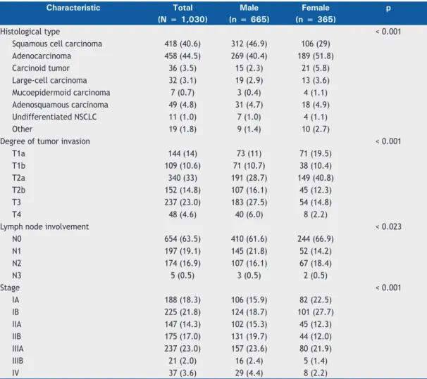
were male. The overall mean age at surgery was 62.8 years for the men and 60.8 years for the women. Table 1 shows the clinical characteristics of the patients, overall and by gender. Overall, the predominant histological type was adenocarcinoma (44.5%), followed by squamous cell carcinoma (40.6%). The histological types differed by gender (p < 0.001), squamous cell carcinoma being more common in men than in women (with a prevalence of 46.9% and 29.0%, respectively), whereas the opposite was found for adenocarcinoma (which had a prevalence of 40.4% and 51.8% among men and women, respectively). Differences between genders were also observed for the degree of tumor invasion (p < 0.001), lymph node classiication (p < 0.023), and staging (p < 0.001), suggesting that the disease was more advanced in the men than in the women (Table 1).
According to two-way ANOVA, there was no evidence of an interaction between gender and period, suggesting that the mean age did not differ between men and women in any of the three periods studied (Figure 1). However, regardless of the period, the adjusted least squares mean ages at surgery were 62.4 and 59.7 years for men and women, respectively, approximately
Table 1. Clinical characteristics of the patients, in the sample as a whole and by gender.a
Characteristic Total Male Female p
(N = 1,030) (n = 665) (n = 365)
Histological type < 0.001
Squamous cell carcinoma 418 (40.6) 312 (46.9) 106 (29)
Adenocarcinoma 458 (44.5) 269 (40.4) 189 (51.8)
Carcinoid tumor 36 (3.5) 15 (2.3) 21 (5.8)
Large-cell carcinoma 32 (3.1) 19 (2.9) 13 (3.6)
Mucoepidermoid carcinoma 7 (0.7) 3 (0.4) 4 (1.1)
Adenosquamous carcinoma 49 (4.8) 31 (4.7) 18 (4.9)
Undifferentiated NSCLC 11 (1.0) 7 (1.0) 4 (1.1)
Other 19 (1.8) 9 (1.4) 10 (2.7)
Degree of tumor invasion < 0.001
T1a 144 (14) 73 (11) 71 (19.5)
T1b 109 (10.6) 71 (10.7) 38 (10.4)
T2a 340 (33) 191 (28.7) 149 (40.8)
T2b 152 (14.8) 107 (16.1) 45 (12.3)
T3 237 (23.0) 183 (27.5) 54 (14.8)
T4 48 (4.6) 40 (6.0) 8 (2.2)
Lymph node involvement < 0.023
N0 654 (63.5) 410 (61.6) 244 (66.9)
N1 197 (19.1) 145 (21.8) 52 (14.2)
N2 174 (16.9) 107 (16.1) 67 (18.4)
N3 5 (0.5) 3 (0.5) 2 (0.5)
Stage < 0.001
IA 188 (18.3) 106 (15.9) 82 (22.5)
IB 225 (21.8) 124 (18.7) 101 (27.7)
IIA 147 (14.3) 102 (15.3) 45 (12.3)
IIB 175 (17.0) 131 (19.7) 44 (12.0)
IIIA 237 (23.0) 157 (23.6) 80 (21.9)
IIIB 21 (2.0) 16 (2.4) 5 (1.4)
IV 37 (3.6) 29 (4.4) 8 (2.2)
2.7 years higher for men (p < 0.001). Similarly, regardless of gender, the adjusted least squares mean ages were 57.7 years for the 1986-1995 period, 62.1 years for the 1996-2005 period, and 63.4 years for the 2006-2015 period, translating to an increase of approximately 5.7 years from the irst period to the last (p < 0.001).
As can be seen in Table 2, there were signiicant differences among the three periods in terms of the histological type (p < 0.001), especially for squamous cell carcinoma, the prevalence of which declined from 49.6% in the 1986-1995 period to 43.0% in the 1996-2005 period and to 34.8% in the 2006-2015 period. However, in those same periods, the prevalence of adenocarcinoma increased from 38.1% to 41.2% and 49.5%, respectively. The most common type of surgery was lobectomy, which was performed in 72.5% of cases in the irst period, compared with 83.6% in the third
period (p < 0.001). The proportion of cases in which pneumonectomy was performed trended down, from 19.7% in the irst period to 9.7% in the third period, as did that in which bilobectomy was performed, from 7.8% in the irst period to 4.1% in the third period. There was also a signiicant difference among the periods in terms of the staging (p < 0.001), with an increase in the proportion of cases classiied as stage I.
The distribution of histological types was determined by gender and period (Figure 2). In the 2006-2015 period, squamous cell carcinoma and adenocarcinoma were the main histological types among men, whereas adenocarcinoma was the predominant histological type among women. Overall, the prevalence of adenocarcinoma increased from 38.1% to 41.2% and 49.5% in the 1986-1995 period, 1996-2005 period, and 2006-2015 period, whereas that of squamous cell carcinoma decreased from 49.6% to 43.0% and 34.8%, respectively. Overall, squamous cell carcinoma was the most common histological type among men, although its prevalence declined from 38.9% in the 1986-1995 period to 23.2% in the 2006-2015 period, being equal to that of adenocarcinoma in the latter. Although the proportion of women with lung cancer was lower than was that of men with lung cancer in all three periods, the prevalence of adenocarcinoma among women seems to be increasing over time. Other histological types were less common in both genders and did not show an apparent trend over the study period.
DISCUSSION
In southern Brazil, the characteristics of lung cancer have changed over the past 30 years. The increase
Table 2. Clinical characteristics of the patients, by period.a
Characteristic Period p
1986-1995 1996-2005 2006-2015
(n = 244) (n = 291) (n = 495)
Histological type < 0.001
Squamous cell carcinoma 121 (49.6) 125 (43.0) 172 (34.8)
Adenocarcinoma 93 (38.1) 120 (41.2) 245 (49.5)
Carcinoid tumor 9 (3.7) 2 (0.7) 25 (5.0)
Large-cell carcinoma 6 (2.5) 12 (4.1) 14 (2.8)
Other NSCLC 15 (6.1) 32 (11.0) 39 (7.9)
Type of surgery
Lobectomy 177 (72.5) 229 (78.7) 414 (83.6) < 0.001
Segmentectomy 0 0 13 (2.6)
Bilobectomy 19 (7.8) 16 (5.5) 20 (4.1)
Pneumonectomy 48 (19.7) 46 (15.8) 48 (9.7)
Stage < 0.001
IA 33 (13.5) 44 (15.1) 111 (22.4)
IB 57 (23.4) 46 (15.8) 122 (24.7)
IIA 38 (15.6) 39 (13.4) 70 (14.1)
IIB 51 (20.9) 56 (19.2) 68 (13.8)
IIIA 55 (22.5) 84 (28.9) 98 (19.8)
IIIB 8 (3.3) 8 (2.8) 5 (1.0)
IV 2 (0.8) 14 (5.0) 21 (4.2)
NSCLC: non-small cell lung cancer. aValues expressed as n (%). Figure 1. Adjusted mean age at surgery, by gender.
68
66 64
62
60
58 56
54
52
Period
Adjusted mean age at surgery
(year)
1986-1995 1996-2005 2006-2015
in the mean age at surgery could be indicative of the aging of the lung cancer patient population, not only at diagnosis but also eligible patients for surgical treatment. Other chronic diseases are now better controlled, leading to an increase in life expectancy and allowing enough time for lung cancer to develop. When compared with that reported for developed nations, the mean patient age at surgery was rather low in the present study, even if we consider only the most recent period, when the mean age was 62.8 years, compared with the 71 years reported for the United States in the Surveillance, Epidemiology and End Results data for the 2004-2008 period.(23)
As has been seen in developed countries,(9,13,24) our
data indicate that the rates of lung cancer in females have risen over the last three decades but have yet to surpass those observed for males. That could be related to the fact that, in historical terms, women took up the practice of smoking later than did men, as well as being related to the latency period. Women started smoking in the 1950s and 1960s, which was also when ilters began to be added to cigarettes because of the link found between lung cancer and smoking. During that same period, the tar content was also a concern and the tobacco industry was forced to reduce the levels of tar in cigarettes. Those factors could explain the higher incidence of adenocarcinoma in women.
The observed increase in the incidence of adenocarcinoma and decrease in that of the squamous cell subtype are in accordance with indings reported for developed countries, such as the United States, Japan, and western European countries.(9,13,24) In
contrast, a study performed in northern India showed no changes in the histology of lung cancer over the past three decades.(25) The diagnosis of adenocarcinoma
is currently extremely important, because it is more frequently associated with particular molecular abnormalities (epidermal growth factor receptor mutations and anaplastic lymphoma kinase fusions), and international guidelines recommend routine testing of adenocarcinoma patients. Current practice requires having the necessary information available in order to make the most appropriate therapeutic recommendation.
The signiicant decrease in pneumonectomy rates observed in the present study relects changes in surgical management techniques and treatment indications. The decrease in the incidence of squamous cell carcinoma is directly related to a lower prevalence of central lesions requiring pneumonectomy.(23) In
addition, the use of sleeve resection allows part of the lung to be spared.
The observed decrease in the incidence of squamous cell lung cancer in Brazil is believed to be attributable to the decline in the number of smokers since 1960, as well as to the increased availability of low-tar and ilter-tipped cigarettes, as also occurred in developed countries.(8) That is probably due to the inability of ilters
to eliminate small particles and to the fact that the smoker tends to increase the time inhaling in order to compensate for the smaller amount of smoke passing through the ilter. The immediate consequence is greater deposition of the smaller carcinogens in the periphery, the most common site for adenocarcinoma.(8,9) The
reported increase in the incidence of adenocarcinoma only among smokers supports that theory. In addition, one multicenter study demonstrated that smokers of ilter-tipped cigarettes are at a lower risk of developing squamous cell carcinoma than are smokers of uniltered cigarettes, although the risk for adenocarcinoma did not differ between the two groups.(13)
It is well known that observational analyses based on clinical data have methodological limitations,(26) such
as the lack of information regarding smoking status or other important clinical variables. Nevertheless, we believe that our indings are relevant. They provide a description of the histological proile of lung cancer in one state in southern Brazil, which has had a higher incidence of lung cancer over the last 30 years than has any other state in the country. Whether or not our results can be generalized to other states in Brazil is a subject for further research. One strength of our study is that all of the slides were analyzed by the same pathology group, according to the most recent staging classiication system, and that the surgical team remained uniform throughout the study period.
In summary, there were signiicant changes in the epidemiology of lung cancer in southern Brazil over the past three decades. The incidence of lung cancer among women in the region has increased. Adenocarcinoma has become the most common histological type, especially among women, and the mean age of patients eligible for lung cancer resection has increased for both genders.
ACKNOWLEDGMENTS
The authors want to thank the Hospital São Lucas of the Pontifícia Universidade Católica do Rio Grande do Sul, especially the Pathology Department, and the Universidade Federal do Rio Grande do Sul, for providing access to information and for supporting this project.
Period
% of histological type
1986-1995 1996-2005 2006-2015 60
50
40
30
20
10
0
Squamous cell (W)
Adenocarcinoma (W) Adenocarcinoma (M) Squamous cell (M)
REFERENCES
1. World Health Organization [homepage on the Internet]. Geneva: WHO; c2016 [cited 2017 Apr 9]. Global status report on noncommunicable diseases 2014 [about 2 screens]. Available from: http://www.who.int/nmh/publications/ncd-status-report-2014/en/ 2. Schmidt MI, Duncan BB, Azevedo e Silva GA, Menezes AM, Monteiro
CA, Barreto SM, et al. Chronic non-communicable diseases in Brazil: burden and current challenges. Lancet. 2011;377(9781):1949-61. https://doi.org/10.1016/S0140-6736(11)60135-9
3. Levy D, de Almeida LM, Szklo A. The Brazil SimSmoke policy simulation model: the effect of strong tobacco control policies on smoking prevalence and smoking-attributable deaths in a middle income nation. PLoS Med. 2012;9(11):e1001336. https://doi. org/10.1371/journal.pmed.1001336
4. World Health Organization; International Agency for Research on Cancer (IARC) [homepage on the Internet]. Lyon: IARC; c2016 [cited 2017 Apr 9]. World Cancer Report 2014 [about 2 screens]. Available from: http://publications.iarc.fr/Non-Series-Publications/World-Cancer-Reports/World-Cancer-Report-2014
5. Shopland DR, Eyre HJ, Pechacek TF. Smoking-attributable cancer mortality in 1991: is lung cancer now the leading cause of death among smokers in the United States? J Natl Cancer Inst. 1991;83(16):1142-8. https://doi.org/10.1093/jnci/83.16.1142 6. Osann KE. Epidemiology of lung cancer. Curr Opin Pulm Med.
1998;4(4):198-204. https://doi.org/10.1097/00063198-199807000-00002
7. Chen K, Wang PP, Sun B, Li Q, Perruccio A, Power D, et al.
Twenty-year secular changes in sex speciic lung cancer incidence rates in an
urban Chinese population. Lung Cancer. 2006;51(1):13-9. https://doi. org/10.1016/j.lungcan.2005.08.013
8. Alberg AJ, Brock MV, Samet JM. Epidemiology of lung cancer: looking to the future. J Clin Oncol. 2005;23(14):3175-85. https://doi. org/10.1200/JCO.2005.10.462
9. Janssen-Heijnen ML, Coebergh JW. Trends in incidence and prognosis of the histological subtypes of lung cancer in North America, Australia, New Zealand and Europe. Lung Cancer. 2001;31(2-3):123-37. https://doi.org/10.1016/S0169-5002(00)00197-5
10. Charloux A, Quoix E, Wolkove N, Small D, Pauli G, Kreisman H. The increasing incidence of lung adenocarcinoma: reality or artefact? A review of the epidemiology of lung adenocarcinoma. Int J Epidemiol. 1997;26(1):14-23. https://doi.org/10.1093/ije/26.1.14
11. Janssen-Heijnen ML, Coebergh JW. The changing epidemiology of lung cancer in Europe. Lung Cancer. 2003;41(3):245-58. https://doi. org/10.1016/S0169-5002(03)00230-7
12. Chang JW, Asamura H, Kawachi R, Watanabe S. Gender difference in survival of resected non-small cell lung cancer: histology-related
phenomenon? J Thorac Cardiovasc Surg. 2009;137(4):807-12. https://doi.org/10.1016/j.jtcvs.2008.09.026
13. Patel JD. Lung cancer in women. J Clin Oncol. 2005;23(14):3212-8. https://doi.org/10.1200/JCO.2005.11.486
14. Tan YK, Wee TC, Koh WP, Wang YT, Eng P, Tan WC, et al. Survival among Chinese women with lung cancer in Singapore: a comparison by stage, histology and smoking status. Lung Cancer. 2003;40(3):237-46. https://doi.org/10.1016/S0169-5002(03)00038-2
15. Rivera MP, Stover DE. Gender and lung cancer. Clin Chest Med. 2004;25(2):391-400. https://doi.org/10.1016/j.ccm.2004.01.006 16. Xie L, Ugnat AM, Morriss J, Semenciw R, Mao Y. Histology-related
variation in the treatment and survival of patients with lung carcinoma in Canada. Lung Cancer. 2003;42(2):127-39. https://doi.org/10.1016/ S0169-5002(03)00283-6
17. Little AG, Gay EG, Gaspar LE, Stewart AK. National survey of non-small cell lung cancer in the United States: epidemiology, pathology and patterns of care. Lung Cancer. 2007;57(3):253-60. https://doi. org/10.1016/j.lungcan.2007.03.012
18. Caldarella A, Crocetti E, Comin CE, Janni A, Pegna AL, Paci E. Gender differences in non-small cell lung cancer: a population-based study. Eur J Surg Oncol. 2007;33(6):763-8. https://doi.org/10.1016/j. ejso.2007.01.001
19. Novaes FT, Cataneo DC, Ruiz Junior RL, Defaveri J, Michelin OC, Cataneo AJ. Lung cancer: histology, staging, treatment and survival. J Bras Pneumol. 2008;34(8):595-600. https://doi.org/10.1590/S1806-37132008000800009
20. Goldstraw P. New TNM classiication: achievements and hurdles.
Transl Lung Cancer Res. 2013;2(4):264-72.
21. Goldstraw P. Updated staging system for lung cancer. Surg Oncol Clin N Am. 2011;20(4):655-66. https://doi.org/10.1016/j.soc.2011.07.005 22. Travis WD. Pathology of lung cancer. Clin Chest Med.
2011;32(4):669-92. https://doi.org/10.1016/j.ccm.2011.08.005
23. Dela Cruz CS, Tanoue LT, Matthay RA. Lung cancer: epidemiology, etiology, and prevention. Clin Chest Med. 2011;32(4):605-44. https:// doi.org/10.1016/j.ccm.2011.09.001
24. Hammond EC, Selikoff IJ, Lawther PL, Seidman H. Inhalation of benzpyrene and cancer in man. Ann N Y Acad Sci. 1976;271:116-24. https://doi.org/10.1111/j.1749-6632.1976.tb23100.x
25. Schottenfeld D. The etiology and epidemiology of lung cancer. In: Pass HI, Carbone DP, Johnson DH, Minna JD, Scagliotti GV, Turrisi AT, editors. Principles and Practice of Lung Cancer. 4th ed. Philadelphia, PA: Lippincott Williams & Wilkins; 2010. p.3-22. 26. Overhage JM, Overhage LM. Sensible use of observational