J Bras Pneumol. 2012;38(3):408-411
Report of two cases of ARDS patients treated with
pumpless extracorporeal interventional lung assist
Relato de dois casos de pacientes com SARA tratados com membrana extracorpórea de troca gasosa sem bomba
Alexandre Peixoto Coscia, Haroldo Falcão da Cunha Ramos, Alessandra Gouvea Longo, Enio Gustavo Schoeder Martins,
Felipe Saddy, Andre Miguel Japiassu
The condition known as ARDS occurs in patients with respiratory failure of various etiologies and results in high morbidity and mortality. Even when a protective ventilation strategy is used,(1) alternative methods of maintaining gas
exchange are necessary. Hypercapnia can be treated with extracorporeal membrane oxygenation (ECMO), which requires a complex apparatus and is associated with side effects, the use of ECMO being therefore limited to referral centers. In contrast with ECMO, interventional lung assist (iLA), which promotes gas exchange for CO2 removal without the use of a pump, is carried out via an artificial shunt, which is placed by means of arterial and venous dissection. The use of iLA is indicated for the control of respiratory acidosis and hypoxemia caused by high CO2 levels (> 80 mmHg) during protective ventilation. The arterial-venous oxygen content difference causes the blood to flow, without the need for extracorporeal circulation. The use of iLA implies a shunt of approximately 30% of the cardiac output. Hemodynamic stability without the use of amines or with the use of low doses of amines and adequate cardiac function are prerequisites. The adequacy of the flow through the shunt and the membrane is measured continuously by means of a Doppler device connected to the iLA system. Because iLA is a recent method, its use has been the subject of several case reports and case series.(2,3) Here, we report two cases of
patients who underwent iLA (Novalung GmbH, Talheim, Germany).
In the first case, a 31-year-old pregnant patient who was at gestational week 24 and who had recently been diagnosed with acute myeloid leukemia (subtype M3) was admitted to the ICU with hypoxemic respiratory failure that progressed to ARDS. She was treated with chemotherapy and required multiple transfusions. The hypothesized causes of ARDS included all
trans-retinoic acid syndrome (secondary to the multiple transfusions) and pneumonia. There was no bleeding from the respiratory tract. Because there was no improvement, tracheal intubation was necessary on post-admission day 4. The patient was started on renal replacement therapy on that same day because of renal failure. Ventilation was set to a plateau pressure < 30 cmH2O, a tidal volume (VT) of 6 mL/kg of body weight, and a positive end-expiratory pressure (PEEP) of 15 cmH2O. Intra-abdominal pressure (IAP)
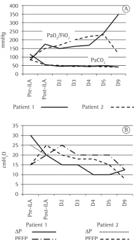
was 20 mmHg, and we attempted to balance it with the preset PEEP. After 24 h, the PaO2/ FiO2 ratio dropped below 90. The inspiratory pressure was then set to achieve a lung opening pressure of 30 cmH2O and a VT of 5 mL/kg. There was hypercapnia and respiratory acidosis (PaCO2 = 115 mmHg and pH = 7.15). Lung
recruitment maneuvers, with a delta pressure of 15 cmH2O and a PEEP of 35 cmH2O for 2 min, were unsuccessful. Because the patient was pregnant, she could not be placed in the prone position. We chose to use iLA without systemic anticoagulation, because the patient had a partial thromboplastin time of 2.3 times the normal levels, as well as thrombocytopenia (57,000 cells/mm3). After 20 min on iLA, with
O2 supplementation at a rate of 9 L/min, the ventilatory parameters were as follows: PEEP, 20 cmH2O; VT, 4 mL/kg; lung opening pressure, 20 cmH2O; and inspiratory/expiratory ratio, 1:1. As a result, the PaO2/FiO2 ratio increased by 100%, whereas CO2 decreased by 50%. There was no need for vasoactive amines, and the flow within the iLA circuit was approximately 1.4 L/min. After 14 h on iLA, the patient was started on airway pressure release ventilation and pressure support ventilation, the lung opening pressure being maintained at 15 cmH2O. After 48 h on iLA, the patient had a spontaneous delivery and, consequently, the IAP decreased
Report of two cases of ARDS patients treated with pumpless extracorporeal interventional lung assist
J Bras Pneumol. 2012;38(3):408-411
409
days, with no improvement. Associated diseases included arterial hypertension and coronary artery disease. The patient was admitted to the ICU with acute hypoxemic respiratory failure caused by ARDS (PaO2 = 51 mmHg and SaO2 = 89%) and required orotracheal intubation. The antibiotic regimen was broadened to piperacillin-tazobactam, azithromycin, and sulfamethoxazole-trimethoprim because there was suspicion of infection with resistant opportunistic pathogens. The patient showed progressive worsening, with a PaO2/ FiO2 ratio of 95 and a PaCO2 of 85 mmHg, at a plateau pressure of 35 cmH2O and a lung
opening pressure of 20 cmH2O. He was started on hemodialysis to control fluids and the severe acidosis (pH = 7.06). His IAP remained below 12 mmHg, and there was no need to use vasopressors. Alveolar recruitment maneuvers, with a delta pressure of 15 cmH2O and a PEEP
of 35 cmH2O for 2 min, were unsuccessful. On post-admission day 3, the patient underwent lung biopsy. Subsequently, he was started on iLA and received systemic anticoagulation. After 24 h, his PaCO2 had decreased to 50 mmHg, with a VT of 5 mL/kg, a plateau pressure of 30 cmH2O, and a lung opening pressure of 15 cmH2O. On
post-admission day 5, iLA was removed because there was improvement (a PaCO2 of 47 mmHg and a PaO2/FiO2 ratio of 225). A diagnosis of
cryptogenic organizing pneumonia was established on post-biopsy day 6. The patient was treated with methylprednisolone for 3 days. The patient improved. He was discharged after 134 days, and, at this writing, he remained functionally independent at home.
We described two cases of patients with ARDS and severe hypercapnia in whom a protective ventilation strategy was impossible. In both cases, because iLA is a novel therapy, the decision to use it was made jointly by the ICU directors, the hospital directors, the team of attending physicians, and the next of kin or guardian of the patient. There was sufficient hemodynamic stability to ensure a pressure gradient between the arterial and venous vasculature.(2) In addition
to improved gas exchange and CO2 removal, the
radiological progression was satisfactory. Regarding gas exchange, the major benefit of iLA is CO2 removal, with little effect on oxygenation.(3) There
have been reports of iLA use in certain conditions, including chest trauma,(4) brain injury,(5) influenza
A (H1N1) infection,(6) obstructive lung disease,(7)
to 7 mmHg. There was a 56% improvement in static compliance and a 64% improvement in the PaO2/FiO2 ratio. On day 8 of iLA, the patient was started on continuous positive airway pressure and pressure support ventilation, PaCO2 being maintained below 40 mmHg and the iLA device being removed (Figure 1). Tracheostomy was required, and ventilator weaning occurred on day 36 of her ICU stay. On post-admission day 53, the patient died from complications of the hematologic disease.
In the second case, a 74-year-old male patient presented with a 6-day history of dry cough and progressive dyspnea on exertion. He was treated as an outpatient with a fluoroquinolone for 3
410 Coscia AP, Cunha HFR, Longo AG, Martins EGS, Saddy F, Japiassú AM
J Bras Pneumol. 2012;38(3):408-411
Felipe Saddy Physician, ICU for Mechanically
Ventilated Patients,
Copa D’Or Hospital, Rio de Janeiro, Brazil
Andre Miguel Japiassu Physician,
Oswaldo Cruz Foundation Evandro Chagas Institute of Clinical Research and the D’Or Institute of Research and
Education, Rio de Janeiro, Brazil
References
1. Amato MB, Barbas CS, Medeiros DM, Magaldi RB, Schettino GP, Lorenzi-Filho G, et al. Effect of a protective-ventilation strategy on mortality in the acute respiratory distress syndrome. N Engl J Med. 1998;338(6):347-54. PMid:9449727. http://dx.doi.org/10.1056/ NEJM199802053380602
2. Brunston RL Jr, Tao W, Bidani A, Alpard SK, Traber DL, Zwischenberger JB. Prolonged hemodynamic stability during arteriovenous carbon dioxide removal for severe respiratory failure. J Thorac Cardiovasc Surg. 1997;114(6):1107-14. http://dx.doi.org/10.1016/S0022-5223(97)70026-6 3. Brederlau J, Muellenbach R, Kredel M, Schwemmer U,
Anetseder M, Greim C, et al. The contribution of arterio-venous extracorporeal lung assist to gas exchange in a porcine model of lavage-induced acute lung injury. Perfusion. 2006;21(5):277-84. PMid:17201082. http:// dx.doi.org/10.1177/0267659106074769
4. Brederlau J, Anetseder M, Wagner R, Roesner T, Philipp A, Greim C, et al. Pumpless extracorporeal lung assist in severe blunt chest trauma. J Cardiothorac Vasc Anesth. 2004;18(6):777-9. PMid:15650994. http:// dx.doi.org/10.1053/j.jvca.2004.08.022
5. Bein T, Scherer MN, Philipp A, Weber F, Woertgen C. Pumpless extracorporeal lung assist (pECLA) in patients with acute respiratory distress syndrome and severe brain injury. J Trauma. 2005;58(6):1294-7. http://dx.doi. org/10.1097/01.TA.0000173275.06947.5C
6. Freed DH, Henzler D, White CW, Fowler R, Zarychanski R, Hutchison J, et al. Extracorporeal lung support for lung transplantation,(8) and major thoracic surgery.
(9) In an attempt to treat acidosis, iLA is often
used in combination with renal replacement therapies.(10) Absolute contraindications to iLA
use include heparin-induced thrombocytopenia, severe septic shock, severe cardiogenic shock, and a body weight of less than 20 kg. To our knowledge, this is the first report of iLA use in a pregnant woman, and the two cases reported here are the first two cases in Brazil in which iLA was used. In the treatment of patients with severe refractory hypercapnia, iLA can be a useful tool (Table 1).
Alexandre Peixoto Coscia Physician, Intensive Care Unit, Quinta D’Or Hospital, Rio de Janeiro, Brazil Haroldo Falcão da Cunha Ramos
Physician, Intensive Care Unit, Quinta D’Or Hospital,
Rio de Janeiro, Brazil Alessandra Gouvea Longo
Physician, Intensive Care Unit, Quinta D’Or Hospital, Rio de Janeiro, Brazil Enio Gustavo Schoeder Martins
Physician, Intensive Care Unit, Quinta D’Or Hospital, Rio de Janeiro, Brazil
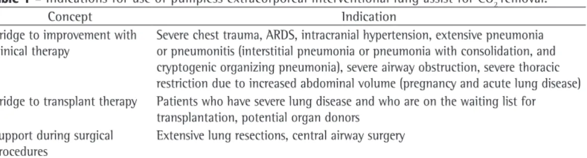
Table 1 - Indications for use of pumpless extracorporeal interventional lung assist for CO2 removal.
Concept Indication
Bridge to improvement with clinical therapy
Severe chest trauma, ARDS, intracranial hypertension, extensive pneumonia or pneumonitis (interstitial pneumonia or pneumonia with consolidation, and cryptogenic organizing pneumonia), severe airway obstruction, severe thoracic restriction due to increased abdominal volume (pregnancy and acute lung disease) Bridge to transplant therapy Patients who have severe lung disease and who are on the waiting list for
transplantation, potential organ donors Support during surgical
procedures
Report of two cases of ARDS patients treated with pumpless extracorporeal interventional lung assist
J Bras Pneumol. 2012;38(3):408-411
411
ASAIO J. 2008;54(1):3-10. PMid:18204308. http:// dx.doi.org/10.1097/MAT.0b013e318161d6ec
9. Wiebe K, Poeling J, Arlt M, Philipp A, Camboni D, Hofmann S, et al. Thoracic surgical procedures supported by a pumpless interventional lung assist. Ann Thorac Surg. 2010;89(6):1782-7; discussion 1788.
10. Kielstein JT, Tolk S, Hafer C, Heiden A, Wiesner O, Kühn C, et al. Effect of acute kidney injury requiring extended dialysis on 28 day and 1 year survival of patients undergoing interventional lung assist membrane ventilator treatment. BMC Nephrol. 2011;12:15. PMid:21489261 PMCid:3096900. http://dx.doi. org/10.1186/1471-2369-12-15
patients who had severe respiratory failure secondary to influenza A (H1N1) 2009 infection in Canada. Can J Anaesth. 2010;57(3):240-7. PMid:20082167. http:// dx.doi.org/10.1007/s12630-009-9253-0
7. Elliot SC, Paramasivam K, Oram J, Bodenham AR, Howell SJ, Mallick A. Pumpless extracorporeal carbon dioxide removal for life-threatening asthma. Crit Care Med. 2007;35(3):945-8. PMid:17255862. http://dx.doi. org/10.1097/01.CCM.0000257462.04514.15
8. Fischer S, Hoeper MM, Bein T, Simon AR, Gottlieb J, Wisser W, et al. Interventional lung assist: a new concept of protective ventilation in bridge to lung transplantation.