Immunophenotyping and extracellular matrix remodeling
in pulmonary and extrapulmonary sarcoidosis*
,**
Imunofenotipagem e remodelamento da matriz extracelular na sarcoidose pulmonar e extrapulmonar
Pedro Henrique Ramos Quintino da Silva, Edwin Roger Parra,
William Sanches Zocolaro, Ivy Narde, Fabíola Rodrigues, Ronaldo Adib Kairalla, Carlos Roberto Ribeiro Carvalho, Vera Luiza Capelozzi
Abstract
Objective: To investigate the significance of cellular immune markers, as well as that of collagen and elastic components of the extracellular matrix, within granulomatous structures in biopsies of patients with pulmonary or extrapulmonary sarcoidosis. Methods: We carried out qualitative and quantitative evaluations of inflammatory cells, collagen fibers, and elastic fibers in granulomatous structures in surgical biopsies of 40 patients with pulmonary and extrapulmonary sarcoidosis using histomorphometry, immunohistochemistry, picrosirius red staining, and Weigert’s resorcin-fuchsin staining. Results: The extrapulmonary tissue biopsies presented significantly higher densities of lymphocytes, macrophages, and neutrophils than did the lung tissue biopsies. Pulmonary granulomas showed a significantly higher number of collagen fibers and a lower density of elastic fibers than did extrapulmonary granulomas. The amount of macrophages in the lung samples correlated with FVC (p < 0.05), whereas the amount of CD3+, CD4+, and CD8+ lymphocytes correlated with the FEV1/FVC ratio
and VC. There were inverse correlations between TLC and the CD1a+ cell count (p < 0.05), as well as between DLCO and collagen/elastic fiber density (r = −0.90; p = 0.04). Conclusions: Immunophenotyping and remodeling both showed differences between pulmonary and extrapulmonary sarcoidosis in terms of the characteristics of the biopsy samples. These differences correlated with the clinical and spirometric data obtained for the patients, suggesting that two different pathways are involved in the mechanism of antigen clearance, which was more effective in the lungs and lymph nodes.
Keywords: Sarcoidosis; Granulomatous disease, chronic; Extracellular matrix; Immunophenotyping; Respiratory function tests.
Resumo
Objetivo: Investigar o significado de marcadores de imunidade celular e de componentes elásticos/colágeno da matriz extracelular em estruturas granulomatosas em biópsias de pacientes com sarcoidose pulmonar ou extrapulmonar. Métodos: Determinações qualitativas e quantitativas de células inflamatórias, de fibras de colágeno e de fibras elásticas em estruturas granulomatosas em biópsias cirúrgicas de 40 pacientes com sarcoidose pulmonar e extrapulmonar foram realizadas por histomorfometria, imuno-histoquímica, e técnicas de coloração com picrosirius e resorcina-fucsina de Weigert. Resultados: A densidade de linfócitos, macrófagos e neutrófilos nas biópsias extrapulmonares foi significativamente maior do que nas biópsias pulmonares. Os granulomas pulmonares apresentaram uma quantidade significativamente maior de fibras de colágeno e menor densidade de fibras elásticas que os granulomas extrapulmonares. A quantidade de macrófagos nos granulomas pulmonares correlacionou-se com CVF (p < 0,05), ao passo que as quantidades de linfócitos CD3+, CD4+ e CD8+ correlacionaram-se com a relação VEF1/CVF e com CV. Houve correlações negativas entre CPT e contagem de células CD1a+ (p < 0,05) e entre DLCO e densidade de fibras elásticas/colágenas (r = −0,90; p = 0,04). Conclusões: A imunofenotipagem e o remodelamento apresentaram características diferentes nas biópsias dos pacientes com sarcoidose pulmonar e extrapulmonar. Essas diferenças correlacionaram-se com os dados clínicos e espirométricos dos pacientes, sugerindo que há duas vias envolvidas no mecanismo de depuração de antígenos, que foi mais eficaz nos pulmões e linfonodos.
Descritores: Sarcoidose; Doença granulomatosa crônica; Matriz extracelular; Imunofenotipagem; Testes de função respiratória.
* Study carried out in the Department of Pathology and in the Department of Respiratory Diseases, Instituto do Coração – InCor, Heart Institute – University of São Paulo School of Medicine Hospital das Clínicas, São Paulo, Brazil.
Correspondence to: Vera Luiza Capelozzi or Edwin Roger Parra. Faculdade de Medicina da Universidade de São Paulo, Avenida Dr. Arnaldo, 455, CEP 01246-903, São Paulo, SP, Brasil.
Tel. 55 11 3061-7427. Fax: 55 11 3064-2744. E-mail: [email protected]
Financial support: This study received financial support from the Brazilian Conselho Nacional de Desenvolvimento Científico e Tecnológico (CNPq, National Council for Scientific and Technological Development) and the Fundação de Amparo à Pesquisa do Estado de São Paulo (FAPESP, São Paulo Research Foundation; Grant no. 2007/56617-5).
Submitted: 19 December 2011. Accepted, after review: 28 February 2012.
took deep breaths in the supine position; for the last 10 cm of the caudal part of the lungs, the scans were taken at 2-3 cm intervals, with the patients in the prone position. Two thoracic radiologists prospectively and independently examined all of the lobes on the HRCT scans for micronodules, opacity, and lymphadenopathy (Table 1).
For all of the patients with pulmonary sarcoidosis, we collected data on the history of smoking and pulmonary function parameters
(FVC, FEV1, FEV1/FVC ratio, RV, TLC, and
single-breath DLCO). We also collected the scores on a six-point dyspnea scale routinely applied to patients treated at our institution, the scores ranging from 0 (no dyspnea) to 6 (very severe dyspnea).(16)
Staining of collagen fibers was performed with a 0.2% solution of Sirius red (Direct Red 80, CI 35780; Aldrich, Milwaukee, WI, USA) dissolved in aqueous saturated picric acid. This dye has been widely used for staining collagen in histological specimens and allows the quantitative analysis of collagen in paraffin sections.(11,17) The enhancement
of collagen birefringence by picrosirius red staining and polarized light microscopy is specific for all collagen structures consisting of aggregates of oriented molecules. Elastic fiber staining was performed with the Weigert’s resorcin-fuchsin
method, after oxidation.(18) This method allows
selective identification of the three types of elastic fibers (oxytalan, elaunin, and fully developed elastic fibers).
The primary antibodies used in the present study were mouse monoclonal antibodies (Dako, Glostrup, Denmark; for all):
• mouse anti-human CD68, clone KP1 (M0814;
dilution, 1:3,200), a marker for macrophages/ histiocytes
• mouse anti-human neutrophil elastase,
clone NP57 (M0752; dilution, 1:800)
• anti-human CD3 (dilution, 1:600), CD4
(dilution, 1:400), CD8 (dilution, 1:100), CD20 (dilution, 1:600), CD1a (dilution, 1:20), and S100 (dilution, 1:600), all of which are markers for T and dendritic cells For the tests, the streptavidin-biotin complex
method was used.(14,19)
Immune cells were quantified by a conventional stereological method, i.e., the point sampled
intercept method.(20) At a magnification of ×400,
8-10 granulomatous structures were evaluated by
Introduction
Sarcoidosis is a systemic granulomatous disease of unknown etiology. The diagnosis is based on clinical, radiographic, and pathological findings. Most patients generally have favorable clinical
courses.(1) Although pulmonary and extrapulmonary
parenchymal involvement improves spontaneously in most patients, progression to fibrosis, leading to permanent functional impairment, occurs in
20-25%, and death occurs in 5-10%.(2,3) In this
context, a large number of clinical, physiological, and radiographic parameters have been investigated
in order to evaluate the outcome of sarcoidosis.(4-8)
The disease activity is marked by the process of extracellular matrix (ECM) remodeling— which involves the balance between immune
cells and tissue remodeling(9,10)—the impact of
histopathological staging on the prognosis of sarcoidosis having never been investigated.
Because cellular immune phenomena and ECM remodeling have been shown to be promising prognostic markers in diffuse interstitial lung
disease,(11-14) we hypothesized that the two have a
similar impact on the prognosis of sarcoidosis. We reviewed medical records and pathology reports over a period of 10 years in order to investigate the significance of cellular immune markers, as well as that of collagen and elastic components of the ECM, within the granulomatous structures found in biopsies of patients with pulmonary
or extrapulmonary sarcoidosis.(15)
Methods
The study group comprised 40 patients who had been diagnosed with sarcoidosis on the
basis of clinical and histological criteria.(1) The
tissue samples examined included pulmonary parenchymal and bronchial biopsy specimens (in 25 patients), mediastinal or peripheral lymph nodes (in 8 patients), liver (in 4 patients), and skin (in 3 patients).
systematic point counting, an eyepiece micrometer and a sampling grid with 100 points and 50 lines having been used in order to count the number of points overlying positively stained cells.
Collagen and elastic fibers were quantified by image analysis. The image analysis system consisted of an Olympus camera coupled to an Olympus microscope (Olympus Optical, Tokyo, Japan), which transmitted the images to a computer equipped with a Pentium 1,330-MHz processor (Intel Corporation, Santa Clara, CA, USA) and a monitor (LG Electronics Brasil, Manaus, Brazil) by means of a digitizing system (Oculus TCX; Coreco Inc., St. Laurent, Quebec, Canada). The images were processed using the program Image Pro-Plus, version 6.0 (Media Cybernetics Inc., Bethesda, MD, USA). For each tissue specimen, a range of 8-10 granulomatous structures were analyzed at ×400 magnification. The density of collagen and elastic fibers was measured and expressed as a ratio between the quantity of fibers and the total area studied. The final results express the area occupied by the various collagen
and elastic fibers in relation to the total area.(21)
One-way ANOVA was used in order to analyze the variation in the mean number of immune cells, collagen fibers, and elastic fibers, as well as the distribution of immune cells, collagen fibers, and elastic fibers within granulomas in the various organ tissues. The means were compared, a priori, by Levene’s test for homogeneity of variance, followed by post hoc analysis with the Bonferroni test for multiple comparisons (in case of homogeneity of variance) or Dunnett’s T3 test (in case of heterogeneity of variance). In addition, a paired t-test and the general linear model were used in order to test the relationships among continuous variables, and the residuals were examined to ensure that they had an approximately normal distribution. All of the analyses were carried out with the Statistical Package for the Social Sciences, version 18.0 (SPSS Inc., Chicago, IL, USA). The threshold for statistical significance was set at p < 0.05.
Results
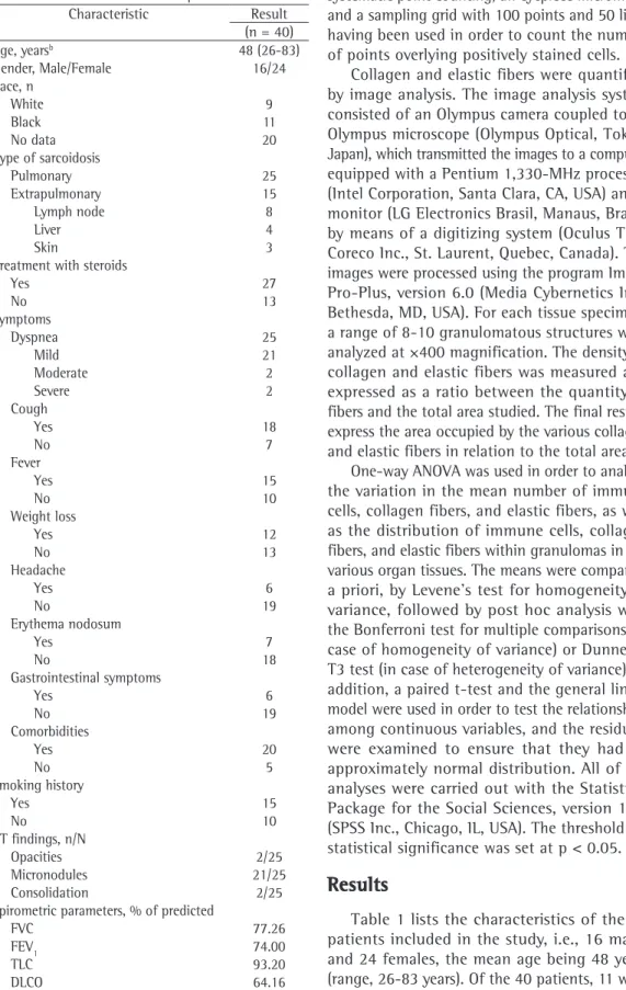
Table 1 lists the characteristics of the 40 patients included in the study, i.e., 16 males and 24 females, the mean age being 48 years (range, 26-83 years). Of the 40 patients, 11 were Black. Most of the 25 patients with pulmonary sarcoidosis showed a restrictive pattern or an
Table 1 - Characteristics of the patients.a
Characteristic Result
(n = 40)
Age, yearsb 48 (26-83)
Gender, Male/Female 16/24
Race, n
White 9
Black 11
No data 20
Type of sarcoidosis
Pulmonary 25
Extrapulmonary 15
Lymph node 8
Liver 4
Skin 3
Treatment with steroids
Yes 27
No 13
Symptoms
Dyspnea 25
Mild 21
Moderate 2
Severe 2
Cough
Yes 18
No 7
Fever
Yes 15
No 10
Weight loss
Yes 12
No 13
Headache
Yes 6
No 19
Erythema nodosum
Yes 7
No 18
Gastrointestinal symptoms
Yes 6
No 19
Comorbidities
Yes 20
No 5
Smoking history
Yes 15
No 10
CT findings, n/N
Opacities 2/25
Micronodules 21/25
Consolidation 2/25
Spirometric parameters, % of predicted
FVC 77.26
FEV1 74.00
TLC 93.20
DLCO 64.16
lymphocytes, in all of the patients. Staining for fungi and AFB gave negative results. In all of the patients with extrapulmonary sarcoidosis, the granulomas were characterized by severe architectural remodeling and variable amounts of fibrous tissue. In particular, the cortical and obstructive pattern, and 15 were current or
former smokers. Twenty-seven patients received corticosteroid therapy.
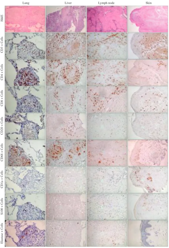
Histological examination revealed noncaseating granulomas (Figure 1), composed of multinucleated giant cells, epithelioid cells, macrophages, and
Table 2 - Summary of morphometric results.a
Parameter Lung Lymph node Liver Skin Extrapulmonary (total)
CD4 7.48 16.89 10.08 8.18 11.71
CD8 5.03 14.92 12.10 4.57 10.53
CD3 7.73 20.36 11.81 12.37 14.84
CD20 2.75 11.38 1.51 2.72 5.20
CD1a 0.36 0.28 0.10 1.95 0.77
S100 1.10 1.83 1.71 1.68 1.74
CD68 9.64 19.22 12.08 11.27 14.19
Elastase 0.40 1.30 2.71 1.35 1.78
Collagen 25.62 15.08 6.30 5.15 8.84
Elastic fiber 3.75 48.39 49.82 29.79 42.66
aThe units of “% of points” indicate the number of points overlying the phenomenon of interest divided by the total
number of points overlying the tumor. In morphometry, this is called a point fraction and is often symbolized as Pp, which has been shown to approximate the volume fraction.(21)
medullary areas of the lymph nodes were mostly replaced by epithelioid and sclerosing granulomas that were peripherally surrounded by lymphocytes.
Figure 1 shows granulomas stained with picrosirius and viewed under polarized light. The granulomas in the samples obtained from the patients with extrapulmonary sarcoidosis showed a homogeneous orange-red birefringence, coincident with minor distortions of the ECM architecture. In contrast, the granulomas in the lung samples obtained from the patients with pulmonary sarcoidosis showed distortions of the ECM architecture, as well as a strong and heterogeneous birefringence. Large, sparse, and fragmented bundles of elastic fibers were more commonly observed in the granulomas in the extrapulmonary samples than in those in the lung samples. This correlates with the epithelioid and sclerosing areas seen in H&E preparations.
Table 2 shows the measurements of collagen and elastic fibers in the pulmonary and extrapulmonary granulomas. The morphological changes in the collagen and elastic fibers coincided with the variations in quantity and density in the two fiber systems. Pulmonary granulomas showed a significantly higher number of collagen fibers and a lower density of elastic fibers than did extrapulmonary granulomas (p = 0.01 and p = 0.003, respectively), pulmonary sarcoidosis being therefore characterized by fibroelastosis.
Figure 2 shows the immune cells in the pulmonary and extrapulmonary granulomas stained by immunohistochemistry; the most common cells around the granulomas found in the patients with extrapulmonary sarcoidosis were as follows: CD3+, CD4+, and CD8+ T-lineage cells; CD20+
lymphocytes; elastase-positive neutrophils; and CD68+ macrophages.
term pulmonary function and will prevent the cumulative morbidity associated with the currently available therapy. The question of interest is whether further information can help us define the immune response and tissue remodeling in sarcoidosis on the basis of the histological characteristics and patterns of the granulomas, which will show a better correlation with pulmonary function tests and the natural history of the disease, as well as with the response to therapy. The pathological process in the granulomatous involvement in sarcoidosis undoubtedly includes complex serial and sequential steps; among these, severe immune dysfunction and tissue remodeling are thought to be important, given that both lead to scarring.(10)
The present study showed a higher degree of immunoreactivity to CD4, CD8, CD3, CD68, and neutrophils in extrapulmonary sarcoidosis than in pulmonary sarcoidosis. In addition, the number of CD4, CD8, CD3, and CD20 lymphocytes was significantly higher in the lymph node granulomas, whereas CD1a dendritic cells and neutrophils were more prominent in the skin and lung granulomas, respectively. It is known that CD4+ T lymphocytes play an essential role in creating the microenvironment for B lymphocyte activation and differentiation following antigen exposure. In the lungs, the fact that follicular B lymphocyte aggregates are located almost exclusively in peribronchiolar areas beneath the epithelium of the bronchial branches suggests that they represent the development of bronchus-associated lymphoid tissue. These structures have been shown to be capable of mounting
competent adaptive immune responses.(24) Unlike
typical lymph nodes, they are not dependent on afferent lymphatics for antigen retrieval; rather, they sample antigen directly from the lung lumen. This raises the question of whether an external factor could promote the formation of lymphoid follicles in sarcoidosis, thereby inducing or modulating the disease process. This question is especially interesting in light of the reported association between smoking and increased formation of peribronchiolar B
cell follicles in some pulmonary diseases.(25)
Although CD4+ and CD8+ T lymphocytes are usually present in very low numbers in peripheral blood, increased numbers have been reported in apparently healthy individuals, as well as in various clinical conditions. In a recent study, The quantity of S100+ cells was very similar in
the granulomas in the various organs studied. Positive associations were found between CD4+ and CD8+ cells (r = 0.55; p = 0.001), between CD8+ cells and elastase-positive neutrophils (r = 0.34; p = 0.03), between CD8+ cells and elastic fibers (r = 0.41; p = 0.01), between CD3+ cells and elastic fibers (r = 0.46; p = 0.004), between CD1a+ cells and collagen fibers (r = 0.35; p = 0.05), and between elastase-positive neutrophils and elastic fibers (r = 0.35; p = 0.03). Negative associations were found between CD8+ cells and collagen
fibers (r = −0.46; p = 0.04), as well as between CD1a+ cells and elastic fibers (r = −0.34; p = 0.05).
The cellular immune components and reparative components correlated with clinical, radiological, and pulmonary function findings in the patients with pulmonary sarcoidosis. We found that CD4+ lymphocytes were negatively associated with
smoking history (r = −0.73; p = 0.03), radiological opacities (r = −0.59; p = 0.04), and micronodules (r = −0.54; p = 0.04). Mediastinal lymph node
granulomas were positively associated with CD8+ lymphocytes (r = 0.70; p = 0.004). An FVC of 70% of the predicted value was positively associated
with CD68+ macrophages (p < 0.05). The FEV1/
FVC ratio was positively associated with CD4+, CD8+, and CD3+ lymphocytes. A TLC of 82% of the predicted value was negatively associated with CD1a+ cells (p < 0.05). Finally, there was a negative association between DLCO and collagen/
elastic fibers (r = −0.90; p = 0.04).
Discussion
The pathological hallmark of sarcoidosis is the presence of noncaseating granulomas in various organs, including the lungs, lymph nodes, liver,
skin, heart, and brain.(22) In this chronic systemic
disease, these histologically compact inflammatory lesions containing T lymphocytes and mononuclear phagocytes can appear and disappear insidiously in some patients, whereas, in others, the lesions can cause significant organ dysfunction due to persistent inflammation and obliteration of vital structures, resulting in extensive tissue fibrosis or
cavitation.(10) The most common and devastating
effects occur in the pulmonary system.(23) Therefore,
long-Moller et al. reported finding dominant Th1 cytokine expression, with elevated mRNA and
protein levels of IL-12 and IFN-γ, but not IL-4,
in sarcoid lung cells when compared with those
in normal samples,(26) supporting the notion of an
exaggerated immunological/inflammatory response.
(27) Consistent with these findings, various other
immunological abnormalities have been identified, including tissue accumulation of CD4+ cells with helper-inducer activity, increased in situ production of cell-derived cytokines, B cell hyperactivity with spontaneous production of immunoglobulins, and accumulation of activated monocytic cells. These cells are known to play key roles in active tissue remodeling by releasing soluble mediators, such as cytokines and chemokines with proinflammatory and fibrogenic activity, reactive oxygen species, and proteolytic enzymes that have the capacity
to degrade the connective tissue scaffold.(10)
In our study, the granulomas in the samples obtained from patients with extrapulmonary sarcoidosis (lymph node, liver, and skin involvement) were found to be the prototype of the apposition of elastic fibers in the tissues. In contrast, the granulomas in the lung samples from patients with pulmonary sarcoidosis were found to represent the prototype of an increased number of collagen fibers and a lower degree of elastic apposition, characterizing a fibroelastotic process. The elastic system plays an important role in maintaining organ patency and the elastic recoil; therefore, elastosis might be important
for the understanding of organ function.(11,12,28)
Similar findings were reported by our group when studying the ECM in lung biopsy specimens from patients with idiopathic interstitial pneumonia.
(13,14) We hypothesized that elastosis is related to
the inflammatory elastolysis observed at the initial stage of inflammation, reinforcing the notion that a proinflammatory mechanism is present in granulomatous diseases. The proinflammatory hypothesis is defended by various authors as
a transient form of organizing fibrosis,(29) and
some of our findings in the present study seem to support this theory, given that the highest levels of neutrophils were observed in the patients with pulmonary sarcoidosis.
For all of these reasons, it would not be surprising to learn that cell immunostaining and the determination of collagen/elastic fiber density can provide relevant information on tissue remodeling in sarcoidosis. Our results confirm
the pathogenetic implications of immune cell infiltration and the remodeling state of granulomas in pulmonary and extrapulmonary sarcoidosis. Although only a few studies focusing on the
production of matrix proteins, TGF-β, fibrin, and
matrix metalloproteinases were able to demonstrate a significant association between tissue remodeling and immune response in sarcoidosis,(10,30) our results
suggest that a fibroelastotic process, together with immune cell infiltration, probably contributes to tissue changes in sarcoidosis. More interestingly, our results suggest that antigen clearance is less efficient in pulmonary sarcoidosis than in extrapulmonary sarcoidosis. More specifically, we found that different functional tests corroborated the remodeling and immune cell regulation in pulmonary sarcoidosis, which is suggestive of a reparative effect on the functional and structural disarray found in the lung parenchyma.
The major limitations of the present study are related to the difficulty in comparing our results with those of other studies, given that there are few studies in the literature showing associations of immunophenotyping and tissue remodeling with standard clinical parameters (age, gender, smoking history, and duration of symptoms prior to lung biopsy), physiological tests, and dyspnea scores in sarcoidosis patients.
In summary, we have demonstrated the pathophysiological features of tissue remodeling and immune response in sarcoidosis. Particularly in pulmonary sarcoidosis, there are two different immune response and tissue remodeling pathways, with an impact on physiological tests. The immune and fibroelastotic processes observed in the parenchyma of the affected organs appear to be part of the general processes of inflammation and collagen/elastic fiber deposition, which have an independent course in sarcoidosis. Our findings suggest that there are two mechanisms of injury and repair in sarcoidosis, direct airway injury being more likely to occur in pulmonary sarcoidosis. Regardless of the pathogenetic determinants, the pathway involvement that is likely to progress to uncontrolled fibrosis and therefore shorten the life of patients with sarcoidosis should be identified so that the treatment is effective.
References
16. Evaluation of impairment/disability secondary to respiratory disorders. American Thoracic Society. Am Rev Respir Dis.
1986;133(6):1205-9. PMid:3509148.
17. Montes GS. Structural biology of the fibres of the collagenous and elastic systems. Cell Biol Int.
1996;20(1):15-27. PMid:8936403. http://dx.doi. org/10.1006/cbir.1996.0004
18. Lemos M, Pozo RM, Montes GS, Saldiva PH. Organization of collagen and elastic fibers studied in stretch preparations of whole mounts of human visceral pleura. Ann Anat.
1997;179(5):447-52. http://dx.doi.org/10.1016/
S0940-9602(97)80048-9
19. Parra ER, Kairalla RA, Ribeiro de Carvalho CR, Eher E, Capelozzi VL. Inflammatory cell phenotyping of the pulmonary interstitium in idiopathic interstitial pneumonia.
Respiration. 2007;74(2):159-69. PMid:17108669. http:// dx.doi.org/10.1159/000097133
20. Hsia CC, Hyde DM, Ochs M, Weibel ER; ATS/ERS Joint Task Force on Quantitative Assessment of Lung Structure. An official research policy statement of the American Thoracic Society/European Respiratory Society: standards for quantitative assessment of lung structure. Am J Respir
Crit Care Med. 2010;181(4):394-418. PMid:20130146. http://dx.doi.org/10.1164/rccm.200809-1522ST
21. Parra ER, Kairalla RA, de Carvalho CR, Capelozzi VL. Abnormal deposition of collagen/elastic vascular fibres and prognostic significance in idiopathic interstitial pneumonias. Thorax. 2007;62(5):428-37. PMid:17251318 PMCid:2117177. http://dx.doi.org/10.1136/ thx.2006.062687
22. Stirling RG, Cullinan P, Bois RMD. Sarcoidosis. In: Schwarz MI, King TE, editors. Interstitial lung disease. Hamilton:
B.C. Decker; 1997. p. 279-323.
23. Lynch JP 3rd, Kazerooni EA, Gay SE. Pulmonary sarcoidosis.
Clin Chest Med. 1997;18(4):755-85. http://dx.doi.
org/10.1016/S0272-5231(05)70417-2
24. Moyron-Quiroz JE, Rangel-Moreno J, Kusser K, Hartson L, Sprague F, Goodrich S, et al. Role of inducible bronchus associated lymphoid tissue (iBALT) in respiratory immunity.
Nat Med. 2004;10(9):927-34. PMid:15311275. http:// dx.doi.org/10.1038/nm1091
25. Wallace WA, Roberts SN, Caldwell H, Thornton E, Greening AP, Lamb D, et al. Circulating antibodies to lung protein(s) in patients with cryptogenic fibrosing
alveolitis. Thorax. 1994;49(3):218-24. PMid:8202877 PMCid:1021149. http://dx.doi.org/10.1136/thx.49.3.218
26. Moller DR, Forman JD, Liu MC, Noble PW, Greenlee BM, Vyas P, et al. Enhanced expression of IL-12 associated with Th1 cytokine profiles in active pulmonary sarcoidosis.
J Immunol. 1996;156(12):4952-60. PMid:8648147.
27. Moller DR. Cells and cytokines involved in the pathogenesis of sarcoidosis. Sarcoidosis Vasc Diffuse Lung Dis.
1999;16(1):24-31. PMid:10207939.
28. Rosenbloom J, Abrams WR, Mecham R. Extracellular
matrix 4: the elastic fiber. FASEB J.
1993;7(13):1208-18. PMid:8405806.
29. Fukuda Y, Mochimaru H, Terasaki Y, Kawamoto M, Kudoh S. Mechanism of structural remodeling in pulmonary fibrosis.
Chest. 2001 Jul;120(1 Suppl):41S-43S. PMid:11451909.
http://dx.doi.org/10.1378/chest.120.1_suppl.S41 30. Roman J. Extracellular matrices in the pathogenesis
of injury and repair. In: Schwarz MI, King TE, editors.
Interstitial lung disease. Hamilton: B.C. Decker; 1997.
p. 207-27. the ATS Board of Directors and by the ERS Executive
Committee, February 1999. Am J Respir Crit Care Med. 1999;160(2):736-55. PMid:10430755.
2. Miller BH, Rosado-de-Christenson ML, McAdams HP, Fishback NF. Thoracic sarcoidosis: radiologic-pathologic
correlation. Radiographics. 1995;15(2):421-37. Erratum in: Radiographics 1997;17(6):1610. PMid:7761646.
3. Holmes J, Lazarus A. Sarcoidosis: extrathoracic
manifestations. Dis Mon. 2009;55(11):675-92.
PMid:19857642. http://dx.doi.org/10.1016/j.
disamonth.2009.05.002
4. Mañá J, Salazar A, Manresa F. Clinical factors predicting persistence of activity in sarcoidosis: a multivariate
analysis of 193 cases. Respiration. 1994;61(4):219-25. http://dx.doi.org/10.1159/000196341
5. Sato H, Grutters JC, Pantelidis P, Mizzon AN, Ahmad T, Van Houte AJ, et al. HLA-DQB1*0201: a marker for good prognosis in British and Dutch patients with sarcoidosis. Am J Respir Cell Mol Biol. 2002;27(4):406-12. PMid:12356573.
6. Suzuki K, Tamura N, Iwase A, Dambara T, Kira S. Prognostic value of Ia+ T lymphocytes in bronchoalveolar lavage fluid in pulmonary sarcoidosis. Am J Respir Crit Care
Med. 1996;154(3 Pt 1):707-12. PMid:8810609.
7. Israel HL, Karlin P, Menduke H, DeLisser OG. Factors affecting outcome of sarcoidosis. Influence of race, extrathoracic involvement, and initial radiologic lung
lesions. Ann N Y Acad Sci. 1986;465:609-18. PMid:3460398. http://dx.doi.org/10.1111/j.1749-6632.1986.tb18537.x
8. Sekiya M, Ohwada A, Miura K, Takahashi S, Fukuchi Y. Serum vascular endothelial growth factor as a possible prognostic indicator in sarcoidosis. Lung.
2003;181(5):259-65. PMid:14705769. http://dx.doi.
org/10.1007/s00408-003-1028-8
9. Gal AA, Koss MN. The pathology of sarcoidosis. Curr Opin Pulm Med. 2002;8(5):445-51. PMid:12172451.
http://dx.doi.org/10.1097/00063198-200209000-00018
10. Roman J, Galis ZS. Sarcoidosis: a mysterious tale of inflammation, tissue remodeling, and matrix metalloproteinases. Hum Pathol. 2002;33(12):1155-7. PMid:12514781. http://dx.doi.org/10.1053/
hupa.2002.130397
11. Negri EM, Montes GS, Saldiva PH, Capelozzi VL. Architectural remodelling in acute and chronic interstitial lung disease: fibrosis or fibroelastosis? Histopathology.
2000;37(5):393-401. PMid:11119120. http://dx.doi. org/10.1046/j.1365-2559.2000.00992.x
12. Rozin GF, Gomes MM, Parra ER, Kairalla RA, de Carvalho CR, Capelozzi VL. Collagen and elastic system in the remodelling process of major types of idiopathic interstitial pneumonias (IIP). Histopathology.
2005;46(4):413-21. PMid:15810953. http://dx.doi. org/10.1111/j.1365-2559.2005.02103.x
13. Ranzani OT, Parra ER, de Morais Fernezlian S, Capelozzi VL. Intraluminal plugs in idiopathic and secondary organizing pneumonia: repair or remodelling? Histopathology.
2007;51(5):622-30. PMid:17927583. http://dx.doi. org/10.1111/j.1365-2559.2007.02845.x
14. Parra ER, Noleto GS, Tinoco LJ, Capelozzi VL. Immunophenotyping and remodeling process in small airways of idiopathic interstitial pneumonias: functional and prognostic significance. Clin Respir
J. 2008;2(4):227-38. PMid:20298339. http://dx.doi. org/10.1111/j.1752-699X.2008.00077.x
About the authors
Pedro Henrique Ramos Quintino da Silva
Medical Student. University of São Paulo School of Medicine, São Paulo, Brazil.
Edwin Roger Parra
Postdoctoral Student. University of São Paulo School of Medicine, São Paulo, Brazil.
William Sanches Zocolaro
Medical Student. University of São Paulo School of Medicine, São Paulo, Brazil.
Ivy Narde
Medical Student. University of São Paulo School of Medicine, São Paulo, Brazil.
Fabíola Rodrigues
Attending Physician. Department of Respiratory Diseases, Instituto do Coração – InCor, Heart Institute – University of São Paulo School of Medicine Hospital das Clínicas, São Paulo, Brazil.
Ronaldo Adib Kairalla
Attending Physician. Department of Respiratory Diseases, Instituto do Coração – InCor, Heart Institute – University of São Paulo School of Medicine Hospital das Clínicas, São Paulo, Brazil.
Carlos Roberto Ribeiro Carvalho
Tenured Associate Professor of Pulmonology. University of São Paulo School of Medicine, São Paulo, Brazil.
Vera Luiza Capelozzi