J Bras Pneumol. 2012;38(3):404-407
To the Editor:
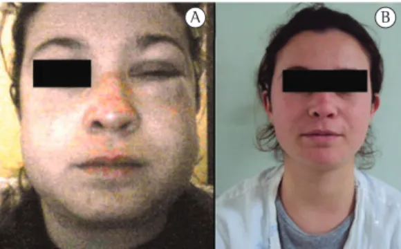
A previously healthy, 21-year-old female nonsmoker sought emergency room treatment complaining of recent and progressive swelling of the face, neck, and chest (Figure 1). The patient reported no pain, dyspnea, or dysphagia at admission. She reported a brief history of odynophagia in the previous week, when, during an episode of cough, she felt intense (dissecting) retrosternal pain, which subsided within a few minutes after its onset. After that episode, she had no symptoms other than the abovementioned swelling.
Physical examination revealed extensive subcutaneous emphysema, which extended from the xiphoid process to the temporal muscle and involved the entire neck and face. Pulmonary percussion and auscultation revealed no abnormalities. Cardiac auscultation revealed the presence of Hamman’s sign (crackles—as was the case in our patient—or bubbling sounds, synchronous with the heartbeat). Oral endoscopy revealed no abnormalities, and the remainder of the physical examination was normal.
A CT scan of the skull, neck, and chest confirmed the presence of pneumomediastinum, pneumothorax (Figure 2A), and extensive soft tissue emphysema in the cervical region (Figure 2B)— involving the face, periorbital tissues, and temporal muscles (Figure 2C)—as well as revealing the presence of pneumorrhachis (Figure 2D).
From postadmission day two onward, the subcutaneous emphysema gradually improved. The patient received conservative treatment, including rest, unrestricted diet, and analgesia, if necessary. The results of routine laboratory tests, including blood workup and urinalysis, were normal. Follow-up chest X-rays revealed a sustained reduction in the abovementioned signs and symptoms. The patient remained under observation for another five days, being asymptomatic and showing nearly complete
resolution of the clinical picture at discharge (Figure 1).
Spontaneous pneumomediastinum, also known as Hamman’s syndrome, is defined as the presence of free air in the mediastinum, being unrelated to trauma or procedures (surgical or otherwise).(1) It is an uncommon entity,(1-6) its
estimated prevalence ranging from 0.001% to 0.01%.(2) Because the course of the disease is
nearly always benign, many patients do not seek medical attention, which is why spontaneous mediastinum often goes undiagnosed. In addition, the presence of trauma, surgical procedures, or mechanical ventilation rules out spontaneous pneumomediastinum, therefore reducing the prevalence of the disease.
Physical exercise, labor, diabetic ketoacidosis, inhaled drug use, cough, and vomiting, among others, have been reported to trigger spontaneous pneumomediastinum.(3) The primary component
of the pathophysiology of Hamman’s syndrome is alveolar rupture, which results from high intra-alveolar pressure, low perivascular pressure, or both. After the initial event, air tracks into the mediastinum during the respiratory cycle, the pressure in the mediastinum decreasing in relation to that in the lung parenchyma.(4) This mechanism
is known as the Macklin effect or phenomenon, having been named after Charles C. Macklin, who described it in detail in 1939.(7)
In two thirds of the cases, as occurred in the case reported here, spontaneous pneumomediastinum can progress and affect the cervical region(5) and, less commonly, facial tissues.
Pneumorrhachis, however, is even rarer, only a few cases having been reported in the literature.
(6) It is believed that, in cases of pneumorrhachis,
air from the posterior mediastinum goes through the neural foramina and into the epidural space.
(6) If the passage of air into the mediastinum
and the previously described anatomical planes is not enough to decrease intra-alveolar pressure, pleural rupture with pneumothorax can occur,(2)
Spontaneous pneumomediastinum (Hamman’s syndrome)
Pneumomediastino espontâneo (síndrome de Hamman)
Giordano Rafael Tronco Alves, Régis Vinícius de Andrade Silva, José Roberto Missel Corrêa, Cassiano Minussi Colpo, Helen Minussi Cezimbra, Carlos Jesus Pereira Haygert
Spontaneous pneumomediastinum (Hamman’s syndrome)
J Bras Pneumol. 2012;38(3):404-407
405
as it did in the case reported here and as it does in 6-30% of patients.(1) Other sites that can
be affected include the pericardium and the peritoneal cavity,(4) which were not affected in
our patient.
Most patients with Hamman’s syndrome eventually present with symptoms, the most common symptoms being dyspnea, chest pain, and cough.(2) We believe that cough was the
triggering factor in our patient, who remained asymptomatic over the clinical course of the disease.
Hamman’s syndrome has been associated with interstitial lung disease, pulmonary emphysema,
Figure 1 - In A, marked swelling of the neck, face, and left eye at admission. In B, nearly complete resolution of the signs and symptoms (photograph taken a few hours before patient discharge).
406 Alves GRT, Silva RVA, Corrêa JRM, Colpo CM, Cezimbra HM, Haygert CJP
J Bras Pneumol. 2012;38(3):404-407
asthma, bronchiectasis, intrathoracic malignancies, and cystic or cavitated lesions, as well as with lung transplant recipients.(1,2) In the case reported
here, the clinical history and CT findings ruled out those diagnoses.
A chest X-ray is usually the first test performed in patients suspected of having pneumomediastinum (spontaneous or otherwise). For spontaneous pneumomediastinum, the sensitivity of the method has been shown to be satisfactory (i.e., 90%, approximately),(1) although
it is known that it depends on the extent of the disease. In the case reported here, chest X-rays were used for follow-up, the diagnosis having been established by CT, which is considered the gold standard for the diagnosis of Hamman’s syndrome.(3,5)
Although endoscopy,(3) bronchoscopy, and
esophagography are recognizably important,(1) some
authors recommend that they be performed only in the presence of dysphagia, vomiting, previous trauma, fever, leukocytosis, pleural effusion, pneumoperitoneum, and gastrointestinal disease,(4)
none of which were found in our patient. In addition, because cervical emphysema is commonly associated with odynophagia, the presence of the latter does not warrant invasive tests.(4)
Hamman’s syndrome treatment remains controversial. Most studies on the topic have limitations, recommending conservative treatment— including rest and analgesia, if necessary—and reporting that Hamman’s syndrome is a benign condition.(2) However, there is no consensus
regarding the management of the syndrome.(4)
Some centers have recommended that invasive tests and antimicrobial agents be used sparingly and dietary restrictions be avoided, given that all of these increase the mean length of hospital stay.(4) In addition, unfamiliarity with the entity
can lead to unnecessary diagnostic tests and inappropriate treatment.(2)
The complications of spontaneous pneumomediastinum vary according to the etiology or the triggering factor. In some cases, delayed diagnosis and failure to detect the primary cause of pneumomediastinum can
lead to esophageal rupture, mediastinitis, and hypertensive pneumothorax, among others.(1,4,5)
Recurrence is rare, long-term follow-up being therefore unnecessary.(2)
Giordano Rafael Tronco Alves Medical Student,
Federal University of Santa Maria School of Medicine, Santa Maria, Brazil
Régis Vinícius de Andrade Silva Resident,
Department of Radiology, Santa Maria University Hospital, Federal University of Santa Maria School of Medicine, Santa Maria, Brazil
José Roberto Missel Corrêa Resident,
Department of Radiology, Santa Maria University Hospital, Federal University of Santa Maria School of Medicine, Santa Maria, Brazil
Cassiano Minussi Colpo Resident,
Department of Surgery, Santa Maria University Hospital, Federal University of Santa Maria School of Medicine, Santa Maria, Brazil
Helen Minussi Cezimbra Resident,
Department of Infectious Diseases, Santa Maria University Hospital, Federal University of Santa Maria School of Medicine, Santa Maria, Brazil
Carlos Jesus Pereira Haygert Assistant Professor,
Department of Diagnostic Imaging, Federal University of Santa Maria,
Spontaneous pneumomediastinum (Hamman’s syndrome)
J Bras Pneumol. 2012;38(3):404-407
407
References
1. Iyer VN, Joshi AY, Ryu JH. Spontaneous pneumomediastinum: analysis of 62 consecutive adult patients. Mayo Clin Proc. 2009;84(5):417-21. PMid:19411438 PMCid:2676124.
2. Ho AS, Ahmed A, Huang JS, Menias CO, Bhalla S. Multidetector computed tomography of spontaneous versus secondary pneumomediastinum in 89 patients: can multidetector computed tomography be used to reliably distinguish between the 2 entities? J Thorac Imaging. 2012;27(2):85-92. PMid:21436744. http:// dx.doi.org/10.1097/RTI.0b013e3182103876
3. Perna V, Vilà E, Guelbenzu JJ, Amat I. Pneumomediastinum: is this really a benign entity? When it can be considered as spontaneous? Our experience in 47 adult patients. Eur J Cardiothorac Surg. 2010;37(3):573-5. PMid:19748792. http://dx.doi.org/10.1016/j.ejcts.2009.08.002
4. Al-Mufarrej F, Badar J, Gharagozloo F, Tempesta B, Strother E, Margolis M. Spontaneous pneumomediastinum: diagnostic and therapeutic interventions. J Cardiothorac Surg. 2008;3:59. PMid:18980688 PMCid:2596119. http:// dx.doi.org/10.1186/1749-8090-3-59
5. Conti-de-Freitas LC, Mano JB, Ricz HM, Mamede RC. A importância da suspeita clínica da síndrome de Hamman na sala de urgência. Rev Bras Cir Cabeça Pescoço. 2009;38(2):122-3.
6. Song Y, Tu L, Wu J. Pneumorrhachis with spontaneous pneumomediastinum and subcutaneous emphysema. Intern Med. 2009;48(18):1713-4. PMid:19755782. http:// dx.doi.org/10.2169/internalmedicine.48.2256 7. Macklin CC. Transport of air along sheaths of pulmonic
blood vessels from alveoli to mediastinum. Arch Intern Med. 1939;64(5):913-26. http://dx.doi.org/10.1001/ archinte.1939.00190050019003