Is there a difference between presence of single stone
and multiple stones in flexible ureterorenoscopy and laser
lithotripsy for renal stone burden <300mm
2?
_______________________________________________
Faruk Ozgor
1, Onur Kucuktopcu
1, Burak Ucpinar
1, Zafer Gokhan Gurbuz
1, Omer Sarilar
1, Ahmet
Yalcin Berberoglu
1, Murat Baykal
1, Murat Binbay
11 Department of Urology, Haseki Training and Research Hospital, Istanbul, Turkey
ABSTRACT
ARTICLE
INFO
______________________________________________________________ ______________________
In this study, we aim to evaluate and compare the effectiveness of flexible ureterore-noscopy (f-URS) for solitary and multiple renal stones with <300 mm2 stone burden.
Patients’ charts who treated with f-URS for kidney stone between January 2010 and June 2015 were reviewed, retrospectively. Patients with solitary kidney stones (n:111) were enrolled in group 1. We selected 111 patients with multiple kidney stones to serve as the control group and the patients were matched at a 1:1 ratio with respect to the patient’s age, gender, body mass index and stone burden. Additionally, patients with multiple stones were divided into two groups according to the presence or abscence of lower pole stones. Stone free status was accepted as complete stone clearence and presence of residual fragments < 2 mm. According to the study design; age, stone bur-den, body mass index were comparable between groups. The mean operation time was longer in group 2 (p= 0.229). However, the mean fluoroscopy screening time in group 1 and in group 2 was 2.1±1.7 and 2.6±1.5 min, respectively and significantly longer in patients with multiple renal stones (P=0.043). The stone-free status was significantly higher in patients with solitary renal stones after a single session procedure (p=0.02). After third month follow up, overall success rate was 92.7% in Group 1 and 86.4% in Group 2. Our study revealed that F-URS achieved better stone free status in solitary renal stones <300 mm2. However, outcomes of F-URS were acceptable in patients with
multiple stones.
Keywords:
Catheters; Kidney Calculi; Urolithiasis
Int Braz J Urol. 2016; 42: 1168-77
_____________________ Submitted for publication: November 29, 2015 _____________________ Accepted after revision: March 25, 2016
_____________________ Published as Ahead of Print: September 01, 2016
INTRODUCTION
Recently, Shock Wave Lithotripsy (SWL) is recommended as one of the first line treatment modalities for renal stones under 300mm2 stone
burden, according to European Urology Associa-tion Guidelines (1). Although, SWL has high suc-cess rates and higher patient compliance, effec-tiveness of SWL tends to decrease in lower pole stone(s), hard stone(s) such as calcium oxalate monohydrate stones and multiple stones (2). On
the other hand, percutaneous nephrolithotomy (PNL) has favourable results for renal stone(s) regardless of stone type, but procedure has po-tential serious complications including bleeding necessitating transfusion, septicaemia and colo-nic injury (3). Additionally, multiple stones may require multiple accesses which may increase the number of complications.
important treatment modality for renal stones(s). Modern flexible ureterorenoscopes may access all pelvicaliecal system including lower pole, and Holmium Laser provides effective stone fragmen-tation regardless of stone type. Additionally, use of ureteral access sheath facilitate the passage of stone fragments, which improves patients quality of life after the procedure (4). Many authors had achieved up to 90% stone free rates with accep-table complications in f-URS procedure in mode-rate sized stones (5, 6).
Although there are many studies showing the effectiveness of f-URS for solitary renal sto-nes, studies evaluating the effectiveness of f-URS in multiple renal stones is limited. In this study, we aimed to evaluate and compare the efficacy and sa-fety of f-URS for solitary and multiple renal stones with <300mm2 stone burden. To our knowledge,
this is the first study comparing the effectiveness of f-URS for solitary and multiple renal stones.
MATERIALS AND METHODS
In a single tertiary academic center, 413 patient’s charts who were treated with f-URS for kidney stone between January 2010 and June 2015 were reviewed, retrospectively. However, patients datas were recorded prospectively. Patients who had kidney stone burden <300mm2 were
enrol-led into the study. Stone burden was calculated according to European Association of Urology guideline’s formula (1). Patients with a solita-ry kidney stone and multiple kidney stones were divided as group 1 and group 2, respectively. At the end of our evaluation, 111 patients enrolled in group 1. We selected 111 patients with multiple kidney stones to serve as the control group and the patients were matched at a 1:1 ratio with respect to the patient’s age, gender, presence of solitary kidney and stone burden. Patients under 18 years of age and patients with renal abnormalities were excluded from the study. Additionally, to evaluate the effect of lower pole stone on f-URS outcomes, patients with multiple stones were divided into two groups according to the presence or absence of lo-wer pole stones.
In all patients, detailed medical history was obtained and physical examination was
per-formed. Preoperatively, renal stone and kidney cha-racteristics were assessed by intravenous urography (IVU) and/or computed tomography (CT). Patients demographic parameters including sex, age, body mass index (BMI), degree of hydronephrosis, sto-ne size, stosto-ne number and location were recorded. Hemoglobin measurements, serum creatinine level, platelet counts and coagulation screen tests were assessed preoperatively. All patients had sterile uri-ne culture prior to procedure. Finally, all patients signed an informed consent form before surgery.
f-URS technique
In all cases, a standardized f-URS procedu-re was performed by well-trained surgeons. After induction of general anesthesia, semirigid urete-roscopy was performed for visual assessment of ureter and facilitate the placing of ureteral access sheath. 8.7F digital flexible ureteroscope (DUR-D Gyrus ACMI, Southborough, MA, USA) or a 7.5F fiber-optic (Storz FLEX-X 2, Tuttlingen, Germany) with a 200 or 273µm laser fiber were used for tre-atment. Stone fragmentation was performed with holmium laser with an energy of 0.8 – 1.5J and a rate of 5 – 10Hz. Stone fragments <2mm were left for spontaneous passage and basket retrieval was performed for >2mm stone fragments. At the end of each procedure, semi-rigid ureteroscopy was perfor-med to check the integrity of the ureter and a 4.8F double-J catheter was routinely placed in each pa-tient. Operation time was accepted as the time pas-sed from insertion of the flexible ureterorenoscope to the completion of double-J catheter placement. Our patients were discharged from the hospital on first postoperative day. In the 3rd week of operation,
double-J catheter was removed. Additional proce-dures were recommended to our patients if residual stone fragments bigger than 4mm were identified.
Initial postoperative stone-free status was evaluated at hospital discharge with a kidney – ure-ter – bladder radiogram. In follow-up, stone-free rates were determined in an outpatient clinic setting at 3 months postoperatively with low-dose spiral CT. Stone free status was accepted as complete sto-ne clearence and presence of residual fragments less than 2mm.
intervals. Numbers and percentages were compared using Chi-square test. Before the comparison of means of values, the values were evaluated for homogenity. Homogenously distributed values were compared using Student T test and heterogenously distributed values were compared using Mann Whitney U test.
RESULTS
According to the study design, gender, age, BMI and stone burden were similar between two groups (p=1, p=0.924, p=0.592, p=0.936, respecti-vely). Additionally, degree of hydronephrosis, sto-ne opacity and presence of solitary kidsto-ney were
comparable between groups (p=0.363, p0.065=, p=0.531, respectively). The mean stone number was 2.38 in Group 2. Preoperative parameters are summarized in Table-1.
The percentage of different scope type usage (fiberoptic or digital) were similar between groups (p=0.299). Although the mean operation time was longer in group 2, there was no statis-tically significant difference (p=0.229). However, the mean fluoroscopy screening time in group 1 and in group 2 was 2.1±1.7 and 2.6±1.5 min, res-pectively and significantly longer in patients with multiple renal stones (P=0.043). Stone relocation from lower pole to the pelvis or to another ap-propriate calyx was performed in 68.8% of our
Table 1 - Pre-Operative Characteristics.
Single stone Multiple stones P value
Number 111 111
Mean age (years) 46.1±14.1 45.9±13.9 0.924
Mean body mass index (kg/m2) 26.6±4.5 26.3±5.2 0.592
Mean stone area(mm2) 155.3±45.2 155.9±60.2 0.936
Gender 1
Male 55.9% 55.9%
Female 44.1% 44.1%
Operation side 0.351
Left 51.5% 53.2%
Right 49.5% 46.8%
The mean stone number <0.001
1 2,38
Hydronephrosis 0.363
0 37 28
1 43 41
2 22 35
3 7 6
4 1 1
Solitary Kidney 6.4% 4.5% 0.531
Stone opacity 0.065
Opaque 100 106
Non opaque 11 5
Previous SWL history 45% 53.2% 0.314
patients. The mean hospitalization time of our patients were 20.6±14.5 and 22.5±16.8 in group 1 and group 2, respectively.
The complications were graded accor-ding to the Clavien-Dindo classification (7). Al-though complication rates were slightly higher in group 2, the difference was not statistically significant. Post operative severe pain was the most common unfortunate event that was seen in 14 (12.6%) and 16 (14.4%) patients in group 1 and group 2, respectively. Post-operative fever requiring the change of antibiotic therapy was seen in 5 patients of group 2. Severe bleeding that resolved spontaneously in follow-up occu-red in one patient in group 1 and in two patients of group 2. None of our patients required blood transfusion. Double-J catheter migration occur-red in five patients (2 patients in group 1 and 3 patients in group 2). The double-J catheter was inserted under local anesthesia in two patients
and under general anesthesia in three patients, respectively (Figure-1).
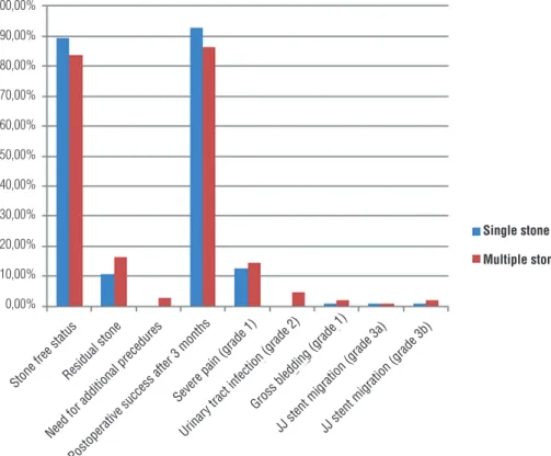
The stone-free status was significantly higher in patients with solitary renal stones when compared to patients with multiple renal stones (89.2% vs. 83.8%) after a single procedu-re (p=0.02). A second f-URS was procedu-requiprocedu-red for 3 patients in group 2. These patients were comple-tely stone free. After three months of follow-up, overall success rate was 92.7% in group 1 and 86.4% in group 2 (Table-2). Additionally, stone compositions are listed in Table-2.
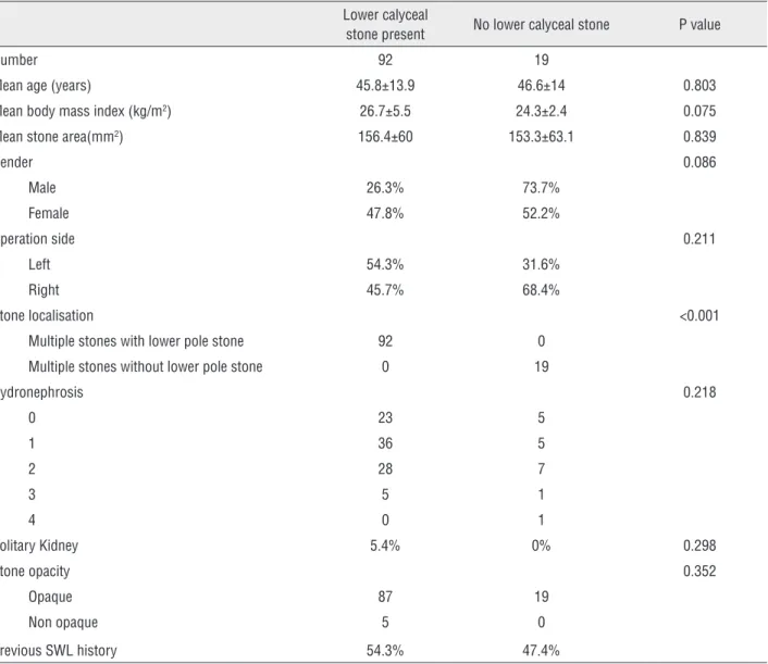
In group 2, 92 patients with lower pole stone and 19 patients without lower pole stone were categorized in group 2a and in group 2b, respectively. Demographic characteristics were well-matched between groups (Table-3). Also, mean operation time and mean fluoroscopy scre-ening time was similar (p=0.431 and p=0.436, respectively). Complication rates were not
signifi-Figure 1 - Comparison of post operative stone free status and complications between patients with single stone and multiple stones.
100,00%
90,00%
80,00%
70,00%
60,00%
50,00%
40,00%
30,00%
20,00%
10,00%
0,00%
Stone free statusResidual stone
Need for additional precedures
Postoperative success after 3 months
JJ stent migration (grade 3a)JJ stent migration (grade 3b)
Single stone
Multiple stones
Severe pain (grade 1)
Urinar
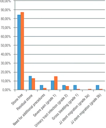
cantly different between groups (p=0.616). Stone free status was 87.0% in group 2a and 84.2% in group 2b after a single procedure (p=0.856) Figu-re-2). Second look f-URS was performed in two patients and one patient in group 2a and in group 2b, respectively (Table-4).
DISCUSSION
The treatment recommendation for kidney stone(s) in urolithiasis guidelines substantially
de-pends on stone burden and location of the stone. However, many studies had demonstrated stone number’s effect on the success and complications of the treatment modality, especially in SWL. We believe that number of stones can effect the de-cision of treatment modality and this variable should be added in urolithiasis guideline’s recom-mendations. Thus, we investigate the efficiency of f-URS in multiple stones, where SWL is recom-mended as the first line treatment option, accor-ding to stone size.
Table 2 - Operative and post-operative characteristics.
Single stone Multiple stones P value
Number 111 111
Type of f-URS 0.299
Flex-X2 (Fiberoptic) 94 87
Dur-D (Digital) 17 24
Mean operation time(minutes) 44.6±16.8 47.8±22.2 0.229
Mean fluoroscopy time(minutes) 2.1±1.7 2.6±1.5 0.043
Mean hospitalisation time(hours) 20.6±14.5 22.5±16.8 0.387
Post operative complications 0.282
Severe pain (grade 1) 14 (12.6%) 16 (14.4%)
Need for antibiotic change due to fever (grade 2) 0% 5(4.5%)
Gross bleeding (grade 1) 1(0.9%) 2(1.8%)
JJ Catheter migration (grade 3a) 1(0.9%) 1 (0.9%)
JJ Catheter migration (grade 3b) 1 (0.9%) 2 (1.8%)
Postoperative success after single session 0.020
Stone free status 99 (89.2%) 93(83.8%)
Residual stone 12(10.8%) 18(16.2%)
Need for additional procedures 0% 3(2.70%) 0.385
Postoperative success after 3 months 103 (92.7%) 97 (86.4%) 0.016
Stone analysis 0.865
Calcium oxalate monohydrate 28 37
Calcium oxalate dihydrate 11 15
Uric acid 4 2
Cystine 4 7
Struvite 1 2
Mixt 8 13
The efficacy and safety of SWL on single kidney stone under 300mm2 is well described,
ho-wever, success rate of the procedure declines as the number of stones increase. Cash et al. reported ≤50% stone free rate after SWL, in the treatment of multiple intrarenal stones (8). McAdams et al. performed SWL in 149 patients and 32 of them had multiple renal stones. The mean number of stones was 1.87 in successfully treated patients and 2.81 in those with treatment failure, in their study (p=0.065) (9). In addition, stone free rates decrease below 70% for single renal stone loca-ted in the lower pole (10). Thus, we believe SWL
is not a suitable option for multiple renal stones, especially if one or more stone(s) are located in the lower pole.
Percutaneous nephrolithotomy is ano-ther treatment alternative for renal stone(s) under 300mm2, with excellent stone free rates up to 95%
in one procedure. However, PNL is associated with increased complication rates when compared to SWL or f-URS (11). Renal parenchymal injury is ine-luctable in PNL procedure and adjacent organ inju-ries mostly occur while performing access. In cases where multiple kidney stones are located in more than one location, one access may not be enough.
Table 3 - Pre-Operative characteristics of patients with multiple renal stones according to absence/presence of lower pole stone.
Lower calyceal
stone present No lower calyceal stone P value
Number 92 19
Mean age (years) 45.8±13.9 46.6±14 0.803
Mean body mass index (kg/m2) 26.7±5.5 24.3±2.4 0.075
Mean stone area(mm2) 156.4±60 153.3±63.1 0.839
Gender 0.086
Male 26.3% 73.7%
Female 47.8% 52.2%
Operation side 0.211
Left 54.3% 31.6%
Right 45.7% 68.4%
Stone localisation <0.001
Multiple stones with lower pole stone 92 0
Multiple stones without lower pole stone 0 19
Hydronephrosis 0.218
0 23 5
1 36 5
2 28 7
3 5 1
4 0 1
Solitary Kidney 5.4% 0% 0.298
Stone opacity 0.352
Opaque 87 19
Non opaque 5 0
Previous SWL history 54.3% 47.4%
Previous studies had demostrated that multiple ac-cess was associated with a greater amount of blood loss, discomfort and renal function deterioration.
We achieved 92.7% and 86.4% stone free status in patients with solitary renal stone and mul-tiple renal stones, respectively. Both Breda et al. and Huang et al. reported 100% success with f-URS in multiple renal stones under 20mm (12, 13). However, their study samples involved less number of patients when compared with our study and included 51 and 25 patients, respectively. Additionally, we performed only 3 (2.3%) second f-URS procedure in patients with multiple renal stones. In Huang’s study, 9 pa-tients (36%) required second f-URS procedure and 2 patients (8%) required third f-URS procedure. Si-milarly, Breda et al. performed 1.4 f-URS procedure while getting a 100% success rate. Additionally, fo-cusing the stone during stone fragmentation is more difficult in hydronephrotic cases, due to increased mobility of the stone. We have encountered more
hydronephrotic cases in group 2 and this might be another reason for lower stone free rates and longer operation time in this group.
Treatment of lower pole stones is challen-ging for urologists because of anatomic variabilities and technical conditions. Nonetheless, we did not shown any significant difference on success in pa-tients with multiple stones, whether papa-tients have lower pole stone(s) or not (p=0.856). Similar to our results, Jacquemet et al. compared 139 and 232 f--URS procedures for stones located in a single renal location other than the lower pole and at least one stone located in the lower pole, respectively. They did not found any impact of lower pole localization on f-URS success (14). Although preoperative cha-racteristics were well matched between our patients with multiple stones, we compared only 92 patients and 19 patients, with and without lower pole stones, retrospectively. We believe studies with high patient volume will clarify this issue.
Figure 2 - Comparison of post operative stone free status and complications between patients with and without lower calyceal stone, in multiple stone group.
100,00%
90,00%
80,00%
70,00%
60,00%
50,00%
40,00%
30,00%
20,00%
10,00%
0,00%
Stone free Residual stone
Need for additional precedures Severe pain (grade 1)
JJ stent migration (grade 3a)JJ stent migration (grade 3b) Urinar
y tract infection (grade 2)Gross bledding (grade 1)
No lower calyceal stone
The mean operation time was longer in pa-tients with multiple renal stones (47.8 min vs. 44.6 min) but the difference was not statistically signi-ficant (p=0.229). We believe that determining the location of the stones in different localizations and focusing with the laser is a time consuming pro-cess. Our operation time was found shorter com-pared to similar studies which evaluate operation time in multiple renal stones. The mean operation time was 65.1 min in Breda’s study but they cal-culated the operation time from the time of cys-toscope insertion to the stent placement, different
from our study. Additionally, their study included patients with 20mm stone size (12). Huang et al. calculated the operation time similar to our study but they informed the mean operation time per patient (81.2 min) including second and third procedures, if necessary (13).
We found that the mean fluoroscopy screening time was significantly longer in pa-tients with multiple renal stones (p=0.043). Pre-vious reports discussed above did not mention about fluoroscopy screening time. However, we are aware that our fluoroscopy screening time
Table 4 - Operative and post-operative characteristics of patients with multiple renal stones according to absence/presence of lower pole stone.
Lower calyceal stone present No lower calyceal stone P value
Number 92 19
Type of f-URS 0.810
Flex-X2 (Fiberoptic) 72 15
Dur-D (Digital) 20 4
Mean operation time(minutes) 47.1±22.1 51.5±23.2 0.431
Mean fluoroscopy time(minutes) 2.6±1.4 2.3±1.8 0.436
Mean hospitalisation time(hours) 21.1±13 29.3±28.5 0.052
Post operative complications 0.616
Severe pain (grade 1) 14(15.2) 2(10.6%)
Need for antibiotic change due to fever (grade 2) 4(4.4%) 1(5.3%)
Gross bleeding (grade 1) 1(1.1%) 1(5.3%)
JJ Catheter migration (grade 3a) 1 (1.1%) 0
JJ Catheter migration (grade 3b) 1 (1.1%) 1 (5.3%)
Peroperative success 0.856
Stone free status 80(87.0) 16(84.2%)
Residual stone 12(13.0) 3(15.8%)
Need for additional procedures 2(2.2%) 1(5.3%) 0.153
Stone analysis 0.977
Calcium oxalate monohydrate 31 6
Calcium oxalate dihydrate 13 2
Uric acid 2 0
Cystine 6 1
Struvite 2 0
Mixt 11 2
was longer when compared to other f-URS stu-dies in the literature. We believe that our ten-dency to frequently determine the location of the stone with retrograde pyelography and aid fluoroscopy screning to find the stone(s) resul-ted in longer fluoroscopy screening time.
Complication rates were 15.3% and 23.4% in patients with solitary renal stone and multiple renal stones, respectively but difference was not statistically significant. We did not face any Clavien 4 and Clavien 5 complications. Severe pain was the most common post operative complication in both groups. Urinary tract infection was seen only in the multiple renal stones group. We deemed that more manipulation in multiple renal stones was related with severe pain and infectious complications. Double-J stent migration was identified in five patients with a kidney – ureter – bladder radiogram postoperatively. The double-J stent was re-inserted under local anesthesia in two patients (Clavien 3a) and under general anesthesia in three patients (Clavien 3b), respectively.
Although with sufficient number of pa-tients, our study has some limitations. First of all, we are aware of the retrospective nature of our study. Secondly, we did not evaluate the need of analgesic requirements postoperatively and cost of our procedures. Also, comparison of the presence of lower pole stones were made be-tween unevenly numbered groups. Additionally, our stone analysis datas are incomplete, which is another limitation of our study. Our Ministry of Health does not provide a free ‘Stone analy-sis’ for every patient, thereby, only 132 of our patients had stone analysis results. Finally, se-condary procedures for residual stone fragments was not commonly applied to our patients, be-cause most of our patients were symptom-free and did not want to undergo a secondary pro-cedure.
CONCLUSIONS
Our study has showed that f-URS achie-ved better stone free status in solitary renal sto-ne than multiple renal stosto-nes under 300mm2.
However, stone free status and complication
rates of f-URS were acceptable in patients with multiple stones. To our knowledge, this is the first study which compares the effect of mul-tiple stones or single stone on the outcomes of f-URS. However, our findings must be supported by further prospective, randomized studies with large patient volume.
ABBREVIATIONS
SWL = Shock Wave Lithotripsy
PNL = Percutaneous Nephrolithotomy
F-URS = Flexible ureterorenoscopy
IVU = Intravenous urography
CT = Computed tomography
BMI = Body mass index
Min = Minutes
CONFLICT OF INTEREST
None declared.
REFERENCES
1. Tuerk C, Knoll T, Petrik A.. Guidelines on urolithiasis, 2010. Available at. http://uroweb.org/wp-content/uploads/ Urolithiasis-2010-pg.pdf
2. Grasso M, Beaghler M, Loisides P. The case for primary endoscopic management of upper urinary tract calculi: II. Cost and outcome assessment of 112 primary ureteral calculi. Urology. 1995;45:372-6.
3. Galvin DJ, Pearle MS. The contemporary management of renal and ureteric calculi. BJU Int. 2006;98:1283-8.
4. Kourambas J, Byrne RR, Preminger GM. Does a ureteral access sheath facilitate ureteroscopy? J Urol. 2001;165:789-93.
5. Miernik A, Wilhelm K, Ardelt PU, Adams F, Kuehhas FE, Schoenthaler M. Standardized flexible ureteroscopic technique to improve stone-free rates. Urology. 2012;80:1198-202.
6. Akman T, Binbay M, Ugurlu M, Kaba M, Akcay M, Yazici O, et al. Outcomes of retrograde intrarenal surgery compared with percutaneous nephrolithotomy in elderly patients with moderate-size kidney stones: a matched-pair analysis. J Endourol. 2012;26:625-9.
8. Cass AS. Comparison of first generation (Dornier HM3) and second generation (Medstone STS) lithotriptors: treatment results with 13,864 renal and ureteral calculi. J Urol. 1995;153:588-92.
9. McAdams S, Kim N, Ravish IR, Monga M, Ugarte R, Nerli R, et al. Stone size is only independent predictor of shock wave lithotripsy success in children: a community experience. J Urol. 2010;184:659-64.
10. Tiselius H-G. Prospective, randomized trial comparing shock wave lithotripsy and ureteroscopy for lower pole caliceal calculi 1 cm or smaller. Eur Urol. 2006; 49:586-7.
11. Matlaga BR, Jansen JP, Meckley LM, Byrne TW, Lingeman JE. Treatment of ureteral and renal stones: a systematic review and meta-analysis of randomized, controlled trials. J Urol. 2012;188:130-7.
12. Breda A, Ogunyemi O, Leppert JT, Schulam PG. Flexible ureteroscopy and laser lithotripsy for multiple unilateral intrarenal stones. Eur Urol. 2009;55:1190-6.
13. Huang Z, Fu F, Zhong Z, Zhang L, Xu R, Zhao X. Flexible ureteroscopy and laser lithotripsy for bilateral multiple intrarenal stones: is this a valuable choice? Urology. 2012;80:800-4.
14. Jacquemet B, Martin L, Pastori J, Bailly V, Guichard G, Bernardini S, et al. Chabannes E, Bittard H, Kleinclauss F. Comparison of the efficacy and morbidity of flexible ureterorenoscopy for lower pole stones compared with other renal locations. J Endourol. 2014;28:1183-7.