Endovascular treatment of late
post-traumatic aortocaval fistula: case report
Tratamento endovascular de fístula aortocaval pós-traumática tardia: relato de caso
Leonardo Pessoa Cavalcante1, Marcos Velludo Bernardes1, Ricardo Dias da Rocha1, Marcos Henrique Parisati1, Juliana Lopes Alfaia1, José Emerson dos Santos Souza1, Patrícia de Souza Lacerda1, Raquel Magalhães Pereira2
Abstract
Aortocaval istulas are rare entities with diferent etiologies. A few are sequelae of trauma, and symptoms may be acute or delayed; the late manifest days, weeks or years after trauma, mainly as congestive heart failure. Treatment may be open surgery or endovascular repair. In the case reported here, a 53-year-old man presented with important signs of congestive heart failure, such as palpitations and dyspnea, paroxysmal atrial ibrillation, in addition to high pulse pressure and bruit in the epigastrium, 27 years after a stab wound in the abdomen. CT angiography conirmed the diagnosis of aortocaval istula, and treatment was endovascular istula repair. Treatment outcome was satisfactory, with signiicant improvement and appropriate control of heart failure three months after the procedure.
Keywords: vascular trauma; arteriovenous istula; abdominal aorta; inferior vena cava; heart failure.
Resumo
As fístulas aortocavais são entidades raras e de etiologia variada. Uma minoria é consequente a eventos pós-traumáticos. As manifestações clínicas, nesses casos, podem ser agudas ou tardias. As tardias manifestam-se dias, semanas ou anos após o trauma, principalmente como quadro de insuiciência cardíaca congestiva. O tratamento de tais fístulas pode ser realizado através do reparo direto por cirurgia aberta ou através da abordagem endovascular. Relatamos o caso de um paciente do sexo masculino, de 53 anos que apresentou, 27 anos após um ferimento por arma branca abdominal, sinais importantes de insuiciência cardíaca congestiva, manifestada como palpitações e dispneia, ibrilação atrial paroxística, além de pressão arterial divergente e sopro em epigástrio. A angiotomograia conirmou o diagnóstico de fístula aortocaval e procedeu-se ao tratamento endovascular para o selamento da fístula. O paciente, segundo acompanhamento após três meses, apresentou evolução satisfatória, com melhora signiicante do quadro e controle adequado da insuiciência cardíaca congestiva.
Palavras-chave: trauma vascular; fístula arteriovenosa; aorta abdominal; veia cava inferior; insuiciência cardíaca.
R E P O R T
1 Service of Vascular Surgery, Hospital Universitário Francisca Mendes, Manaus, AM, Brazil. 2 Universidade Federal do Amazonas - UFAM, Manaus, AM, Brazil.
Financial support: None.
Conlicts of interest: No conlicts of interest declared concerning the publication of this article. Submitted: 26.08.12. Accepted: 07.11.12.
INTRODUCTION
Aortocaval istulas (ACF), irst described by Syme in 1831,1 are rare entities with different etiologies.
Most (80% to 92%) result from the erosion or rupture of abdominal aortic aneurysms (AAA) into the inferior vena cava (IVC), at an incidence of 1% to 6% of complicated aneurysms1-6. The remaining
10% to 20% are sequelae of trauma. Secondary ACF and syphilitic and mycotic aneurysmshave also been described,7 as well as aneurysms associated with rare
entities, such as Ehler Danlos and Marfan syndromes and Takayasu artheritis3,5,6,8.
Patients with ACF may be divided into two groups according to the presentation of post-traumatic ACF: acute and potentially fatal event at the scene of the injury or during emergency surgery; or delayed presentation of symptoms weeks, months or even years after trauma1. In the second group, patients
usually have high-output congestive heart failure (CHF) due to the istula4,6.
Because of their severity and high morbidity and mortality, ACF should be treated as soon as a diagnosis is made. Treatment principles are: 1) close the arterial defect; 2) restore the arterial low; and 3) perform the venous repair (desirable, but not mandatory). These principles guide both open surgery and endovascular repair9.
This study describes and discusses the endovascular treatment of a post-traumatic ACF due to an abdominal stab wound.
CASE REPORT
A 53-year-old man presented to the emergency room with acute paroxysmal atrial ibrillation (AF) and clinical symptoms of palpitation and dyspnea. Physical examination revealed high pulse pressure (180 x 60 mmHg), normal peripheral pulses, systolic-diastolic bruit over the epigastric region and a scar of a median laparotomy performed 27 years earlier in the interior of the state of Amazonas, Brazil, due to a stab wound in the epigastrium.
Echocardiography revealed a moderate increase of the left cardiac chambers, increased left ventricular mass and accelerated turbulent low in the proximal descending thoracic aorta.
CT angiography of the thoracic and abdominal aorta revealed a connection between the abdominal aorta and the inferior vena cava (IVC) that extended from 14 mm below the lower renal artery (left) to about 34 mm below it. At this point, the diameter of the abdominal aorta was 21 mm. IVC had a diffuse increase of its diameter (Figures 1 and 2) and early contrast-enhancement (at arterial phase) (Figure 3).
Erosion of the anterior portion of the body of the second lumbar vertebra was also detected.
After the diagnosis was conirmed, endovascular repair was performed in a cardiovascular unit operating room using left femoral puncture and right femoral dissection. First, digital angiography of the abdominal aorta at the level of the renal arteries was used to conirm the position of the ACF (Figure 4a). Immediately after that, using a road map, a 0.035”
× 260 cm extra-stiff Lunderquist guide wire was
Figure 1. CT angiography of abdominal aorta shows connection between abdominal aorta and inferior vena cava, as well as erosion of anterior portion of L2 body.
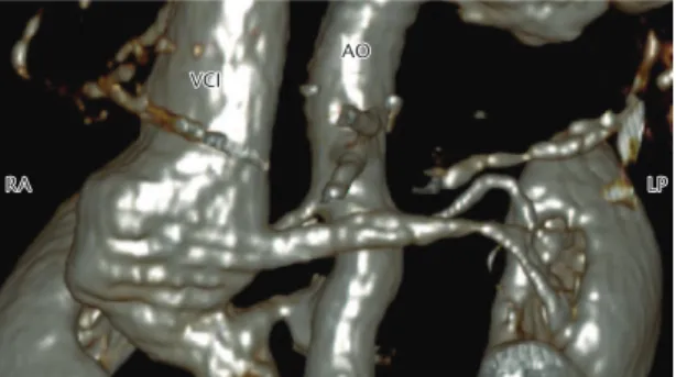
Figure 2. hree-dimensional angiotomography reconstruction shows connection between abdominal aorta (AO) and inferior vena cava (IVC).
used to ascend and deploy the proximal module (aortic extension) of an Endurant abdominal aorta
endovascular prosthesis (Medtronic®)
measuring
25 × 49 mm. This device was chosen because of its proximal free low and controlled release (proximal neck smaller than 15 mm) and the anchorage components, which prevent distal migration of the device since only an aortic module was used. A reliant stent graft balloon (Reliant,Medtronic®) was used to
accommodation of the device, and the inal digital angiography indings were satisfactory (Figure 4b).
During postoperative recovery, the patient had discrete transient blood urea nitrogen elevation. He was discharged on the ifth postoperative day, when levels were already normal.
After three months, a control CT scan showed stent integrity and that the aortic istula had been completely closed (Figure 5). There was not early IVC contrast-enhancement, and IVC caliber was substantially reduced (Figure 6). Anticoagulation agents were discontinued by the cardiologist because the patient did not have paroxysmal AF anymore.
DISCUSSION
Post-traumatic ACF accounts for less than 20% of all ACFs. In this group, 90% result from penetrating injuries (gunshot or stab wounds) or iatrogenic causes (spinal surgery,10 arterial catheterization), while the
other 10% are secondary to blunt trauma11-13.
The low incidence of this type of istula after trauma may be explained by the fact that in most cases, several not documented, the vascular lesion leads to death by exsanguination and severe
hypovolemic shock at the scene of the injury. After being taken to the operating room, many patients (40% to 50%) do not survive the procedure because of the dificulty in surgical exposure, intraoperative hemorrhage, hypothermia and coagulopathies due to the prolonged surgery duration11-12.
Late symptoms of this type of lesion, such as the ones presented by the patient described here, are, therefore, even rarer, and may be diagnosed weeks to years later. Sigler et al.14 reported 5 cases of
post-traumatic ACF and CHF symptoms 2 days to 6 months after trauma. Spencer et al.1 described two
cases of ACF with delayed presentation, 20 and 30 years after a gunshot wound to the abdomen; both underwent exploratory laparotomy at the time of injury. Galvão et al.10 also described a late diagnosis
(30 months) of an ilio-iliac arteriovenous istula after iatrogenic trauma (left hemilaminectomy).
The patient in this report presented with important signs of CHF 27 years after a penetrating injury (stab wound) and exploratory laparotomy. There were clinical signs present (palpitations and dyspnea due to paroxysmal AF) and compatible tests results (echocardiogram showing high cardiac output). Physical examination detected a high pulse pressure (180 x 60 mmHg) and a systolic-diastolic bruit over the epigastrium.
Other typical signs of ACF are abdominal pain, pulsatile abdominal mass, abdominal bruit and acute dyspnea.3,6 Signs of peripheral venous congestion,
such as lower limb edema, deep vein thrombosis and pelvic venous hypertension with anuria, hematuria, edema or scrotal hematoma, have also
been described2,4-6,8. In large ACFs, clinical signs
of pulmonary edema, central venous congestion, hepatomegaly and ascites may also be detected, in addition to echocardiographic indings of high-output CHF3,4.
CHF occurs as a result of the deviation of blood low from the aorta to the IVC, which leads to an
important drop of arterial resistance and activation of the renin-angiotensin-aldosterone system and the sympathetic autonomous nervous system10,15.
Therefore, a hyperdynamic circulatory state occurs in about 30% to 50% of the cases15. If the ACF is
large or if the heart cannot increase its output due to a previous heart disease, for example, heart failure or refractory shock ensues15.
Although clinical history and careful physical examination may suggest the diagnosis, classical signs and symptoms are absent in about 50% of the cases, when there is a partial istula obstruction, for example,5 which makes the diagnosis dificult.
Therefore, complementary tests are required. In stable patients, Doppler ultrasound of the abdominal aorta may conirm the diagnosis. For surgical planning, the imaging study of choice is CT angiography of the abdominal, iliac and femoral arteries1,3,8,15. Some CT signs that suggest the
presence of ACF are loss of fat tissue between the IVC and aorta, IVC early contrast enhancement (in the arterial phase)8,15 (Figure 3) and the visualization
of a istula (Figures 1 and 2). Other signs are IVC dilatation, retrograde opaciication of the renal veins during the arterial phase, poor renal enhancement, increase of kidney size and retroperitoneal and pelvic venous congestion15. In the case described here,
echocardiography detected CHF and CT angiography
Figure 5. hree-dimensional angiotomography reconstruction shows full stent, patency of both renal artery and complete closure of aortic istula.
conirmed the diagnosis of a high output istula. The surgery was then performed.
ACF treatment is based primarily on the surgical closure of the istula during open surgery or an endovascular repair9. Blood and blood components
should be readily available because bleeding due to manipulation of the lesion may be catastrophic1. In
the literature, operative mortality during ACF open repair ranges from 16 to 66%2,5. Cinara et al.2 found
a mortality rate of 19% in a study with 26 patients. Lower rates (about 7%) were found in surgical treatment of ACF after spine surgeries due to the lower age of the patients and the smaller diameter of the istulas10.
Open surgery is often indicated for patients with acute symptoms that need emergency repair. In chronic cases (delayed presentation), this technique is indicated for those with an aortic-iliac-femoral anatomy unfavorable for endovascular repair, and results are better in young and previously healthy patients9. As very few of the chronic ACF patients
meet this last two requirements, since diagnosis is usually made because of the negative heart repercussion (CHF), this approach turns out to be of little advantage for this group of patients.
Endovascular repair, therefore, has become the treatment of choice. The irst case of AAA treated with an endovascular prosthesis via femoral artery was published by Parodi et al.16 in 1991, and the use
of similar prostheses has been growing since then. When endovascular repair is chosen, laparotomy is avoided, and the endovascular prosthesis is implanted through an inguinal incision. This procedure is less invasive because it avoids aortocaval clamping and the risk of serious intraoperative hemorrhage is lower, which, consequently, speeds up patient recovery and provides an early return to family life and work17.
Therefore, endovascular repair is extremely beneicial, particularly for those with a favorable aortic-iliac-femoral anatomy and high surgical risk, because it exposes these patients, usually older and with other comorbidities, to a less serious aggression (less risk of hemorrhage and/or lesion to neighboring organs) than open surgery9,17.
The success of the endovascular repair in the present case was conirmed when, in the 3rd month post-operative follow-up, our patient’s cardiologic and hemodynamic condition was stable, the paroxysmal AF had disappeared and CT scans revealed an important reduction of IVC caliber and patency of the abdominal aorta and renal arteries without contrast extravasation (Figures 5 and 6).
Studies in the literature conirm that, after vascular repair of the lesions, the clinical condition of patients with arteriovenous istulas improves signiicantly, and their recovery is usually satisfactory2,8.
CONCLUSION
This case demonstrates that the treatment of aortocaval istulas using an endovascular approach is safe and effective.
REFERENCES
1. Spencer T, Smyth S, Wittich G, Hunter G. Delayed presentation of traumatic aortocaval istula: A report of two cases and a review of the associated compensatory hemodynamic and structural changes. J Vasc Surg. 2006;43(4):836-840. PMid:16616246. http:// dx.doi.org/10.1016/j.jvs.2005.12.003
2. Cinara I, Davidovic L, Kostic D, Cvetkovic S, Jakovljevic N, Koncar I. Aorto-Caval Fistulas: A Review of Eighteen Years Experience. Acta Chir Belg. 2005;105:616-620. PMid:16438071.
3. Zuniga C, Rodriguez J, Caceres P. Fístula aortocava como complicación de aneurisma aórtico abdominal. Rev Chilena Cirugía. 2011;63(6):623-626. http://dx.doi.org/10.4067/ S0718-40262011000600013
4. Kafejian O, Ricci JA, Nem Junior JT, Roberti T, Bolanho E. Aneurisma de aorta abdominal: diagnóstico, tratamento cirúrgico e resultados. Cir Vasc Ang. 1988;4(1):8-15.
5. Oliveira LAV, Leão PP, Barbatto HA, Malheiros FD. Aneurisma de aorta abdominal com fístula espontânea aorto-cava. Cir Vasc Ang. 1992;8(3):15-18.
6. Timi JRR, Góes Junior DCA, Oliveira A, Abrão E. Aneurisma de aorta abdominal roto para veia cava inferior – Relato de caso e revisão de literatura. Cir Vasc Ang. 1992;8(3):21-23.
7. Devoto M, Fabiani A , Secin F, Zaefferer P, Bracco A . Aneurisma micotico de aorta abdominal complicado con fístula aortoduodenal y aorto cava simultâneos. Actas Cardiovasc. 1996;7(2):119-23.
8. Pinto DM, Bez LG, Dias Junior JO, Lopes CS, Mandil A. Aneurisma ilíaco associado a fístula arteriovenosa. J Vasc Br a s . 2 0 0 7 ; 6 ( 3 ) : 2 9 9 - 3 0 2 . http : / / d x . d o i . o rg / 1 0 . 1 5 9 0 / S1677-54492007000300016
9. Hunter GC. Acquired Arteriovenous Fistulae. In: Cronenwe JL, Johnston KW, editors. Rutherford’s Vascular Surgery. Philadelphia: WB Saunders; 2010. p. 1087-102. http://dx.doi.org/10.1016/ B978-1-4160-5223-4.00071-8
10. Galvão GE, Zorn WGW, Bellen B. Fístula arteriovenosa pós-laminectomia. Cir Vasc Ang. 1991;7(1):17-20.
11. Feliciano DV, Burch JM, Graham JM. Abdominal vascular injury. In: Feliciano DV, Moore EE, Mattox KL, editors. Trauma. Norwalk: Appleton & Lange; 1996. p. 615.
12. Carrillo EH, Bergamini TM, Miller FB, Richardson JD. Abdominal vascular injuries. J Trauma. 1997;43:164-71. PMid:9253935. http:// dx.doi.org/10.1097/00005373-199707000-00043
14. Sigler L, Gutiérrez-Carreño R, Martínez-López C, Lizola RI, Sánchez-Fabela C. Aortocava fistula: experience with five patients. J Vasc Surg. 2001;35(3):207-12. http://dx.doi. org/10.1177/153857440103500308
15. Herrero M, García V, Escudero A, et al. Insuficiencia renal aguda como forma de presentación de una fístula aortocava asociada a um aneurisma de aorta abdominal. Cartas al director. Nefrologia. 2011;31(1):124-6.
16. Parodi JC, Palmaz JC, Barone HD. Transfemoral intraluminal graft implantation for abdominal aortic aneurysms. Ann Vasc Surg. 1991;5:491-9. PMid:1837729. http://dx.doi.org/10.1007/ BF02015271
17. Mertens R, Krämer A, Valdés F, et al. Uso de endoprótesis en el tratamiento de lesiones no oclusivas del territorio ilíaco. J Vasc Bras. 2006;5(2):89-94. http://dx.doi.org/10.1590/ S1677-54492006000200003
Correspondence
Raquel Magalhães Pereira Rua homé de Sousa, 443 – Dom Pedro I CEP 69040-190 – Manaus (AM), Brazil E-mail: [email protected]
Author information
LPC, RDR, MHP, and JLA are vascular surgeons at the Service of Vascular Surgery of Hospital Universitário Francisca Mendes. MVB is chief of the Service of Vascular Surgery Hospital Universitário
Francisca Mendes. JESS and PSL are resident physicians of the Service of Vascular Surgery of Hospital Universitário Francisca Mendes. RMP is a medical student at Universidade Federal do Amazonas (UFAM).
Author contributions