Rigid Bronchoscopy in Airway Foreign Bodies:
Value of the Clinical and Radiological Signs
Kunjan Acharya
11Department of ENT-Head and Neck Surgery, Tribhuvan University
Teaching Hospital, Maharajgunj, Kathmandu, Nepal
Int Arch Otorhinolaryngol 2016;20:196–201.
Address for correspondenceDr. Kunjan Acharya, Department of ENT-Head and Neck surgery, Tribhuvan university Teaching Hospital, Maharajgunj Kathmandu Kathmandu 3578, Nepal
(e-mail: [email protected]; [email protected]).
Introduction
Airway foreign body (FB) is one of the common emergencies an ENT surgeon must attend to. Its clinical presentation is variable, ranging from a clinically asymptomatic state to dire state of respiratory failure needing urgent attention and intervention. The gold standard for management is rigid bronchoscopy (RB) under general anesthesia.
Rigid bronchoscopy, performed under General Anesthe-sia (GA), exposes the patient to the risk of GA and the complication of the procedure itself. According to Righini,1
unnecessary rigid bronchoscopy performed under general anesthesia can expose the child to the risk of perioperative procedure complication (8–17%) like bronchospasm, desa-turation and bleeding, or airway edema. Hence, the right decision to perform RB is based on clinicalfindings and radiological support. My study aims at correlating the clini-cal presentation and X-ray chestfindings with the result of RB (positive versus negative RB).
According to Hsu,2the usual age group of presentation is between 1–3 years old. However it can occur in older children as well. The crucial decision of performing rigid bronchoscopy
Keywords
►
foreign bodies
►
bronchoscopy
►
airway obstruction
Abstract
Introduction
Foreign body in airway is a common emergency in ENT practice. As we
know, Rigid Bronchoscopy is the method of choice for removing it, although at times it
leads to specialists performing unnecessary bronchoscopy, exposing patients to hazards
of general anesthesia.
Objective
The objective of my study is to calculate sensitivity, speci
fi
city, positive
predictive value, odds ratio from the clinical and radiological signs, comparing with the
gold standard, the rigid bronchoscope procedure.
Method
This is a prospective analytical study designed at University Teaching Hospital
and conducted over a period of 18 months, from March 2011 to August 2012. Data
collection was broadly classi
fi
ed into three different categories: (1) Symptomatology,
such as presence or absence of choking, cyanosis, and dif
fi
culty in breathing; (2) Clinical
signs, such as the presence or absence of air entry, crackles, and rhonchi 3. Chest X-ray
fi
ndings were suggestive of a foreign body.
Results
There were a total of 40 rigid bronchoscopies performed under general
anesthesia for the diagnosis and therapeutic reasons. Among 40 patients who
under-went rigid bronchoscopy, 32 (80%) were found to have varieties of foreign bodies in their
airway while 8 patients (20%) had negative bronchoscopy. The history of choking is the
only clinical symptoms which came out to be statistically Signi
fi
cant (
p
¼
0.043) with
odds ratio of 5.
Conclusion
Rigid bronchoscopy is the gold standard technique for diagnosis and
procedure of choice to remove FB from airway. Regardless, it still presents a small
chance of negative result, especially when there is no history of aspiration.
received February 16, 2016 accepted May 1, 2016 published online May 30, 2016
DOI http://dx.doi.org/ 10.1055/s-0036-1584293. ISSN 1809-9777.
Copyright © 2016 by Thieme Publicações Ltda, Rio de Janeiro, Brazil
is made on clinical backgrounds. This includes history and clinicalfindings. It is very important to ask for the history of choking or aspiration from the parents or care-taker. Upon clinical examination, a foreign body in the airway may reveal diminished breath sounds on the affected side or presence of rhonchi or crept. At the same time, two or morefindings is possible. Radiological signs can be collapsed lung, radiopaque shadow, or area of consolidation.
The hypothesis of my study is to show the relation of clinical and radiological signs and in cases with presence of FB in the airway. It helps us understand the association of radiological and clinicalfindings to the results of bronchos-copy to avoid negative bronchosbronchos-copy.
Objectives
The objective of my study is to calculate sensitivity, specifi ci-ty, positive predictive value, and odds ratio of the clinical and radiological signs and to compare them with the rigid bron-choscopyfindings.
Method
This is a prospective analytical study done over a period of 18 months, from March 2011 to August 2012. All faculties were involved in performing rigid bronchoscopy and foreign body removal. It was done at a tertiary level hospital.
The institutional review board approved the study prior to its undertaking study and the subjects gave their formal consent. The inclusion criteria were all patients with strong clinical suspicion of foreign body in airway with a history of
choking, cyanosis, difficulty in breathing, along with the presence of clinical signs, like decreased air entry, cyanosis, or crept. Patients of any age and sex who qualified for the inclusion criteria were included. Before the procedure, the ENT residents examined all patients and noted findings in Performa. After which all patients underwent chest X-ray and at times CT scan of chest whenever indicated. The chest X-Ray was looked for radiological signs, presence of foreign body like collapsed segment, consolidation, trapped air or some-times the foreign body itself when the foreign body was radio opaque.
Data has been collected broadly into three different cate-gories: (1) Symptomatology, such as the presence or absence of choking, cyanosis, and difficulty in breathing; (2) Clinical signs, such as the presence or absence of air entry, crept, and rhonchi; (3) Radiological signs, such as plain chest X-ray findings.
After proper clinical evaluation and chest X-ray, all pa-tients underwent rigid bronchoscopy under general anesthe-sia. We used bronchoscopes of the rigid type to perform bronchoscopy. We determined the size of the bronchoscope according to the child’s age. After induction of intravenous anesthesia, we performed direct laryngoscopy and inserted the bronchoscope with the help of the laryngoscope in a rotating manner and used a 0-degree telescope to locate the foreign body. Once identified, we used optical forceps to hold and to remove the foreign body.
However, at times it was not possible to remove FB in the first attempt, and the procedure was repeated after 2–3 days. The decision to continue or to abandon the procedure de-pended upon the intraoperative bleeding and oxygen
Table 1 Frequency distribution of clinical & radiological signs
Present Absent
Symptoms H/o choking 27 (67.5%) 13 (32.5%)
H/o cyanosis 8 (20%) 32 (80%)
H/o SOB 24 (60%) 16 (40%)
Clinical Signs Decreased/absent air entry Total 31 (77.5%) 9 (22.5%)
Bilateral absent: 2
Unilateral absent: 3
Unilateral decreased: 26
Crepitation Total 10 (25%) 30 (75%)
Unilateral crepitation: 8
Bilateral crepitation: 2
Rhonchi Total 6 (15%) 34 (85%)
Unilateral Rhonchi: 5
Bilateral Rhonchi: 1
Radiological sign Chest X-ray Normal 19 (47.5%) Abnormal 21(52.5%)
Radiopaque shadow: 8
Consolidation: 3
Collapsed Lung: 10
saturation. Any amount of bleeding obscuring the surgical procedure and oxygen saturation compromising the anes-thetic state of patient led to us to postpone the procedure to the next setting.
After extraction of the foreign body, we repeated bron-choscopy to check for any remaining foreign bodies as well as to examine the tracheobronchial tree for any trauma.
Rigid bronchoscopicfindings were correlated with clinical signs and symptoms using SPSS 16. We calculated sensitivity, specificity positive predictive value. We analyzed the statis-tical relation of clinical and chest X-rayfindings with rigid bronchoscopy.
Results
There were total of 40 RB performed under general anesthesia for the diagnosis and therapeutic reasons. The age of the
patient ranged from 8 months to 13 years. Of the 40 patients, 29 (72.5%) were men and 11 (27.5%) were women.
Upon clinical examination, the history of choking was the most common symptom and decreased air entry was the most commonfinding, as displayed in►Table 1.
Among 40 patients who underwent RB, 32 (80%) had foreign body in their airway, while 8 patients (20%) had negative RB results.
When the different clinical manifestations were cross-tabulated with the rigid bronchoscopy findings, history of choking was the only manifestation, which came out to be statistically significant (p¼0.043) with odds ratio of 5. The detailed of cross tabulation between RBfindings and clinical manifestation are shown in►Table 2.
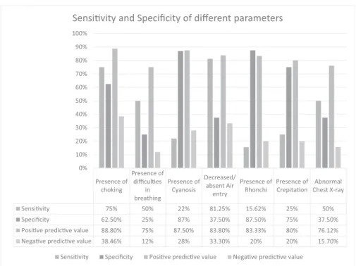
Similarly, I used data to calculate sensitivity, specificity, positive and negative predictive value for each of the clinical manifestations, as shown in►Fig. 1.
Table 2 Analysis between RBfindings and clinical manifestation
RB Findings Clinical Manifestation Pvalue (Chi square test)
Foreign Body History of choking
Absent Present 0.043
Odds ratio: 5
No FB: 8 5 3
FB present: 32 8 24
Difficulties in breathing
Absent Present 0.439
(using Fisher’s Exact Test)
No FB: 8 2 6
FB present: 32 14 18
History of Cyanosis
Absent Present 0.553
No FB: 8 7 1
FB present: 32 25 7
Air entry
Normal Decreased
No FB: 8 3 5 0.256
FB present: 32 6 26
Crepitation
Absent Present
No FB: 8 6 2 1.00
FB present: 32 24 8
Rhonchi
Absent Present
No FB: 8 7 1 0.82
FB present: 32 27 5
Chest X-ray
Normal Abnormal
No FB:8 3 5 0.52
FB present: 32 16 16
Discussion
The most common symptom of foreign body aspiration was choking (67.5%), which was the only statistically significant result in this study. Similarly, decreased air entry was present in 77% of the patients, but was not significant in our study. However, Righini et al1revealfindings contrary to our study. In their study, the FB aspiration as well as unilateral decreased air entry was statistically significant to the presence of FB in the airway. Similarly, a study done by Gursu Kiyan a (2009)3et al in 192 patients over period of 5 years (2003–2007) showed history of aspiration and decreased air entry both were statistically significant, p value was<0.0001 in both variable. History of aspiration is important clinical parameter to decide for RB as shown from our study. It is supported by study done by Martinot et al.4 He conducted comparative study between Rigid and Flexible Bronchoscopy tofind the negative bronchoscopy rate by two different techniques. He adopted a treatment algorithm where he used the child who had history of aspiration were directly channelized to under-go rigid bronchoscopy avoiding theflexible one. This shows asking the history alone is very important and must not be ignored at any cost.
Similarly, sensitivity and specificity of different clinical and radiological signs are displayed in►Fig. 1. However none of the clinical parameters like choking, difficulty in breathing, cyanosis, decreased air entry, rhonchi and crept individually have high sensitivity and specificity together. This might be one of the important factor responsible for predicting the outcome of rigid bronchoscopy. In my study the unilateral decrease in air entry had highest sensitivity of 81% whereas presence of cyanosis and presence of rhonchi on auscultation had very high specificity of 87%.
The study done by Kiyan et al3 on 192 patients who underwent rigid bronchoscopy, with the presence of wheeze as a symptom, had highest sensitivity at 87.7%, followed by unilateral decreased air entry on auscultation (i.e., 78.3%). This result slightly varies from this study, as I have not considered wheeze as a symptom during data collection but the sensitivity of unilateral decreased air entry is practi-cally similar. In the same study, the presence of cyanosis had very high specificity, at 98.1%, which was also the highest indicator in our analysis, reaching 87% specificity. In the Kiyan et al3study, the presence of productive cough had also very high specificity, at 96.3%, which was not taken into consider-ation during my study design. However, the presence of rhonchi had highest specificity in my study, but was not the highest in the study by Kiyan et al,3although the value was similar (i.e., 85%).
The above results signify that there is always a chance of negative bronchoscopy exposing the patients to the hazards of general anesthesia and possible complication of rigid bronchoscopy during the procedure and afterwards. This is because of the inability to identify the single clinical param-eter that can almost always predict the presence of foreign in the airway in every case. The rate of negative rigid bronchos-copy was 20% in my study, which is similar to the other studies done by different authors.
The literature on negative bronchoscopy includes the study done by Oren Cavel et al5displaying different rates on negative bronchoscopy and its complication, shown in►Table 3. The rate of negative rigid bronchoscopy ranges from 18% to 43%, with most of them around 20%.
Hence, the alternative modality of investigation is prefer-able, especially if the clinician does not find a history of aspiration. Also, as suggested by Martinot et al4in the study Presence of
choking
Presence of difficules
in breathing
Presence of Cyanosis
Decreased/ absent Air entry
Presence of Rhonchi
Presence of Crepitaon
Abnormal Chest X-ray
Sensivity 75% 50% 22% 81.25% 15.62% 25% 50% Specificity 62.50% 25% 87% 37.50% 87.50% 75% 37.50%
Posive predicve value 88.80% 75% 87.50% 83.80% 83.33% 80% 76.12% Negave predicve value 38.46% 12% 28% 33.30% 20% 20% 15.70%
0% 10% 20% 30% 40% 50% 60% 70% 80% 90% 100%
Sensivity and Specificity of different parameters
Sensivity Specificity Posive predicve value Negave predicve value
on a patient scheduled to undergoflexible bronchoscopy who had negative history of aspiration before doing rigid bron-choscopy. Theflexible bronchoscopy is an alternative tool to detect FB in airway when there is no definite history of aspiration. However, it is difficult to perform in very young
children and also has a chance of negative bronchoscopy: 12% (Righini et al)1and 38% (Martinot et al).4
The other investigating tool in patients with suspicious history of FB in the airway is CT thorax, which for being noninvasive is an alternative to diagnose FB in suspicious Table 3 Studies showing different rates of negative RB
Article No. of
Bronchoscopies
Negative RB rate Complications Remarks, authors’
comments
Maddali 175 20% 47% of cases, mostly minor
anesthetic complications. Risk factors IV anesthesia RB time>30 minute
Retrospective,‘‘to avoid unwarranted RB, clinicians should be aware of the clinical presentations of FBA’’
Kiyan et al3 207 26% 0.5% major
2.9% minor
Retro,‘‘with the help of our low complication rate, we claim that even a slight doubt of FBA using these criteria re-quires RB to avoid fur-ther complications of missed FBA’’
Righini et al1 54 16% 4% bronchospasm
4% subglottic edema
Prospective, propose an algorithm for choosing betweenflexible and RB
Cohen et al8 142 (flexible and RB under general anesthesia)
57% 8.5% Prospective,
recom-mend the use offlexible or RB depending on suspicion
Even et al9 98 43% – Prospective,‘‘medical
history is the key for the diagnosis of FBA. If FBA is suspected, bronchos-copy should be performed’’
Kadmon et al10 91 47%, 14%in
Obvious cases (8/59)
4% Retrospective, propose
computerized scoring system to determine the need for bronchoscopy
Ciftci et al11 663 16% 5%, risk factors–emergency bronchoscopy, prolonged procedure, delayed diagno-sis, type of foreign body (prayer beads, ball point pen lid)
Retrospective, no fur-ther imaging suggested
Martinot et al4 28, all‘‘obvious’’cases 18% – Prospective, RB in ur-gent or clinically and radiologically obvious cases,flexible bron-choscopy for all the rest
Hoeve et al12 115 26% – Retrospective,‘‘if
aspi-ration of FB is consid-ered, RB is mandatory’’
Rizk, Rassi13 106 23% 15% required switch to as-sisted ventilation due to bra-dycardia and desaturations
Retrospective, 12% of RB were preceded by flex due to low suspension
cases. The study done by Bhat et al6showed high sensitivity and specificity of CT scan up to 92.3 and 85.5%, respectively, in 20 patients. Similarly, a study done by Bai et al7in 45 patients had 100% of sensitivity and specificity in detecting the FB in the airway by using CT scan. Besides the high rate of detection of FB by CT scan, it also locates FB in the airway and gives the number of FB, which definitely helps the surgeon to perform RB in a planned manner.
Conclusion
Rigid bronchoscopy remains the gold standard technique of diagnosis and management of airway FB, however it has a small chance of negative result. Similarly, the history of choking is the only clinical parameter which is significantly associated with the presence of FB in airway. Hence, in cases with negative history of choking but with clinical suspicion of FB in the air way, subsequent management should be tailored. Alternative tools for confirming the presence of FB should be utilized, such as like CT scan of thorax orflexible bronchoscopy.
References
1 Righini CA, Morel N, Karkas A, et al. What is the diagnostic value of flexible bronchoscopy in the initial investigation of children with suspected foreign body aspiration? Int J Pediatr Otorhinolaryngol 2007;71(9):1383–1390
2 Hsu Wc, Sheen Ts, Lin Cd, Tan Ct, Yeh Th, Lee Sy. Clinical experi-ences of removing foreign bodies in the airway and esophagus
with a rigid endoscope: a series of 3217 cases from 1970 to 1996. Otolaryngol Head Neck Surg 2000;122(3):450–454
3 Kiyan G, Gocmen B, Tugtepe H, Karakoc F, Dagli E, Dagli TE. Foreign body aspiration in children: the value of diagnostic criteria. Int J Pediatr Otorhinolaryngol 2009;73(7):963–967
4 Martinot A, Closset M, Marquette CH, et al. Indications forflexible versus rigid bronchoscopy in children with suspected foreign-body aspiration. Am J Respir Crit Care Med 1997;155(5):1676–1679 5 Cavel O, Bergeron M, Garel L, Arcand P, Froehlich P. Questioning the
legitimacy of rigid bronchoscopy as a tool for establishing the diagnosis of a bronchial foreign body. Int J Pediatr Otorhinolar-yngol 2012;76(2):194–201
6 Bhat KV, Hegde JS, Nagalotimath US, Patil GC. Evaluation of com-puted tomography virtual bronchoscopy in paediatric tracheobron-chial foreign body aspiration. J Laryngol Otol 2010;124(8):875–879 7 Bai W, Zhou X, Gao X, Shao C, Califano JA, Ha PK. Value of chest CT in the diagnosis and management of tracheobronchial foreign bodies. Pediatr Int 2011;53(4):515–518
8 Cohen S, Avital A, Godfrey S, et al. Suspected foreign body inhala-tion in children: what are the indicainhala-tion for bronchoscopy? J Pediatr 2009;155:276–280
9 Even L, Heno N, Talmon Y, et al. Diagnostic evalution of Foreign body aspiration in Children: a prospective study. J Pediatr Surg 2005;40:1122–1127
10 Kadmon G, Stern Y, Bron E, et al. Computerise scoring system for the diagnosis of foreign body aspiration in children. Ann Otol Rhinol Laryngol 2008;117:839–843
11 Ciftci AO, Bingol-Kologlu M, Senocak ME, et al. Bronchoscopy for evalution of foreign body aspiration in children. J Pediatrl Surg 2003;38:55–57
12 Hoeve LJ, Rombout J, Pot DJ, et al. Foreign body aspiration in children . J Pediatr Surg 2003;38:1170–1176