Preventable causes of death and factors associated with
newborn survival at a university hospital in Curitiba,
Paraná, Brazil
Causas evitáveis de morte e fatores associados à sobrevida de recém-nascidos em um
hospital universitário da cidade de Curitiba, Paraná, Brasil
Mona A. Simões1; Francisco Cesar Pabis1; Ana Karyn E. Freitas2; Patricia K. Watanabe2; Rafael M. Kayano2; Lúcia de Noronha1
1. Pontifícia Universidade Católica do Paraná (PUCPR), Paraná, Brazil. 2. Universidade Federal do Paraná (UFPR), Paraná, Brazil.
First submission on 09/04/16; last submission on 28/08/16; accepted for publication on 06/09/16; published on 20/10/16
ABSTRACT
Introduction: The analysis of deaths occurred in the neonatal period and the association of these data to necropsy data are crucial to reduce infant mortality rate worldwide. Objective: To analyze the preventable causes of death and the factors associated with a higher
risk of early newborn death. Methods: A cross-sectional and descriptive study was performed with data about newborns that died during the neonatal period at a university hospital located in Curitiba; 314 cases of pediatric necropsies were selected, and preventable causes of death, survival time, sex, weight, gestational age, irst- and ifth-minute Apgar score, cyanosis, acidosis, meconium aspiration, the need for oxygen resuscitation, cause of death and baseline disease were analyzed. Results: When considering only the cause of death, 300 cases
(95.54%) would have preventable causes, but when analyzing the underlying disease, the number of cases decreased to 209 (66.56%). The most frequent cause of death was hypoxia (85%), and the main baseline disease was diffuse alveolar damage (52.9%). There was a positive association between these variables with survival time: cyanosis (p = 0.02), gestational age (p = 0.012), cause of death (p <
0.001), Apgar score < 6 (p < 0.001) and pH value (p < 0.001). Conclusion: The incidence of preventable causes of death is probably
lower when analyzed concurrently with the underlying disease. Cyanosis, gestational age, cause of death, Apgar < 6 and arterial blood pH are associated with survival time of newborns.
Key words: newborn; premature; cause of death; autopsy; fetal hypoxia.
INTRODUCTION
Reduction of infant mortality rate is a goal in all countries in the world, including Brazil(1). From January to June 2012,
according to the Ministry of Health, 11,089 deaths occurred in the neonatal period (from birth up to 28 days of life), with 10,889 (98%) considered deaths from preventable causes(2).
Analysis of clinical, epidemiological, demographic features and etiopathogenesis of deaths occurred in the neonatal period, and the association of these data to those of necropsies will be able to bring relevant information to help in the prevention of fatal outcomes.
This study was aimed at observing the preventable causes of death, as well as the clinical and laboratory factors associated with higher risk of early neonatal death.
METHODS
A descriptive cross-sectional study was conducted with an active survey of records and iles of the necropsy bank of the pediatric and perinatal pathology unit of Hospital de Clínicas da Universidade Federal do Paraná (HC/UFPR), between January 1992 and December 2007. This study was approved by the Research Ethics Committee of the university under report nº. 2533.140/2011-06.
Among the 1,837neonatal necropsies of the necropsy bank, 483 (26.3%) were analyzed from newborns who died within 28 days postpartum (neonatal death). Cases with complete necropsies (analysis, macroscopic and microscopic indings), whose records presented all the data analyzed in this study, were included. Thus, 314 cases were selected, from which were collected: birth weight, gestational age, sex, irst-minute and ifth-minute Apgar scores, presence or absence of acidosis, presence or absence of cyanosis, signs of meconium aspiration, need for oxygen resuscitation, cause of death and baseline disease.
The Brazilian Ministry of Health considers as avoidable causes of death those comprised in the list of preventable causes of death for children under 5 years of age(3):
1. Preventable causes of death 1.1. Reducible by immunosuppression
1.2. Reducible by adequate assistance to women during pregnancy and childbirth and to the newborn
1.2.1. Reducible by adequate assistance to women during pregnancy
1.2.2. Reducible by adequate assistance to women during childbirth
1.2.3. Reducible by adequate assistance to the newborn 1.3. Reducible by adequate diagnostic and treatment actions 1.4. Reducible by adequate actions of health promotion, associated with adequate actions of attention to health
2. Ill-deined causes
3. Other causes (not clearly avoidable)
Postneonatal survival time was determined by age at death and divided the sample in two groups. The irst group comprised the newborns who survived up to seven days; the second, those whose death occurred more than seven up to 28 days after birth.
Data such as sex, birth weight, gestational age, irst-minute Apgar score, ifth-minute Apgar score, presence or absence of cyanosis, signs of meconium aspiration, need for oxygen resuscitation, presence or absence of acidosis, cause of death, and arterial blood pH level were associated with survival time in neonatal period, in order to analyze possible risk factors. Data on acidosis were classiied in two groups, whether present or absent. Acidosis was considered present when arterial blood pH was ≤ 7.2.
Gestational age of newborns was determined by ultrasound age (or chronological age, in the absence of the irst) and was divided into three groups: 1) gestational age of 24-33 weeks;
2) gestational age of 33-36 weeks and 6 days; 3) gestational age > 37 weeks.
In order to compare groups in relation to newborn survival, Student’s t-test was considered for independent samples or the
analysis of variance (Anova) with a factor. For assessment between quantitative variables and newborn survival, Pearson’s correlation coeficient was estimated. In order to describe survival time, Kaplan-Meier curves were built. Comparison between groups in relation to survival time was drawn using the Log-rank test. Values
of p < 0.05 indicated statistical signiicance. Data were analyzed
using Statistica v.8.0 software.
RESULTS
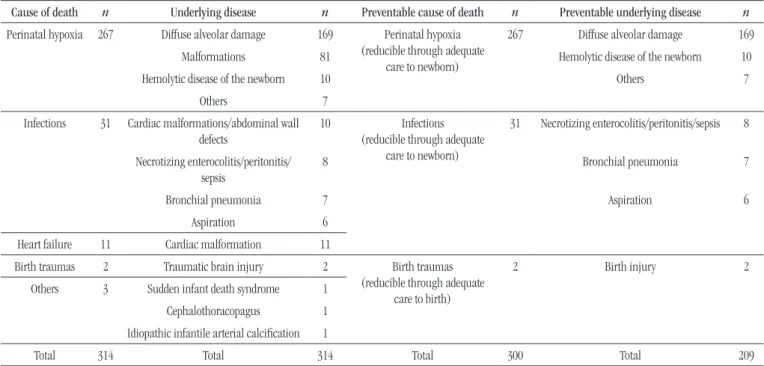
Using just annotations of necropsy reports about cause of death (irst column of Table 1) to classify these causes as preventable or not, according to the list of avoidable death causes in children under 5 years published by the Brazilian Ministry of Health (previously speciied), we observed that 300 (95.54%) cases (third column of Table 1) would be considered preventable causes of death. However, analyzing the annotations of death causes together with those of the underlying disease (second column of Table 1), probably only 209 (66.56%) cases would be avoidable causes (fourth column of Table 1).
The results obtained about survival, birth weight, gestational age, irst- and ifth-minute Apgar score, presence or absence of acidosis, presence or absence of cyanosis, presence or absence of meconium aspiration signs, and need for oxygen resuscitation are shown in Table 2.
The group with gestational age > 37 weeks presented longer survival (> 7 days, on average) than the group of 24-33 weeks of gestational age, with an average of less than 7 days of life (p = 0.012).
An Apgar score < 6 in the irst minute and/or ifth minute was another signiicant factor in the reduction of newborns’ life time
(p < 0.001), as demonstrated in the Figure.
The presence of cyanosis was statistically higher in the group of shorter survival, that is, in the group of those who presented less than seven days of postneonatal life (p = 0.02).
Perinatal hypoxia was the main death cause in this study and was associated with shorter survival of newborns (p < 0.001).
TABLE 1 − Causes of death and underlying diseases found in the study (n = 314), as well as preventable causes of death
Cause of death n Underlying disease n Preventable cause of death n Preventable underlying disease n Perinatal hypoxia 267 Diffuse alveolar damage 169 Perinatal hypoxia
(reducible through adequate care to newborn)
267 Diffuse alveolar damage 169
Malformations 81 Hemolytic disease of the newborn 10
Hemolytic disease of the newborn 10 Others 7
Others 7
Infections 31 Cardiac malformations/abdominal wall defects
10 Infections (reducible through adequate
care to newborn)
31 Necrotizing enterocolitis/peritonitis/sepsis 8
Necrotizing enterocolitis/peritonitis/ sepsis
8 Bronchial pneumonia 7
Bronchial pneumonia 7 Aspiration 6
Aspiration 6
Heart failure 11 Cardiac malformation 11
Birth traumas 2 Traumatic brain injury 2 Birth traumas
(reducible through adequate care to birth)
2 Birth injury 2
Others 3 Sudden infant death syndrome 1
Cephalothoracopagus 1 Idiopathic infantile arterial calciication 1
Total 314 Total 314 Total 300 Total 209
TABLE 2 − Clinical, epidemiological and laboratory profile of the studied population (n = 314)
Variable Median Average Standard deviation Minimum value Maximum value
Weight Gestational age (weeks)
Survival time (days)
1,490 g 33
1
1,680 g 32
4
919 g 4.5
5
400 g 24
1
5,700 g 42 28
n %
Sex
Total
Male Female Undetermined
142 171 1 314
45.2 54.5 0.3
First-minute Apgar
Total
< 6 > 6
211 75 286
73.8 26.2
Fifth-minute Apgar
Total
< 6 > 6
146 140 286
51 49
Total pH
Total
< 7.1 > 7.1
177 70 247
71.7 28.3
Cyanosis
Total
Present Absent
276 24 300
92 8
Signs of meconium aspiration
Total
Present Absent
42 246 288
14.6 85.4
Oxygen resuscitation
Total
Necessary Not necessary
263 38 301
There was no statistically signiicant difference when newborn survival was compared with sex (p = 0.488), birth weight
(p = 0.79), presence or absence of clinical signs of meconium
aspiration (p = 0.629), need for oxygen resuscitation (p = 0.19),
and presence or absence of acidosis (p = 0.221).
DISCUSSION
According to the Brazilian Ministry of Health, from January to June 2012, 98% of death causes in the neonatal period were considered preventable(3). In our study we observed that 95.54%
of deaths would be considered preventable causes by the Ministry of Health when we took into consideration just the immediate cause of death, according to recommendation of the list of preventable causes of death for children under ive, published by the Ministry
of Health(3). However, if we consider the immediate cause of
death together with the baseline disease found in the necropsy reports, just 66.56% could be considered preventable causes. This fact is particularly important when it comes to public health statistics collected from death certiicates. These discrepancies may be associated with the illing of death certiicates by health professionals, because death certiicates generally bring information on the cause of death in the irst line, and the underlying and correlate diseases in the three subsequent lines. Moreover, information from death certiicates tend to be more
concise, and, not rarely, imprecise. If statistics were produced based on diagnoses described in the irst line of death certiicates, that is, just with the immediate death causes, we would have a large number of preventable causes of death, as already reported by the Ministry of Health and found in this study. However, these data can be distorted by inadequate interpretations of the true event that led the patient to death. For this reason, there is a need to alert city and state professionals that work in mortality committees to consider not only the immediate cause of death, but also the
baseline disease.
Another important factor is that practically all statistics on neonatal mortality are based on death certiicates, which not always contain the ideal information for processing of these data. There are several reports in scientiic literature pointing to signiicant discrepancies between information collected from death certiicates and information collected from necropsy reports(4).
Because of this, postmortem histopathological examination is important. Necropsy studies are necessary to identify death-causing factors, and serve as quality control of diagnostic procedures and the provided treatment(5).
In order to interfere in the death process, it is necessary to know the factors that cause neonatal death and, thus, identify
avoidable deaths(4). According to Malta et al. (2007), avoidable
causes of death can be partially or totally prevented by health services and depend on the technology available at that moment. The Brazilian reality demonstrates that neonatal mortality is
FIGURE − Relationship between first- and fifth-minute Apgar score < 6 and newborn survival
A B
Apgar 1’ Apgar 5’
Accumulated proportion Accumulated proportion
< 6
≥ 6
≥ 6
< 6 1
0.9
0.8
0.7
0.6
0.5
0.4
0.3
0.2
0.1
0
1
0.9
0.8
0.7
0.6
0.5
0.4
0.3
0.2
0.1
0
0 5 10 15 20 25 30 35 0 5 10 15 20 25 30 35
considered relevant, although studies on avoidable death become important tools to improve assistance and consequently, reduction
of these deaths(3).
Preterm birth has been identiied as major contributor to
neonatal mortality(6). In our results, there was predominance of
preterm babies in the necropsy indings, representing 77.5% of the sample. Studies demonstrate that preterm newborns present higher susceptibility to adverse events. The consequences of prematurity relect both immaturity of organs and systems and intensive interventions necessary to survival(7-9). Approximately 12% of all
births in the United States are preterm, and approximately 2% are less than 32 weeks’ gestation. Prematurity is one of the leading causes of newborns’ hospitalization, morbidity and mortality. Among these causes, we may cite neurological, cardiovascular, ophthalmological, hematologic, nutritional, metabolic, gastrointestinal, kidney, immune system, temperature regulation and respiratory problems. These last ones are the most common causes of hypoxia and death in newborns(8-10).
Among all the stresses to which the newborn is subject, probably the most important and clinically relevant is hypoxia. This can be deined as an inadequate oxygen supply at cell level, most of the times indicated by the presence of cyanosis. The criteria used to characterize hypoxia, besides the presence or absence of cyanosis, are Apgar scores ≤ 6, need of continuous resuscitation, severe acidosis (pH < 7 or base deicit ≥ 16 mmol/l), and evidence of hypoxic-ischemic encephalopathy in the neurologic examination (lethargy, stupor, coma, hypotonia, or abnormal relexes)(11-14).
As a result of this study, in agreement with the literature, we present the predominance of diffuse alveolar damage as basic cause, and hypoxia as the main cause of death. These results demonstrate the importance of prematurity in the development of pulmonary lesions, which can be responsible for hypoxia just after birth or in the long term. In the histopathological study, the main indings of hypoxia are congestion, edema, and petechial hemorrhages in internal organs, with the most common being brain edema, ecchymoses and hemorrhages in lungs, heart, and thymus. The causes of perinatal hypoxia of major importance are those related to failures in the natural adaptation of the cardiovascular and respiratory system of the newborn; the most common are: apnea at birth, transient tachypnea of the newborn, hyaline membrane disease (or respiratory distress syndrome), among others. Any of these situations can cause a hypoxemic condition that may bring dangerous consequences to the baby, with serious death risk(10-12).
Sequelae of a hypoxemic event depend on its intensity and duration. In some cases, the brain may be the only affected organ.
In a series with 57 children, hypoxic-ischemic encephalopathy isolated occurred in 14 (24.5%). In another retrospective study with 130 hypoxic newborns, there were also dysfunction in the renal (70%), cardiovascular (62%), pulmonary (86%), and hepatic (85%) systems(8). Early monitoring, measures that minimize
hypoxia, and the establishment of clinical and laboratory criteria to guide this care are of utmost importance for these patients’
survival.
Quantifying hypoxia in newborns is a dificult task. The Apgar score is one of the most used parameters, and it can be low even if the newborn has not presented fetal acidosis. In most of our sample, Apgar score was < 6, both in the irst minute and in the ifth minute(15).
The deinition of hypoxia or asphyxia is confusing, because of that the American Academy of Pediatrics determined that the term asphyxia must be used in case of profound metabolic or mixed acidemia (pH < 7) in umbilical cord arterial blood, Apgar score 0-3 for longer than ive minutes, neonatal neurological manifestations (seizures, coma, hypotonia), and multisystem organ dysfunction (for example, cardiovascular system). Another concept for deinition of asphyxia is that formulated by Buonocuore et al. (2002). It uses the following parameters: umbilical cord pH < 7.2, ifth-minute Apgar of 4-6, and FiO2 ≥ 0.4 to saturate 86%. There is no consensus on the use of these criteria on neonatology services(16).
In our sample, factors such as sex, need for resuscitation, presence of meconium and acidosis were not associated with newborn survival time. Factors such as the presence of cyanosis, gestational age, cause of death, irst- and/or ifth-minute Apgar < 6, and pH value were associated with death in the irst week of life. The Brazilian Network on Neonatal Research demonstrated that factors such as gestational age, ifth-minute Apgar of 0-6, and presence of respiratory distress are associated with early neonatal death, besides presenting that fetal maturity has been the dominant predictive
variable. Geib et al. (2010) indicated male sex, prematurity,
low birth weight, and irst- and ifth-minute Apgar score < 7 as determinant of neonatal mortality. In another study carried out with 13,399 newborns, the ifth-minute Apgar score of 4-6 demonstrated a neonatal death risk 13 times bigger. The irst-minute Apgar value was not considered useful as a death risk predictor(17-19).
RESUMO
Introdução: A análise de óbitos ocorridos no período neonatal e a associação desses dados aos de necrópsias são fundamentais no auxílio à redução da taxa de mortalidade infantil no mundo. Objetivos: Observar as causas evitáveis de morte e os fatores associados ao maior risco de óbito neonatal precoce. Métodos: Foi realizado estudo transversal e descritivo de recém-nascidos que foram a óbito em um hospital da Universidade Federal do Paraná (UFPR). Foram selecionados 314 casos de necrópsias e analisadas as causas evitáveis de morte, tempo de sobrevida, gênero, peso, idade gestacional, índice de Apgar do primeiro e do quinto minuto, cianose, acidose, aspiração meconial, necessidade de reanimação com oxigênio, causa de morte e doença básica. Resultados: Quando se analisa apenas a causa de morte, 300 casos (95,54%) seriam de causas evitáveis, porém, quando se analisa a doença básica, o número de casos diminui para 209 (66,56%). A causa de morte mais frequente foi hipóxia (85%), e a doença básica principal foi dano alveolar difuso (52,9%). Houve associação
positiva das seguintes variáveis com o tempo de sobrevida: cianose (p = 0,02), idade gestacional (p = 0,012), causa do
óbito (p < 0,001), valor de Apgar < 6 (p < 0,001) e valor do pH (p < 0,001). Conclusão: A incidência de causa evitável de morte é provavelmente menor quando analisada concomitantemente com a doença básica. A cianose, a idade gestacional, a causa do óbito, o Apgar < 6 e o valor do pH do sangue arterial estão associados ao tempo de sobrevida de recém-nascidos.
Unitermos: recém-nascido; prematuro; causas de morte; autópsia; hipóxia fetal.
Mortality causes within this age group in Brazil, in the investigated literature, were prematurity, congenital malformations, infections, asphyxia and hypoxia. It is important to highlight that a proportionally higher death rate for infection was found in the North and Northeast Regions of Brazil; and more records of congenital malformations, in the South and Southeast Regions(22).
A study in Fortaleza indicates low birth weight and prematurity as determining factors for neonatal death(23). Another research
conducted in capitals of the Brazilian Northeast Region, in which irst-day mortality was analyzed, high neonatal mortality in this period is associated with weight, sex, vitality at birth and the worst structure of the hospital where the childbirth took place(24).
In the world scenario, a multicenter study involving India, Guatemala, Pakistan, Zambia, and Kenya, the main mortality causes found were: trauma at birth, congenital anomaly, infection, asphyxia, and prematurity complications(25).
Even in countries such as the United States of America and Canada, prematurity and its consequences are one of the main causes of neonatal mortality(26).
We have not found, in the researched literature, a Brazilian study that uses necropsy analyses to clarify the mortality cause in this age group.
CONCLUSION
The incidence of preventable causes of death is probably lower when analyzed concomitantly with the underlying disease.
Cyanosis, gestational age associated with prematurity, cause of death, Apgar score < 6, and arterial blood pH value are associated with survival time of newborns in the studied sample.
Regardless of the region of the world, prematurity, as this study demonstrates, is one of the leading neonatal mortality causes.
The study of preventable causes of death and factors that lead to early neonatal death is fundamental to reduce infant mortality rate in the world.
REFERENCES
1. Silveira MF, Santos IS, Barros AJD, et al. Increase in preterm birth in Brazil: review of population-based studies. Rev Saúde Pública. 2008; 42(5): 274-82. PubMed PMID: 18833394.
2. Brasil. Ministério da Saúde. Datasus. Informações de saúde – painel de
monitoramento de mortalidade infantil e neonatal. Available at: http://
www.datasus.gov.br. [Accessed on: 20 Dec. 2012].
3. Malta DC, Duarte EC, Almeida MF, et al. Lista de causas de mortes evitáveis por intervenções do Sistema Único de Saúde do Brasil. Epidemiol Serv Saúde. 2007; 16(4): 233-44. Available at http://scielo.iec.pa.gov.br/ scielo.php?script=sci_arttext&pid=S167949742007000400002&lng=pt. http://dx.doi.org/10.5123/S1679-49742007000400002.
CORRESPONDING AUTHOR
Lúcia de Noronha
Laboratório de Patologia Experimental; Centro de Ciências Biológicas e da Saúde, Campus 1; PUCPR; Rua Imaculada Conceição, 1.155; Prado Velho; CEP: 80215-901; Curitiba-PR, Brasil; e-mail: [email protected].
5. Laurenti R, Mello J, Prado MH; Lebrão ML; Gotlieb SLD. Estatísticas de Saúde. São Paulo: DU/EDUSP; 1985.
6. Noronha L, Martins VDM, Bones RB, et al. Mortalidade intra-uterina e perinatal: análise comparativa de 3.904 necropsias do Hospital de Clínicas de Curitiba no período de 1960 a 1995. J Pediatr (Rio de Janeiro). 2000; 76(3): 213-21.
7. Lawn JE, Cousens S, Zupan J; Lancet Neonatal Survival Steering Team. 4 million neonatal deaths: When? Where? Why? Lancet. 2005; 365(9462): 891-900. PubMed PMID: 15752534.
8. Cloherty JP, Eichenwald EC, Stark AR. Manual of neonatal care. 6 ed. Lippincott Williams & Wilkins; 2007.
9. McComick MC, Litt JS, Smith VC, Zupancic JAF. Prematurity: an overview and public health implications. Annu Rev Public Health. 2011; 32: 367-79. DOI: 10.1146/annurev-publhealth-090810-182459. 10. Reynolds EOR. Hypoxia in the newborn infant. J Clin Path Suppl (R Coll Pathol). 1977; 11: 134-41. PubMed PMID: PMC1522205. 11. Gluckman PD, Wyatt JS, Azzopardi D, et al. Selective head cooling with mild systemic hypothermia after neonatal encephalopathy: multicentre randomised trial. Lancet. 2005; 365: 663.
12. Wigglesworth S, Singer DB. Textbook of fetal and perinatal pathology. 2 ed. Massachusetts: Blackwell Science; 1998.
13. Zhang L, Xue Q. Prenatal hypoxia causes a sex-dependent increase in heart susceptibility to ischemia and reperfusion injury in adult male offspring: role of protein kinase C epsilon. J Pharmacol Exp Ther. 2009; 330(2): 624-32. PubMed PMID: 19470841.
14. Rohan AJ, Golombek SG. Hypoxia in the term newborn: part one-cardiopulmonary physiology and assessment. MCN Am J Matern Child Nurs. 2009; 34(3): 144-52. PubMed PMID: 19262264
15. Procianoy RS, Silveira RC. Síndrome hipóxico-isquêmica. J Pediatr (Rio de Janeiro). 2001; 77(supl.1): S63 -S70.
16. Cruz ACS, Ceccon MEJ. Prevalência de asixia perinatal e encefalopatia hipóxico-isquêmica em recém-nascidos de termo considerando dois critérios diagnósticos. Rev Bras Crescimento e Desenvolvimento Humano. 2010; 20(2): 302-16. Available at: http://pepsic.bvsalud.org/scielo.php?script=sci_ arttext&pid=S010412822010000200013&lng=pt&tlng=pt.
17. Almeida MFB, Guinsburg R, Martinez FE, et al. Fatores perinatais associados ao óbito precoce em prematuros nascidos nos centros da
Rede Brasileira de Pesquisas Neonatais. J Pediatr (Rio de Janeiro). 2008; 84(4): 300-7. Available at: http://dx.doi.org/10.1590/S0021-75572008000400004.
18. Geib LTC, Freu CM, Brandão M, Nunes ML. Determinantes sociais e biológicos da mortalidade infantil em coorte de base populacional em Passo Fundo, Rio Grande do Sul. Ciência & Saúde Coletiva. 2010; 15(2): 363-70. Available at: http://dx.doi.org/10.1590/S1413-81232010000200011.
19. Casey BM, McIntire DD, Leveno KJ. The continuing value of the Apgar score for the assessment of newborn infants. N Engl J Med. 2001; 344: 467-71. PubMed PMID: 11172187.
20. Huseman D, Metze B, Walch E, Buhrer C. Laboratory markers of perinatal acidosis are poor predictors of neurodevelopmental impairment in very low birth weight infants. Early Hum Dev. 2011; 87(10): 677-81. PubMed PMID: 21658869.
21. Zorzy PM, Madi JM, Rombaldi RL, et al. Fatores perinatais associados a recém-nascidos à termo com pH < 7,1 na artéria umbilical e índice de Apgar < 7 no 5º minuto. Rev Bras Ginecol Obstet. 2012; 34(8): 381-5.
Available at: http://dx.doi.org/10.1590/S0100-72032012000800007.
22. Lansky S, Friche AAL, Silva AAM, et al. Pesquisa nascer no Brasil: peril da mortalidade neonatal e avaliação da assistência a gestante e ao recém-nascido. Cad Saúde Publica, Rio de Janeiro. 2014; 30: S192- S207.
Available at: http://dx.doi.org/10.1590/0102-311x00133213.
23. Nascimento RM, Leite AJMS, Almeida NMGS, Almeida PC, Silva CF. Determinantes da mortalidade neonatal: estudo caso-controle em Fortaleza, Ceará, Brasil. Cad Saúde Pública, Rio de Janeiro. 2012; 28(3): 559-72.
24. Castro ECM, Leite AJM, Guinsburg R. Mortalidade com 24 horas de vida de recém-nascidos pré-termo de muito baixo peso da região Nordeste do Brasil. Rev Paul Pediatr. 2016; 34(1): 106-13.