Usefulness of the second heart sound for predicting pulmonary
hypertension in patients with interstitial lung disease
Utilidade da segunda bulha cardíaca na predição de hipertensão pulmonar em
portadores de doenças intersticiais pulmonares
Sandra de Barros Cobra
I, Rayane Marques Cardoso
II, Marcelo Palmeira Rodrigues
IIIUniversidade de Brasília (UnB), Brasília, Distrito Federal, Brazil
ABSTRACT
CONTEXT AND OBJECTIVE: P2 hyperphonesis is considered to be a valuable inding in semiological di-agnoses of pulmonary hypertension (PH). The aim here was to evaluate the accuracy of the pulmonary component of second heart sounds for predicting PH in patients with interstitial lung disease.
DESIGN AND SETTING: Cross-sectional study at the University of Brasilia and Hospital de Base do Distrito Federal.
METHODS: Heart sounds were acquired using an electronic stethoscope and were analyzed using phono-cardiography. Clinical signs suggestive of PH, such as second heart sound (S2) in pulmonary area louder than in aortic area; P2> A2 in pulmonary area and P2 present in mitral area, were compared with Doppler echocar-diographic parameters suggestive of PH. Sensitivity (S), speciicity (Sp) and positive (LR+) and negative (LR-) likelihood ratios were evaluated.
RESULTS: There was no signiicant correlation between S2 or P2 amplitude and PASP (pulmonary artery systolic pressure) (P = 0.185 and 0.115; P= 0.13 and 0.34, respectively). Higher S2 in pulmonary area than in aortic area, compared with all the criteria suggestive of PH, showed S = 60%, Sp= 22%; LR+ = 0.7; LR- = 1.7; while P2> A2 showed S= 57%, Sp = 39%; LR+ = 0.9; LR- = 1.1; and P2 in mitral area showed: S= 68%, Sp = 41%; LR+ = 1.1; LR- = 0.7. All these signals together showed: S= 50%, Sp = 56%.
CONCLUSIONS: The semiological signs indicative of PH presented low sensitivity and speciicity levels for clinically diagnosing this comorbidity.
RESUMO
CONTEXTO E OBJETIVO: Hiperfonese de P2 tem sido considerada como achado valoroso no diagnóstico semiológico de hipertensão pulmonar (HP). O objetivo foi de avaliar a acurácia do componente pulmonar da segunda bulha cardíaca em predizer HP nos pacientes portadores de doenças intersticiais pulmonares.
TIPO DE ESTUDO E LOCAL: Estudo transversal na Universidade de Brasília e Hospital de Base do Distrito Federal.
MÉTODOS: Os sons cardíacos foram adquiridos com estetoscópio eletrônico e analisados por fono-cardiograia. Os sinais clínicos sugestivos de HP, como B2 mais intensamente audível em área pulmonar que aórtica, P2 > A2 na área pulmonar e P2 presente em área mitral foram confrontados com parâmetros cardiográicos no exame de Doppler sugestivos de HP. Sensibilidade (S), especiicidade (E), razões de verossimilhança positiva (RV+) e negativa (RV-) foram avaliados.
RESULTADOS: Não houve correlação signiicativa entre amplitude de B2 e P2 e a PSAP (pressão sistólica arterial pulmonar) (P = 0,185 e 0,115; P = 0,13 e 0,34; respectivamente). A análise da presença de B2 mais intensa na área pulmonar que aórtica, quando comparada a todos os critérios sugestivos de HP, mostrou S = 60%; E = 22%; RV+ = 0,7; RV- = 1,7; enquanto P2 > A2 mostrou: S = 57%; E = 39%; RV+ = 0,9; RV- = 1,1; e P2 no foco mitral mostrou: S = 68%; E = 41%; RV+ = 1,1; RV- = 0,7. Todos os sinais juntos mostraram S = 50%; E = 56%.
CONCLUSÃO: Os sinais semiológicos indicativos de HP apresentam baixos valores de especiicidade e sensibilidade para diagnóstico clínico dessa comorbidade.
IMD, MSc. Cardiologist, Hospital de Base do Distrito Federal (HBDF), Brasília, Federal District, Brazil. IIMD. Resident in General Surgery, Universidade de Brasília (UnB), Brasília, Federal District, Brazil. IIIMD, MSc, PhD. Professor, School of Medicine, Universidade de Brasília (UnB), Brasília, Federal District, Brazil.
KEYWORDS:
Hypertension, pulmonary. Lung diseases, interstitial. Heart auscultation. Echocardiography. Phonocardiography.
PALAVRAS-CHAVE:
Hipertensão pulmonar. Doenças pulmonares intersticiais. Auscultação cardíaca.
INTRODUCTION
Interstitial lung diseases are a heterogeneous group of disor-ders that afect the lung parenchyma. However, despite their diferences, they all share chronic evolution associated with functional and structural deterioration of the pulmonary
paren-chyma.1 his process is oten also accompanied by pulmonary
hypertension (PH), caused either by hypoxic pulmonary vaso-constriction or direct vascular impairment of vascular function,
such as occurs in sarcoidosis.2 he presence of PH is a
predic-tor of mortality.3
Detecting the presence of PH is important because this dis-order is a determining factor for various therapeutic measures,
among them lung transplantation.3 Moreover, a inding of PH
may signal inapparent hypoxemia, such as occurs repeatedly during sleep or upon physical efort. herefore, prompt and easy identiication of factors that can provide additional informa-tion about the evoluinforma-tion of the disease is extremely important.
In 1970, Harris4 considered that both the intensity of the
sec-ond heart sound (S2) and its behavior during breathing deserved
attention during routine auscultation. A change in its
character-istics could be an early clinical sign of PH.5
In this scenario, splitting of S2 mainly occurs because of
delays in the pulmonary component (P2), although there is
a slight advance of the aortic component (A2). This occurs
even during expiration, with a delay of 0.02 to 0.03 seconds, and 0.02 in only 2% of the population, especially in
chil-dren and young adults.6
Hyperphonesis of P2 has traditionally been acknowledged
in all semiology books as indicative of PH. However, there is little evidence to support this. It is deined as more
accentu-ated presence of S2 in the pulmonic area than in the aortic area5
or, more speciically, as P2 > A2 in the pulmonic area. It shows
highly variable sensitivity (S) (96% to 58%) and speciicity (Sp)
(46% to 19%).5,7
In fact, it is not uncommon for a semiological tradition to be established based on pathophysiological deductions, with-out proper clinical validation of the inding, including its per-ceived variability, which has a direct relationship with the cred-ibility and routine application of this knowledge on a daily basis. For instance, the reliability of cardiac auscultation indings is
rarely evaluated. Regardless of these issues, hyperphonesis of P2
is still included in the guidelines for PH as an indicator of this
condition.8
If, on the one hand, the beneits arising from a useful clini-cal inding, as a means for diagnosis that is doubly accessible in terms of both cost and speed of recognition, are enormous; on the other hand, acceptance of unproven validity can be harmful to the same extent. herefore, it is increasingly important to deter-mine the accuracy and real reliability of these clinical indings.
OBJECTIVE
In this study, we aimed to evaluate the pulmonary component of
S2 as a predictor of PH in patients with interstitial lung diseases.
We also attempt to determine the pulmonary artery systolic
pres-sure (PASP) value at which the pulmonary component of S2
would be a more useful predictor of PH.
METHODS
his was a cross-sectional study from March to November 2011, in which 69 patients with various interstitial lung diseases seen in the outpatient care of a tertiary-level hospital were
consecu-tively examined. his number was deined a priori, assuming an
efect size of 0.4 w for the outcome, which represents a
moder-ate to great efect, in addition to 80% power and an α value of
5%.9 he study protocol was approved by our institution’s ethics
committee and all the participating subjects signed an informed consent form.
Each participating patient underwent cardiac auscultation in a quiet environment, in the supine position, with spontane-ous breathing. he sounds were recorded using a 3MM Littmann electronic stethoscope, model 3200 (St. Paul, MN, USA) for fur-ther analysis. Next, the patient underwent color Doppler echo-cardiography carried out by a single examiner who was unaware of any of the clinical data. Electrocardiographic monitoring was done during the test.
We evaluated 69 patients aged between 21 and 86 years, with a mean age of 58 ± 16.6 years. Twenty-eight subjects (40.6%) were male and 41 (59.4%), female. Regarding the distribution of diseases, 15 patients (21.7%) had idiopathic pulmonary ibro-sis, 22 (32%) had idiopathic interstitial diseases, 11 (16%) had interstitial lung disease associated with collagen-vascular disease, nine (13%) had sarcoidosis, seven (10.1%) had chronic hypersen-sitivity pneumonia and ive (7.2%) presented other difuse inter-stitial lung diseases.
Phonocardiogram
Phonocardiograms corresponding to heart sounds obtained by means of an electronic stethoscope were recorded in the aortic, pulmonic, mitral and tricuspid areas. he recording was done during spontaneous and continuous breathing.
he pulse tracings were transformed into signals by means of the Zargis Cardioscan heart sound analysis sotware (Princeton, NJ, USA) and were adjusted for reading in accordance with the
same measurement scale. he amplitude of S2 was measured
(with or without splitting) and the amplitude of its P2 component
was measured separately; both measurements were obtained in the pulmonic area.
he parameters subsequently evaluated were the relative
P2 of greater amplitude than A2 (P2 > A2); P2 in the mitral area;
absence of splitting of S2; and, inally, simultaneous occurrence
of all the parameters.
he analyses were performed by three independent exam-iners. hey took into consideration the sounds, the pulse trac-ings and the additional features of the sotware, which made it possible to view the spectrums of the phonographic wave forms, among other things. Decisions were then based on the consensus reached among the examiners.
Phonocardiogram results were also compared with PASP mea-surements by means of Doppler echocardiography, using Doppler and additional criteria for diagnosing PH, as described below.
Transthoracic Doppler echocardiography
For transthoracic Doppler echocardiography evaluations, the patients were examined in the let lateral decubitus position, using standard echocardiographic projections. We used an ultra-sound machine (model Vivid S5, General Electric. Milwaukee, WI, USA) with a multifrequency transducer and a frequency range from 2.5 to 3.5 MHz.
Measurements of variables relating to the heart chambers and ventricular function were obtained as established by the
American Society of Echocardiography.10
Doppler analyses were performed in real time. Doppler color low mapping in multiple views was used in order to more accu-rately measure tricuspid regurgitation. We used continuous wave Doppler ultrasound at a sweep speed of 50-100 mm/sec. hree to ive measurements per pulse tracing were taken.
To calculate PASP by measuring tricuspid regurgitation, we used the modiied Bernoulli equation. We then obtained the pressure gradient between the right ventricle (RV) and right atrium (RA). he estimated right atrial pressure was added to
this parameter,11,12 given that there was no right ventricular
out-low tract obstruction.
Right atrial pressure was obtained by assessing the percent-age collapse and the diameter of the inferior vena cava during spontaneous breathing. If the inspiratory collapse was greater than 50% and the diameter was less than 2.1 cm, the pressure added was 5 mmHg; if the inspiratory collapse was less than 50% and the diameter was greater than 2.1 cm, the pressure added was 10 mmHg; in patients where the inferior vena cava plethora was markedly greater than 2.1 cm and collapse was less than 50%, the
pressure added was 20 mmHg.13-15
Pulmonary hypertension criteria
Pulmonary hypertension was considered “probable” when PASP
was greater than 50 mmHg.15,16 It was considered “possible” when
PASP luctuated between 37 and 50 mmHg, or when it was below 37 mmHg and accompanied by additional echocardiographic
variables of PH, including the existence of dilation and/or hyper-trophy of the right chambers, paradoxical movement of the interventricular septum or right ventricular dysfunction (ana-lyzed in accordance with the recommendations of the American
Society of Echocardiography for evaluating the right chambers).15
Data analysis
Continuous variables were described as the mean plus or minus standard deviation, along with the amplitude. Categorical vari-ables were expressed as percentages. We conducted analyses
on the correlations of the amplitudes of S2and P2 in relation
to PASP with the aim of assessing the inluence of one vari-able on another. Since these varivari-ables did not show normal distribution according to the Shapiro-Wilk test, the Spearman correlation coeicient was used.
A receiver operating characteristic (ROC) curve with its components of sensitivity (S), speciicity (Sp) and positive (LR+) and negative (LR-) likelihood ratios was constructed in order to determine the discriminatory power of each parameter studied. he area under the curve was expressed in terms of the 95% con-idence interval (95% CI).
he indings were considered statistically signiicant when the probability P for two-tailed tests was P < 0.05. he data were analyzed using the Statistical Package for the Social Sciences (SPSS) sotware version 20 and Excel, both for the Mac OS X operating system.
RESULTS
he prevalence of PH in the sample, when all the echocardio-graphic criteria were taken into consideration, was 73% in patients with idiopathic pulmonary ibrosis, 41% in those with idiopathic diseases, 27% in those with collagen-vascular dis-ease, 22% in those with sarcoidosis, 25% in those with chronic hypersensitivity pneumonia and 25% in those with other difuse lung diseases. In these patients, the forced vital capacity (FVC) showed a mean of 67 ± 22.7%, with a minimum value of 18%
and maximum of 110%. Hemoglobin oxygen saturation (SpO2)
in ambient air showed a mean of 93.4 ± 4.8% and a minimum value of 70% and maximum of 99%.
PASP estimated by means of Doppler echocardiogra-phy (which was feasible in all tests) was normal in 41 patients (59.4%), while 17 (24.7%) had additional echocardiographic cri-teria that, together with PASP, were suggestive of PH (possible PH). In 11 patients (15.9%), the PASP values measured by means of Doppler indicated PH (probable PH). herefore, the combina-tion of all the criteria measured through the examinacombina-tion led us to observe PH in 28 patients (40.6%).
Table 1 shows the frequencies of the clinical indings
all phonocardiographic parameters, in comparison with Doppler
echocardiographic criteria, as shown in Tables 2 and 3.
From observing the behavior of the maximum amplitude
of S2 on phonocardiography (with or without splitting) and
the amplitude of P2, in relation to the variation of PASP, we
obtained a weak and not statistically signiicant correlation. he
correlation index ρ was 0.185 for S2 (P = 0.13) and 0.115 for P2
(P = 0.34).
In assessing the ROC curve, the best cutof point for PASP was deined as 53 mmHg. At this pressure, simultaneous pres-ence of the three clinical signs studied showed LR+ = 2.32 and LR- = 0.88. he area under the curve was 0.518 (95% CI: 0.376 to 0.659; P = 0.80). his value was very close to the limit set for probable PH. Considering the pretest probability to be the preva-lence of PH above 53 mmHg within the sample studied (which was 13%), the post-test probability would increase to 26%. For each clinical sign isolated, there were no points on the curve that yielded LR+ greater than 2 or LR- less than 0.5.
DISCUSSION
his study included patients with several types of interstitial lung diseases, with diferent FVC values and degrees of hypoxemia at the time of evaluation. We observed a range of situations: normal PASP values, mild degrees of PH and also markedly elevated levels of the disease, which constituted the later stages of this comorbidity. he prevalence of PH in these diseases varies widely accord-ing to the diagnosis and pulmonary involvement. It is also a
pre-dictor of morbidity and mortality.17-19
Doppler echocardiography has been used in other clini-cal studies to trace PH, in which the prevalence of this disorder was between 5.7% and 73.8% when pulmonary involvement was
due to sarcoidosis.20 In interstitial diseases relating to
collage-nosis, especially scleroderma, the prevalence of PH was around
18.1%.21 In idiopathic pulmonary ibrosis, these data are not yet
well deined, with the possibility of reaching 84% in patients with
advanced degrees of pulmonary involvement.22 Other authors
have also demonstrated its occurrence in one third of patients
with IPF (interstitial pulmonary ibrosis).23 In the present study,
the prevalence rates of PH were in agreement with the range of values previously reported.
Since the recognition of inspiratory splitting of the second
heart sound by Potain24 100 years ago, numerous studies have
tried to explain how these heart sound variations occur and whether these changes can be attributed to various disorders. Analyses have been conducted with the aim of comparing tra-ditional phonocardiograms with intracardiac pressure measure-ments made through cardiac catheterization, in order to relate pressure values to semiological indings.
here are no studies comparing intracardiac pressure mea-surements obtained using Doppler echocardiography with digi-tal phonocardiogram pulse tracings obtained using an electronic stethoscope, in which patients with interstitial lung disease were speciically targeted. However, the reasons that would lead to increased PASP and possible semiological changes would be sim-ilar to those found in other diseases.
he relative intensities of heart sounds are still an integral part of auscultation. In cases of PH, the explanation for indings that the pulmonary component of the second heart sound presents greater intensity than that of the aortic component is believed to be associated with hemodynamic concepts and factors
relat-ing to the anatomy of the pulmonary artery.25 However, there is
still controversy about the exact mechanism of this phenomenon.
Earlier studies25-27 indicated that the amplitude of the P
2
component in PH may not difer signiicantly from that of A2.
his would be explained by the fact that although the diastolic pressure gradient in the right ventricle is elevated in this condi-tion, it would not exceed the gradient of the let chamber. In this
regard, increased amplitude of the P2 component could only be
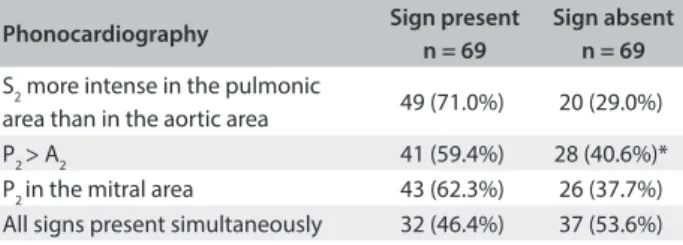
Table 1. Frequencies of clinical indings surveyed
*Splitting was not observed in 9 patients (13%).
Phonocardiography Sign present
n = 69
Sign absent n = 69 S2 more intense in the pulmonic
area than in the aortic area 49 (71.0%) 20 (29.0%)
P2 > A2 41 (59.4%) 28 (40.6%)*
P2 in the mitral area 43 (62.3%) 26 (37.7%)
All signs present simultaneously 32 (46.4%) 37 (53.6%)
Table 2. Comparison between clinical indings predictive of pulmonary hypertension (probable)
S Sp LR+ LR- P
S2 more intense in the pulmonic
area than in the aortic area 63% 27% 0.9 1.3 0.50
P2 > A2 63% 41% 1.0 0.9 0.90
P2 in the mitral area 63% 37% 1.0 0.9 0.90
All signs present simultaneously 63% 57% 1.4 0.6 0.20
S = sensitivity; Sp = speciicity; LR+ = positive likelihood ratio; and LV- = negative likelihood ratio.
Table 3. Comparison between clinical indings predictive of pulmonary hypertension (possible and probable)
S = sensitivity; Sp = speciicity; LR+ = positive likelihood ratio; and LV- = negative likelihood ratio.
S Sp LR+ LR- P
S2 more intense in the pulmonic
area than in the aortic area 60% 22% 0.7 1.7 0.12
P2 > A2 57% 39% 0.9 1.1 0.70
P2 in the mitral area 68% 41% 1.1 0.7 0.40
expected in those few patients with PH in the later stages of the disease, in which the rate of increase of this gradient would be extremely high. Nevertheless, the analysis on this component did
not show statistical signiicance.25
One anatomical factor that could also contribute towards
greater amplitude of P2 in cases of PH would be greater surface
area of the pulmonary valve and higher pulmonary artery disten-sibility, which would produce intense vibration of the semilunar valves, in comparison with the aortic valve. he combination of
these factors was signiicant.26,27 he data from our study were
consistent with the facts previously described and also showed no
relationship between higher amplitude of the P2 component
mea-sured by means of phonocardiography and elevated PASP levels measured by Doppler echocardiography.
he PASP values estimated by means of color Doppler echo-cardiography showed a good correlation with invasive measure-ments (r = 0.92). he S and Sp values for predicting PH ranged from 79 to 100% for S and from 60 to 98% for Sp, in a study
show-ing high prevalence of PH.28
hrough evaluating the presence of clinical indings sugges-tive of PH and comparing the data with measurements of PASP by means of Doppler echocardiography, we noted that our values for S and Sp and the ratios for LR+ and LR- were of low clinical relevance, even when the pulmonary pressure levels were high. he indings from clinical studies that did not report any relationship between the
relative intensities of the components of S2 found through
phonocar-diography and measurements of pulmonary pressure through
cath-eterization29 are in agreement with these data. Other clinical indings
such as P2 with higher amplitude than A2 and the presence of P2 in
the mitral area were also compared in other studies in which pres-sure meapres-surements were made by means of catheterization. here was no relationship between elevated measurements and the exis-tence of these signs. In this context, the S and Sp values for
high-amplitude P2 components were respectively 58-96% and 19-46%,
thus demonstrating a wide variation.5,7
So far, the results from rigorous analysis on the S and Sp of semiological indings predictive of PH that were associated with the second heart sound have not been conclusive. However, our results showed that the discriminatory power of each of the clini-cal parameters evaluated was not very important for the diagnos-tic suspicion of PH “at the bedside”.
It should be noted that even the data from the NIH registry, which was a relevant prospective study, refer to the existence of an accentuated pulmonary component of the second heart sound, seen on clinical examination in more than 90% of the patients
with PH, irrespective of its cause.30 However, the NIH study aimed
to investigate factors associated with survival in this population. he only concern was to report the clinical indings, without deter-mining the S and Sp of these semiological indings.
hus, considering a context in which the prevalence of PH is high, indings of physical signs with high Sp would increase the likelihood of the disease post-test. Absence of signs showing high S would practically dismiss this possibility, and this would be useful for tracing. Our data demonstrated that these signs do not have the capacity to conirm the presence or absence of the disease. Other methods such as Doppler echocardiography are required in order to diagnose this complication.
CONCLUSIONS
herefore, we can conclude that, in the context of symptomatic evaluation for predicting PH in patients with interstitial diseases, clinical signs are not useful. heir pathophysiological concepts would only be useful for academic thinking. hese signs cannot take on the function of reaching a diagnosis.
REFERENCES
1. Ryu JH, Daniels CE, Hartman TE, Yi ES. Diagnosis of interstitial lung
diseases. Mayo Clin Proc. 2007;82(8):976-86.
2. Leslie KO. Pathology of interstitial lung disease. Clin Chest Med.
2004;25(4):657-703, vi.
3. Behr J, Ryu JH. Pulmonary hypertension in interstitial lung disease.
Eur Respir J. 2008;31(6):1357-67.
4. Harris A. The second heart sound in health and in pulmonary
hypertension. Am Heart J. 1970;79(2):145-8.
5. Fowler NO, Noble WJ, Giarratano SJ, Mannix EP. The clinical estimation
of pulmonary hypertension accompanying mitral stenosis. Am Hear
J. 1955;49(2):237-49.
6. Harris A, Sutton G. Second heart sound in normal subjects. Br Heart J.
1968;30(6):739-42.
7. Whitaker W. Clinical diagnosis of pulmonary hypertension in
patients with mitral stenosis. Quarterly Journal of Medicine.
1954;23(89):105-12. Available from: http://qjmed.oxfordjournals.org/
content/23/1/105. Accessed in 2015 (Sep 15).
8. Task Force for Diagnosis and Treatment of Pulmonary Hypertension
of European Society of Cardiology (ESC); European Respiratory
Society (ERS); International Society of Heart and Lung Transplantation
(ISHLT),et al. Guidelines for diagnosis and treatment of pulmonary
hypertension. Eur Respir J. 2009;34(6):1219-63.
9. Faul F, Erdfelder E, Lang AG, Buchner A. G*Power 3: a lexible statistical
power analysis program for the social, behavioral, and biomedical
sciences. Behav Res Methods. 2007;39(2):175-91.
10. Lang RM, Bierig M, Devereux RB, et al. Recommendations for
chamber quantiication: a report from the American Society of
Echocardiography’s Guidelines and Standards Committee and the
Chamber Quantiication Writing Group, developed in conjunction
with the European Association of Echocardiography, a branch
of the European Society of Cardiology. J Am Soc Echocardiogr.
11. Berger M, Haimowitz A, Van Tosh A, Berdof RL, Goldberg E.
Quantitative assessment of pulmonary hypertension in patients with
tricuspid regurgitation using continuous wave Doppler ultrasound. J
Am Coll Cardiol. 1985;6(2):359-65.
12. Fisher MR, Foria PR, Chamera E, et al. Accuracy of Doppler
echocardiography in the hemodynamic assessment of pulmonary
hypertension. Am J Respir Crit Care Med. 2009;179(7):615-21.
13. Ommen SR, Nishimura RA, Hurrell DG, Klarich KW. Assessment of right
atrial pressure with 2-dimensional and Doppler echocardiography:
a simultaneous catheterization and echocardiographic study. Mayo
Clin Proc. 2000;75(1):24-9.
14. Currie PJ, Seward JB, Chan KL, et al. Continuous wave Doppler
determination of right ventricular pressure: a simultaneous
Doppler-catheterization study in 127 patients. J Am Coll Cardiol. 1985;6(4):750-6.
15. Rudski LG, Lai WW, Ailalo J, et al. Guidelines for the echocardiographic
assessment of the right heart in adults: a report from the American
Society of Echocardiography endorsed by the European Association
of Echocardiography, a registered branch of the European Society of
Cardiology, and the Canadian Society of Echocardiography. J Am Soc
Echocardiogr. 2010;23(7):685-713; quiz 786-8.
16. Simonneau G, Galiè N, Rubin LJ, et al. Clinical classiication of pulmonary
hypertension. J Am Coll Cardiol. 2004;43(12 Suppl S):5S-12S.
17. Olschewski H, Ghofrani HA, Walmrath D, et al. [Inhaled prostacyclin
and iloprost in severe pulmonary hypertension secondary to
pulmonary ibrosis]. Pneumologie. 2000;54(3):133-42.
18. King TE Jr, Tooze JA, Schwarz MI, Brown KR, Cherniack RM. Predicting
survival in idiopathic pulmonary ibrosis: scoring system and survival
model. Am J Respir Crit Care Med. 2001;164(7):1171-81.
19. Lee P, Langevitz P, Alderdice CA, et al. Mortality in systemic sclerosis
(scleroderma). Q J Med. 1992;82(298):139-48.
20. Handa T, Nagai S, Miki S, et al. Incidence of pulmonary hypertension
and its clinical relevance in patients with sarcoidosis. Chest.
2006;129(5):1246-52.
21. Chang B, Wigley FM, White B, Wise RA. Scleroderma patients with
combined pulmonary hypertension and interstitial lung disease. J
Rheumatol. 2003;30(11):2398-405.
22. Nadrous HF, Pellikka PA, Krowka MJ, et al. Pulmonary hypertension in
patients with idiopathic pulmonary ibrosis. Chest. 2005;128(4):2393-9.
23. Lettieri CJ, Nathan SD, Barnett SD, Ahmad S, Shorr AF. Prevalence and
outcomes of pulmonary arterial hypertension in advanced idiopathic
pulmonary ibrosis. Chest. 2006;129(3):746-52.
24. Potain C. Note sur les dédoublements normaux des bruits de coeur.
Bull Mem Soc Méd Hop Paris. 1866;3:138.
25. Sabbah HN, Stein PD. Investigation of the theory and mechanism of
the origin of the second heart sound. Circ Res. 1976;39(6):874-82.
26. Stein PD, Sabbah HN, Anbe DT, Khaja F. Hemodynamic and anatomic
determinants of relative diferences in amplitude of the aortic and
pulmonary components of the second heart sound. Am J Cardiol.
1978;42(4):539-44.
27. Kusukawa R, Bruce DW, Sakamoto T, Mac Canon DM, Luisada AA.
Hemodynamic determinants of the amplitude of the second heart
sound. J Appl Physiol. 1966;21(3):938-46.
28. McGoon M, Gutterman D, Steen V, et al. Screening, early detection,
and diagnosis of pulmonary arterial hypertension: ACCP
evidence-based clinical practice guidelines. Chest. 2004;126(1 Suppl):14S-34S.
29. Sutton G, Harris A, Leatham A. Second heart sound in pulmonary
hypertension. Br Heart J. 1968;30(6):743-56.
30. D’Alonzo GE, Barst RJ, Ayres SM, et al. Survival in patients with primary
pulmonary hypertension. Results from a national prospective
registry. Ann Intern Med. 1991;115(5):343-9.
Sources of funding: None
Conlict of interest: None
Date of irst submission: April 12, 2015
Last received: June 7, 2015
Accepted: July 12, 2015
Address for correspondence:
Sandra de Barros Cobra Negreiros
SQSW 305 — Bloco G — apto 609
Setor Sudoeste — Brasília (DF) — Brasil
CEP 70673-427
Cel. (+5561) 9972-1013
Tel. (+55 61)3361-7744