ABSTRACT
http://dx.doi.org/10.1590/1679-775720130023
The sealing ability of MTA apical plugs exposed
to a phosphate-buffered saline
Josiane de ALMEIDA1, Ana Maria Hecke ALVES2, Roberto Ferreira de MELO3, Mara Cristina Santos FELIPPE2, Eduardo Antunes BORTOLUZZI2, Cleonice da Silveira TEIXEIRA2, Wilson Tadeu FELIPPE2
1- DDS, MSc, PhD student in Endodontics, Department of Endodontics, School of Dentistry, Federal University of Santa Catarina, Florianópolis, SC, Brazil. 2- DDS, MSc, PhD, Associate Professor of Endodontics, Department of Endodontics, School of Dentistry, Federal University of Santa Catarina, Florianópolis, SC, Brazil.
3- PharmD, MSc, PhD, Associate Professor of Clinical Analisys, Department of Clinical Analisys, School of Pharmacy, Federal University of Santa Catarina, Florianópolis, SC, Brazil.
Corresponding address: Josiane de Almeida - Rua Madre Maria Villac, 76 - apto 402 - Canasvieiras - Florianópolis - SC - Brazil - 88054-000 - Phone: + 55 (0) 48 37219549 - e-mail: [email protected]
6XEPLWWHG-DQXDU\0RGL¿FDWLRQ0DUFK$FFHSWHG-XQH
O
trioxide aggregate (MTA) – with and without calcium chloride (CaCl2) – to phosphate-buffered saline (PBS) on the apical microleakage using a glucose leakage system. Material and Methods: Sixty root segments were randomly divided into 4 experimental groups (n=15). After resecting the apical segments and enlarging the canals with Gates-Glidden !!2 and the root canals were dressed with a moistened cotton pellet or PBS, as follows: 1) MTA/cotton pellet; 2) MTA/PBS; 3) MTA+10%CaCl2/cotton pellet; 4) MTA+10%CaCl2/PBS. All root segments were introduced "#$&'* evaluate the glucose leakage along the apical plugs. The amount of glucose leakage was *67&8 roots were used as controls. The data were analyzed using Kruskal-Wallis and Mann-Whitney tests (p<0.05). Results: There were no differences between groups 1 and 2 (p>0.05), and 3 and 4 (p>0.05). The addition of CaCl2** ability (p<0.05). Conclusion: The interaction with PBS did not improve the MTA sealing ability. The addition of CaCl2*&Key words:$*&&9&
INTRODUCTION
Most endodontic failures result from the passage of irritating substances from infected root canals to the periapical tissues24. Thus, any material used to seal communications between root canal and periodontium should offer an effective marginal seal7,11-13,15,21.
The sealing ability of mineral trioxide aggregate (MTA)7,11-13,15,21 is superior to other materials5,11. Notwithstanding, few studies demonstrate total **
**1,20.
The interaction of MTA with phosphate-buffered saline (PBS) positively influences its sealing ability15,16. In PBS, the MTA releases some
components, triggering the formation of carbonated apatite. This spontaneous precipitation promotes a biomineralization process that leads to the formation of an interfacial layer with tag-like structures at the cement-dentin interface18.
It has been demonstrated that the addition of calcium chloride (CaCl2) to MTA may improve its sealing ability2& E the biomineralization process since it promotes a great release of calcium ions and contributes to carbonated apatite formation at the cement-dentin interface18.
MATERIAL AND METHODS
Sixty-four extracted, human, single-rooted teeth were used. The study was approved by the Ethics Committee for Research with Human Beings of the Federal University of Santa Catarina (protocol number 2128).
The procedures were performed as described by Reyes-Carmona, et al.19 (2010). The crowns were sectioned, and a 2 mm root tip resection was performed with a high-speed bur under cooling water, so that all root segments were about 12 mm long. The canals were cleaned and shaped using #1-5 Gates-Glidden drills in a crown-down fashion, and 1% sodium hypochlorite (NaOCl) was used for irrigation. A standardized open apex was created by retrograde preparation of the canal with a #6 9V9XYZ&[\]& canal rinse was performed with 17% EDTA followed by 1% NaOCl.
$SH[L¿FDWLRQSURFHGXUHV
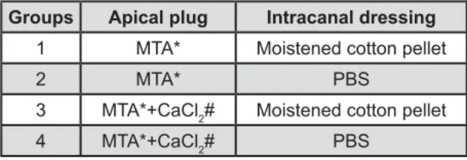
The root sections were randomly divided into 4 experimental groups (n=15). Then, the apical described in Figure 1.
MTA cement was mixed following the manufacturer’s recommendations, and MTA+CaCl2 was mixed according to Bortoluzzi, et al.2 (2009): 1 g of MTA with 0.1 g of CaCl2 mixed with 0.18 mL of H2O.
The cement mixture was introduced into the canal, condensed with moistened paper points, and compacted with pluggers (Dentsply, Tulsa Dental, Tulsa, OK, USA) to create a 4 mm thick apical plug. Radiographs were taken from all root segments to ensure void-free MTA placement and plug thickness.
In groups 1 and 3, a cotton pellet moistened with distilled water was placed in the cervical region of each root segment, which was replaced by a dry pellet after 24 h. In groups 2 and 4, the remaining "#$X`8j Dermatológica e Coméstica Ltda, Florianópolis, SC, Brazil; pH=7.2) as an intracanal dressing (Figure 1).
All access openings were covered with cotton X! Septodont Brasil Ltda, São Paulo, SP, Brazil). Thereafter, the root segments were introduced in * 20 mL PBS and stored for 2 months at 37°C.
Assembled double chamber and glucose leakage measuring
The root segments were fixed in a device designed to test glucose leakage [adapted from Leal, et al.13 (2011)]. The cervical portion of each root segment was fastened in a 2 mL Eppendorf tube with the apical 7 mm protruding through the
end. The upper portion of the Eppendorf tube was connected to a screw device through which 0.75 mL of 1 mol L-1 of glucose solution was injected. The Eppendorf tube was attached to a bucket containing 0.75 mL of deionized water, so that the apical 3 mm of the root were immersed in the water. Low-viscosity cyanoacrylate adhesive (Araldite, Brascola, Joinville, SC, Brazil) was used to seal all interfaces and connections.
For the positive control group (n=2), root segments without apical plugs were used. Two teeth with intact crowns, to which two layers of nail varnish were applied over the root surface, were used as negative control group (n=2).
A pressure of 103 KPa (15 psi) was created by a compressed air pump (Inalar Compact, NS Indústria de Aparelhos Médicos, São Paulo, SP, Brazil), which was connected to a system constituted by a manometer, a valve to control the pressure and a cannula in which the screw device, connected to the ~&* was forced into the tube for 60 min. A system was developed to run six root segments simultaneously.
A 10 μL aliquot of solution contained in the bucket (sample) was drawn using a micropipette, *** kit (Glicose Pap Liquiform, Labtest Diagnóstica, Lagoa Santa, MG, Brazil).
Each sample was analyzed using a UV/VSI spectrophotometer (BIO-2000, Bioplus 2004R, Barueri, SP, Brazil) at 505 nm wavelength to obtain converted to glucose concentration. All readings were taken in duplicate, and the mean value was considered for statistical analysis.
Statistical analysis
The Kruskal-Wallis test assessed overall differences between groups, represented by the mean values of glucose concentration. Since overall differences were observed, pairwise comparisons were performed using the Mann-Whitney U test *[&
Groups Apical plug Intracanal dressing
1 MTA* Moistened cotton pellet
2 MTA* PBS
3 MTA*+CaCl2# Moistened cotton pellet
4 MTA*+CaCl2# PBS
Figure 1- Groups, materials used to form the apical plug and intracanal dressing
*MTA Branco - Angelus Soluções Odontológicas, Londrina, PR, Brazil
RESULTS
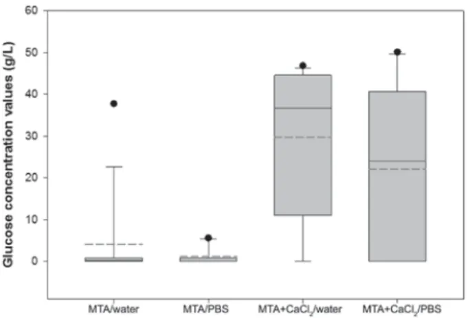
The graph in Figure 2 displays the mean, median and standard deviations and data distribution for each experimental group.
Control groups
In the negative control group no trace of glucose solution was detected, whereas in the positive control group the mean value of glucose concentration was 79 g x L-1.
Experimental groups
The number and percentage of samples that showed traces of solution, as well as the mean value of glucose concentration, are shown in Table Z& with PBS as an intracanal dressing was evaluated, no difference was observed between the results of groups 1 and 2 (p>0.05), as well as between groups 3 and 4 (p>0.05). Nevertheless, the root segments that received PBS as an intracanal dressing showed the lowest number of samples with traces of the solution and the lower concentration mean value.
!!2 to
6 * observed between the results of groups 1 and 3
(p<0.05), as well as between the results of groups 2 and 4 (p<0.05). The root segments that received MTA+CaCl2 had a higher number of samples with traces of the solution and the higher mean glucose concentration.
DISCUSSION
This method is reliable, since it has a high degree
27, overcoming most
limitations observed in other leakage tests27; and it is useful for quantitative leakage evaluation13. Besides, it has already been proven that, until up to two hours of contact between MTA and glucose,
**13.
Among the different groups, some rootsegments did not exhibit leakage. In teeth in which the apical plug was made with MTA, the sealing ability may be explained by a slight expansion that occurs after setting, which provides a better adaptation to the dentin walls22,23. As in all segments the external surface of the MTA contacted the PBS contained in 6 process in the apical third of the plugs19 additionally contributed to leakage reduction.
Conversely, some samples from the experimental groups presented glucose traces, that is, the plug allowed leakage. Other authors found that, though achieving superior results compared with other materials7, the sealing provided by MTA is not totally 16,28. The occurrence of leakage suggests the presence of through-and-through voids in the cement mass or in the cement-dentin interface13. The presence of interconnected pores in the material’s body4 may also allow glucose leakage.
In this study, the segments that received PBS as intracanal dressing achieved better results. Other authors presented similar results when MTA was kept in contact with PBS15,16. Martin, et al.15 (2007) suggested that the continuous release of calcium and hydroxyl ions from MTA, and the later interaction with PBS, results in the deposition of carbonated apatite, which partially obliterates the spaces between MTA and the dentin, reducing the leakage. Besides, the formation of apatite on the external surface of the material contributes to the expansion of the cement9*of the surface porosities10, improving the MTA adaptation to the canal walls. Reyes-Carmona, et al.19 (2010) showed that the use of PBS as intracanal dressing in association with the PBS that diffused through the apical barrier promotes the biomineralization process in MTA apical plugs. As the crystalline precipitates that are formed on the cement surface are porous25, studies are being conducted to determine whether the interaction of the MTA with PBS promotes changes in MTA porosity or not.
Contrasting previous studies3,12, the addition Groups n (%) Glucose (g x L-1)
1 (MTA/water) 9 (60) 4.07
2 (MTA/PBS) 5 (33.33) 1.14
3 (MTA + CaCl2/
water)
13 (86.66) 27.27
4 (MTA + CaCl2/
PBS)
9 (60) 22.18
Table 1- Groups, number and percentage of samples with traces of glucose solution and concentration mean value
of CaCl2 to MTA reduced its sealing ability. When combined with CaCl2, MTA needs a smaller amount of water for mixture3, due to the hydration of the silicate17 and the hygroscopic action of CaCl
22. The immediate contact of MTA+CaCl2 with PBS or moistened cotton may have promoted greater water absorption, which changed the powder-liquid proportion and increased its porosity8,14. Above the ideal proportion, water favors the formation of many capillary pores, increasing shrinkage and cracking, with consequent loss on the sealing ability14. It must be mentioned that Dreger, et al.6 (2012) with Portland cement+CaCl2 Portland cement alone. According to the authors, the lower bioactivity was a result of addition of calcium chloride, which reduced the setting time of the cement and consequently reduced the dissolution of its components.
There were large discrepancies in the glucose concentration values when samples of a same group were compared. The high leakage values indicate the existence of wide through-and-through voids in the material, while lower values indicate the presence of a narrow through-and-through void26.
The use of PBS as intracanal dressing may improve MTA sealing. However, due to the controversies still found in microleakage studies, the results of this research must be validated by other methodologies such as the bacterial leakage test, currently in progress.
CONCLUSION
The interaction with PBS did not improve the MTA sealing ability. The addition of CaCl2 to the MTA *&
ACKNOWLEDGEMENTS
The authors would like to thank Angelus Soluções Odontológicas for kindly providing the MTA for this study. The authors deny any potential &
REFERENCES
1- Aqrabawi, J. Sealing ability of amalgam, super EBA cement, **&#`& 2000;188:266-8.
2- Bortoluzzi EA, Broon NJ, Bramante CM, Felippe WT, Tanomaru 8~& setting time, solubility, disintegration, and pH of mineral trioxide ***"&~& 2009;35:550-4.
3- Bortoluzzi EA, Broon NJ, Bramante CM, Garcia RB, Moraes IG, Bernardineli N. Sealing ability of MTA and radiopaque Portland V *& Endod. 2006;32:897-900.
4- Camilleri J, Mallia B. Evaluation of the dimensional changes of mineral trioxide aggregate sealer. Int Endod J. 2011;44:416-24. [V`#`#~`9&! V* in a standardized and reproducible way. J Endod. 2006;32:206-9. 6- Dreger LAS, Felippe WT, Reyes-Carmona JF, Felippe GS, Bortoluzzi EA, Felippe MCS. Mineral trioxide aggregate and Portland cement promote biomineralization in vivo. J Endod. 2012;38:324-9.
V8$8$E*$ $&!*V** polymicrobial marker. J Endod. 2008;34:201-3.
8- Fridland M, Rosado R. Mineral trioxide aggregate (MTA) solubility and porosity with different water-to-powder ratios. J Endod. 2003;29:814-7.
V99"`$`*~" Prati C. Kinetics of apatite formation on a calcium-silicate cement V*****V solutions. Clin Oral Invest. 2009;14:659-68.
Z\V99""!&V* (bioactivity) of ProRoot MTA. Int Endod J. 2010;43:917-29. 11- Gondim E Jr, Kim S, Souza-Filho FJ. An investigation of *V** * 7&$* Med Oral Pathol Oral Radiol Endod. 2005;99:755-60.
12- Hong ST, Bae KS, Baek SH, Kum KY, Lee W. Microleakage of accelerated mineral trioxide aggregate and Portland cement in an in vitro&~&'\\ [V&
13- Leal F, De-Deus G, Brandão C, Luna AS, Fidel SR, Souza EM. Comparison of the root-end seal provided by bioceramic repair cements and White MTA. Int Endod J. 2011;44:662-8.
14- Manual técnico: aditivos para congreto e argamassas. 39th ed.
Salvador: Vedacit; 2003.
15- Martin RL, Monticcelli F, Bracket WW, Loushine RJ, Rockman RA, Ferrari M, et al. Sealing properties of mineral trioxide ******in vitro &~&'\\ ''V[&
16- Parirokh M, Askarifard S, Mansouri S, Haghdoost AA, Raoof M, Torabinejad M. Effect of phosphate buffer saline on coronal leakage of mineral trioxide aggregate. J Oral Sci. 2009;51:187-91. 17- Ramachandran VS. Concrete admixtures handbook. New Jersey: Noyes Publications; 1984.
18- Reyes-Carmona JF, Felippe MS, Felippe WT. Biomineralization ability and interaction of mineral trioxide aggregate and white V* & Endod. 2009;35:731-6.
19- Reyes-Carmona JF, Felippe MS, Felippe WT. A phosphate-buffered saline intracanal dressing improves the biomineralization ability of mineral trioxide aggregate apical plugs. J Endod. 2010;36:1648-52.
20- Scheerer SQ, Steiman HR, Cohen J. A comparative evaluation V* in vitro leakage study using Prevotella nigrescens. J Endod. 2001;27:40-2.
21- Shahi S, Rahimi S, Hasan M, Shiezadeh V, Abdolrahimi M. Sealing ability of mineral trioxide aggregate and Portland cement for furcal perforation repair: a protein leakage study. J Oral Sci. 2009;51:601-6.
22- Sluyk SR, Moon PC, Hartwell GR. Evaluation of setting properties and retention characteristics of mineral trioxide aggregate when used as a furcation perforation repair material. J Endod. 1998;24:768-71.
23- Storm B, Eichmiller FC, Tordik PA, Goodell GG. Setting expansion of gray and white mineral trioxide aggregate and Portland cement. J Endod. 2008;34:80-2.
25- Weller RN, Tay KCY, Garret LV, Mai S, Primus CM, Gutmann JL, et al. Microscopic appearance and apical seal of root canals *V"~$ V*&E~&'\\Z V& 26- Wu MK, van der Sluis LW, Ardila CN, Wesselink PR. Fluid * V * by three different gutta-percha techniques. Int Endod J. 2003;36:533-40.