ABSTRACT
Effect of lipstick on composite resin color at
different application times
Avilmar Passos GALVÃO1 2, Luciana DANTAS34!""5
1- DDS, MSc, Private practice, Salvador , Bahia, Brazil.
2- DDS, MSc, PhD, Assistant Professor, Department of Restorative Dentistry, Dental School, Federal University of Santa Maria, Santa Maria, RS, Brazil. 3- DDS, Postgraduate student, Dental School, Piracicaba, SP, Brazil.
4- DDS, MSc, PhD, Assistant Professor, Department of Dental Practice, Dental School, Federal University of Bahia, Salvador, BA, Brazil. 5- DDS, PhD, Assistant Professor, School of Dentistry, Bahiana Foundation for Science Development, Salvador-BA, Brazil.
Corresponding address: André Mallmann - Rua Venâncio Aires, 1795 / sala 71 - Centro - Santa Maria, RS - Brasil - 97010-003 - Phone: + (55) 55 96539692 / Fax: + (55) 55 32239506 - e-mail address: [email protected]
# $%&'('(()* + %- )'(/(*-%0'/'(/(
O
resin immediately, 30 min and 24 h after photoactivation. Material and Methods: Ninety specimens were prepared with a composite resin, Filtek-Z350. Specimens were polished ! "#$%& ! "'( immediately; B- 30 min; C- 24 h) and the contact with lipstick (UF- lipstick with ultra +/( &1! that did not have any contact with lipstick (C- without lipstick). Color measurements of the specimens were carried out using a spectrophotometer (Easyshade – CIE L* a* b* system). For UF and F groups, the baseline color of the specimens was measured immediately before pigmentation and the lipsticks were applied dry after 1 hour. The excess lipstick ! ! !18 calculated and data were analyzed statistically by the Kruskal-Wallis test at 5%. Results: The means between the differences of color values were: AUF: 16.0; AF: 12.4; AC: 1.07; BUF: 9.51; BF: 8.3; BC: 0.91; CUF: 17.7; CF: 12.41; CC: 0.82. Conclusion: Groups where lipstick was applied showed greater staining than the control group at the three evaluation 1 1 groups that received lipstick application.
Key words: Composite resins. Pigmentation. Color.
INTRODUCTION
Technological advances of composite resins have resulted in improvements in their physical and chemical properties, increasing their longevity. However, despite this evolution, problems related to the intrinsic and extrinsic discoloration of composite resins still remain. Among the intrinsic factors, the most commonly reported are intimately linked to the chemical mechanism of tertiary amines, and are mainly associated with the self-cured composite resins1. These tertiary amines aid staining, leading to a change in tone from a whitish to a yellowish aspect3.
In the current light-cured composite resins,
color alteration is usually related to extrinsic factors and depends on several factors, such as staining agents2,9,16,17,20,32, surface roughness5,22,24; contact time with, or immersion in, coloring e nv i r o n m e n t s1 9 , 2 0, a n d t y p e o f c o m p o s i t e resin2,8,15,22,26.
in restorations.
There are several methods known for color evaluation of teeth and dental materials. Visual techniques measure color by subjective comparison using different color scales, from acrylic resin scales to ceramic scales. On the other hand, instrumental techniques are objective measurements obtained by devices, such as spectrophotometers, colorimeters and computerized image analysis4,12,14. Instrumental color perception has been preferred over visual methods because the instrumental process is @ 7. Digital instruments, such as colorimeters and spectrophotometers, combined with computers, generate a numeric description of colors. The spectrophotometer ! light of an object and has been used to measure the visible light of vital or extracted teeth and restorative materials12,14. Regardless the equipment used, the (CIE) L*a*b* color space system is commonly employed in studies of composite resin color stability. This system consists of the following parameters: L*, which refers to luminosity (white to black); a*, which refers to the red-green color axis and b*, which refers to yellow-blue axis.
Due to the high staining index of coloring agents on composite resins, patients are recommended to avoid contact with coloring substances for at least 24 h after completion of composite resin restorations. This is indicated because water sorption by resinous materials10 can continue for several days and depends on the composite resin matrix27.
Although several studies evaluating coloring substances, such as coffee, tea, cola-soft drinks, chlorhexidine, silver nitrate, dental bacterial plaque disclosing solutions, and oral antiseptics, no reports were found with regards to lipstick’s power of staining, although there is clinical evidence that this substance causes alterations in composite resin color. Taking into account that lipstick is a differentiated coloring agent, presenting their duration on lips, and also considering the contact time with esthetic dental materials, it is included as a pigmenting agent. This clinical situation is commonly observed when a patient receives composite resin restorations on the buccal surface of anterior teeth. However, there is no staining effect of lipstick and the necessary waiting period until it has no effect on the dental restorative materials.
stained with two types of lipstick (one with a common &J% min and 24 h after composite photoactivation,
considering composite resin without any lipstick contact as the control group.
The hypotheses tested were that 1) the composite resin presents color alteration when in contact with lipstick; 2) the composite resin presents less color alteration after longer time lapses following polymerization when compared to contact with lipstick after shorter post-polymerization time; 3) ! alteration of composite resin than lipstick with a 1
MATERIAL AND METHODS
Specimen Preparation:
Q composite resin (Filtek Z350 Shade A3 - 3M ESPE, X1 Y [Q \X'& ! internal dimensions of 6 mm (diameter) x 2 mm (thickness). Composite resin was placed in one increment and a polyester band was placed over 1 Using a glass slide to apply pressure to the polyester matrix extruded excess composite resin. The specimen was photoactivated for 20 s using a LED light source (Optilight LD MAX; Gnatus, Ribeirão Preto-SP, Brazil) with an approximate intensity of 350 mW/cm2 as measured with a radiometer (L.E.D. Radiometer, Demetron-Kerr, Danbury, CT, USA). The ( aluminum oxide abrasive disks (Sof-Lex Pop-On; 3M ESPE, St. Paul, MN, USA). After, specimens were ! "#$%& ! elapsed between composite resin photoactivation and contact with the pigmenting agent, as shown in Figure 1.
Two red lipsticks with hydrating characteristics represented pigmenting agents. One lipstick had "Y _ ` \ FPS 12; Avon, São Paulo, SP, Brazil) and other had "{ |! ` + Avon). The compositions of both lipsticks are described in Figure 2.
Color evaluation:
to blue.
Before analyzing the color, the spectrophotometer was calibrated according to the manufacturer’s recommendations, using a white calibration standard provided with the equipment. A mortise @ which was positioned over the specimens to allow the contact of tip of the spectrophotometer with specimen surface at 90° angle, standardizing all readings. To calculate the differences between the "}~& was calculated according to the following equation: }~#""}`&2"}&2"}&2)½+}`#$%" !!&+}#$%"! !&+}#$%"! reading).
Sequence of tests
For the immediately after photoactivation (0 h) groups (G1, G4 and G7): 1) surface was cleaned with absorbent paper; 2) the initial color of the specimen was recorded (average of 3 readings); 3) control group (G1) was not stained and test groups "( ( &
lipsticks; 4) after 1 h in a dry condition, the excess lipstick was removed with dry absorbent paper; 5) ! (average of 3 readings).
For the 30 min after photoactivation groups (G2, G5 and G8): 1) the specimens were immersed in deionized water for 30 min; 2) surface was cleaned with absorbent paper; 3) the initial color of the specimen was recorded (average of 3 readings); 4) control group (G2) was not stained, and test groups "( ( & lipsticks; 5) after 1 h in a dry condition, the excess lipstick was removed with dry absorbent paper; 6) ! (average of 3 readings).
For the 24 h after photoactivation groups (G3, G6 and G9): 1) the specimens were immersed for 24 h in deionized water; 2) surface cleaning with absorbent paper; 3) the initial color of the specimen was taken (average of 3 readings); 4) control group (G3) was not stained and test groups (G6- lipstick ( & were stained with the respective lipsticks; 5) After 1 h in a dry condition, the excess lipstick was removed
Composition
Composite resin Filtek Z 350 (Batch - 8NU)
Silane treated ceramic; dimethacrylate of bisphenol-A; polyethylene glycol diether; diurethane dimethacrylate; silane treated silica; methacrylate of bisphenol-A;
diglycidyl ether; dimethacrylate of 2,2'; ethylenedioxydiethylo, water.
(Batch - B2417)
Cyclomethicone; diphenyl; dimethicone; polyethylene; ethylhexyl
methoxycinnamate; acrylates/stearyl acrylate/dimethicone methacrylate copolymer; cera microcristalina; isopropryl isostearate; phenyl trimethicone; titaniumdioxide; acrylates/dimethocone copolymer; polymethilsilsequioxisano; glycerin; water; PVP/
hexadecenecopolymer; sucrose distearate; acrylatescopolymer; perfume; PVM/
triethoxysilane; lecithin/C12-15 alkyl benzoate/tocopheryl acetate/ascorbyl palmitate/betacarotene/retinyl palmitate; retinyl palmitate.
(Batch - B2327)
Ricinus communis; oil; petrolium; isopropyl palmitati; caprylic/capric triglyceride; !"# hydroxystearato; polyethyene; triisostearyl citrate; myristyl lactate; perfume;
caprylyl glycol; tocopherylacetato; acrylates copolymer; silica.
Figure 2- Composition and batch numbers of studied materials
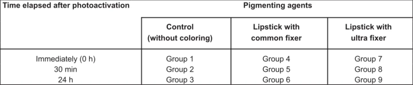
Time elapsed after photoactivation Pigmenting agents Control
(without coloring)
Lipstick with @@+B
Lipstick with +B
Immediately (0 h) Group 1 Group 4 Group 7
30 min Group 2 Group 5 Group 8
24 h Group 3 Group 6 Group 9
+&! of the specimen was taken (average of 3 readings).
Analysis of data
Data were calculated to obtain the difference "}~& and means of the 10 specimens of each group were calculated. Data were submitted to analysis by Kruskal-Wallis test for comparison at 5% !1
RESULTS
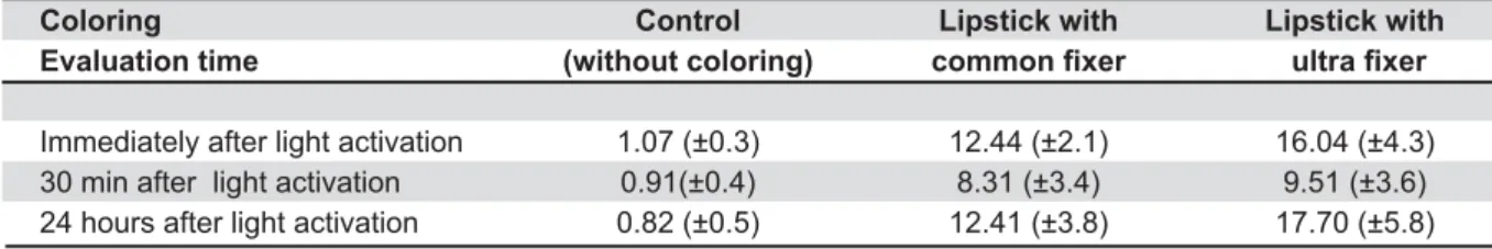
Means and standard deviations are presented $1 ! differences between the control groups (p>0.05) at the three evaluated times. The composite resin color !! all time periods with both lipsticks than the control groups did. Groups that received the lipstick with ultra fixer stained more than the groups that J% min of application. However, there was no statistical difference observed for this color difference. The common-fixer and ultra-fixer lipsticks showed greater staining values at “0 h” and after “24 h” J% ! differences between “0 h” and “24 h”. Table 2 presents the statistical analysis (Kruskal-Wallis) of ! 1
DISCUSSION
Color determination in dentistry is frequently measured by measuring reflected light by instrumental and visual means21. The instrumental method reduces the subjective potential of error on the color analysis, increases the reliability of color reading and presents great acceptance in the literature2,7,25,26. Based on these facts, the present study used the instrumental technique with a spectrophotometer, recording the color values with the CIE (Comission International l´Eclairage) L* a* b* system. This system has the ability to determine "}~& applying an equation using three co-ordinates: L* - the luminosity of a color (from white to black), a* - the color position on the red-green axis and b* - the color position on the yellow-blue axis.
Despite the fact that instrumental methods of color reading register absolute numbers, there is the possibility of clinical extrapolation of these values, as described by Johnston and Kao13 (1989). !! }~ ! than 3.3 indicate a clinically visual perception of color change. The current study observed that, on composite resins where lipstick with common ! ! was present when compared to composite resins ! hypothesis. It is interesting to point out that the alteration of the composite resin color by this pigmenting agent at different times after " !}~ 8.31 to 17.70), exceeded the clinically perceptive "}~#J1J& Kao13 (1989) and the values that are considered as a clinically unacceptable color match, according to Douglas, et al.6 (2007). Additionally, when the composite resin was not in contact with lipstick, independent of water immersion, there was little ! (from 0.82 to 1.07). The alteration of color between ! were only stored in water might have been caused by dehydration of composite resin during the "$& readings, which was used to standardize the control groups and the groups in which the material was in contact with lipstick (1 h). For the “immediate”
Coloring Control Lipstick with Lipstick with
Evaluation time (without coloring) @@+B +B
Immediately after light activation 1.07 (±0.3) 12.44 (±2.1) 16.04 (±4.3)
30 min after light activation 0.91(±0.4) 8.31 (±3.4) 9.51 (±3.6)
24 hours after light activation 0.82 (±0.5) 12.41 (±3.8) 17.70 (±5.8)
Table 1-$%&"*<$=<&%" >
0 h 30 min 24 hours
Control X Ultra 0.1 0.1 0.1
Control X Common 0.1 0.1 0.1
Common X Ultra 5 ns 5
Control Common Ultra
0h X 30min ns 1 0.1
0h X 24h ns ns ns
30min X 24h ns 1 0.1
Table 2-%?EH
statistical analysis
group (without water), this alteration was accepted as normal and can be explained as a continuation 1 discoloration that occurred with the control groups }~ were much lower at the three evaluated periods of }~ !! for clinical visualization.
Among the possible reasons for the increased staining caused by lipstick are the susceptibility of the polymer for water sorption and the hydrophilic nature of its matrix29. This water sorption is important because, if the composite resin can absorb water, it is also capable of absorbing other ! which would result in the resin discoloration. The methodology used in the current study applied lipsticks after different time periods after photoactivation was proposed because it has been suggested that, due to composite resin present initially high sorption10, the absorption of pigment is initially higher. However, the discoloration of composite resin was high even when lipstick was applied after 24 h had past after light activation, similar to the color alteration noted immediately after photoactivation. The second hypothesis of the present study was rejected, as staining by ! the conclusion of restorative procedure, but also after extended times from photoactivation. It is believed that hydration of the composite resin occurred from 24 h immersion in water. Thus, when water was removed for the application of lipstick, dehydration could have occurred in that 1 h period that the Sp stayed in the dry condition and in contact with the lipstick. Due to the hydration capacity of the lipsticks, the dehydrated composite resin could have absorbed pigments from these 1' !! composite resins were hydrated during the 30 min after photoactivation and had surface painted with 1 8 ! alterations in the 30 min groups, they presented less staining than the groups where lipstick was applied immediately or 24 h after photoactivation.
This reduced staining of composite resin after “30 min”, for both lipsticks, is most likely explained by the existence of no difference in color alteration ! ! this time. Composite resins showed higher color alteration both immediately and after “24 h” when ( 1 ! third hypothesis of the current study that the excess ( to evaluation.
It is already known that composite resins present color alteration when subjected to a coloring
environment11. Several studies have shown the !!! frequently utilized by patients. In a great number of studies, drinks such as coffee, tea and wine2,11,23,28,32, oral antiseptics, such as chlorhexidine19, water aging14,26,30 or whitening agents action are commonly used18,31. Another aspect observed in the literature is the evaluation of contact time with coloring agents and which products produce the greatest amount of staining2,32.
According to the results of this study and considering the close contact of the lips with the buccal surface of the maxillary anterior teeth, lipstick wearers should be warned to avoid the ! receiving aesthetic composite resin restorations in these teeth.
Studies analyzing possible color alteration caused by lipstick were not found in the current literature, a fact that indicated the necessity of more studies for a greater understanding of the effect of this substance on composite resins. It is valid to point out the importance of evaluating other considerable aspects of composite resins as related to staining with lipstick, such as polishing methods, application times and contact duration, as well as different kinds of composite-resin and lipstick combinations, and different brands and formulations of lipsticks. Other clinical variables should be further analyzed, such as the frequency of reapplication of lipstick, the influence of saliva and the action of the tongue in terms of cleaning the stained resin restoration surface, and others. Eventually, laboratory studies with clinical evaluations will enable the determination of parameters and protocols to reduce staining promoted by the use of products for esthetic restorations of composite resins.
CONCLUSION
Groups where lipstick was applied showed greater staining than the control group at the three evaluated time periods. It was also observed that ( ! ! ( 1' ! 1
ACKNOWLEDGEMENTS
REFERENCES
1- Asmussen E. Factors affecting the color stability of restorative resins. Acta Odontol Scand. 1983;41:11-8.
( ! [/ [1 ( ! ! restorative materials. J Dent. 2005;33:389-98.
3- Bowen RL, Argentar H. A stabilizing comonomer: I. Synthesis 18 1$+$$%$(1 4- Cal E, Sonugelen M, Guneri P, Kesercioglu A, Kose T. Application of a digital technique in evaluating the reliability of shade guides. J Oral Rehabil. 2004;31:483-91.
({!|1~ ! ! surface texture of resin composites. Dent Mater. 1994;10:325-30. 6- Douglas RD, Steinhauer TJ, Wee AG. Intraoral determination of the tolerance of dentists for perceptibility and acceptability of shade mismatch. J Prosthet Dent. 2007;97:200-8.
7- Douglas WH. Precision of in vivo colorimetric assessments of teeth. J Prosthet Dent. 1997;77:464-70.
8- Douglas WH, Craig RG. Resistance to extrinsic stains by hydrophobic composite resin systems. J Dent Res. 1982;61:41-3. (~~'\'{|~1{ of resin composites after immersion in different drinks. Dent Mater J. 2006;25:371-6.
10- Ferracane JL. Hygroscopic and hydrolytic effects in dental polymer networks. Dent Mater. 2006;22:211-22.
11- Fontes ST, Fernández MR, Moura CM, Meireles SS. Color 1 J Appl Oral Sci. 2009;17:388-91.
12- Ishikawa-Nagai S, Ishibashi K, Tsuruta O, Weber HP. Reproducibility of tooth color gradation using a computer color-matching technique applied to ceramic restorations. J Prosthet Dent. 2005;93:129-37.
13- Johnston WM, Kao EC. Assessment of appearance match by visual observation and clinical colorimetry. J Dent Res. 1989;68:819-22.
14- Joiner A. Tooth colour: a review of the literature. J Dent. 2004;32:3-12.
15- Kolbeck C, Rosentritt M, Lang R, Handel G. Discoloration of facing and restorative composites by UV-irradiation and staining food. Dent Mater. 2006;22:63-8.
$( ` [X { {~1 X composites. J Prosthet Dent. 1988;60:151-4.
17- Mair LH. Staining of in vivo subsurface degradation in dental composites with silver nitrate. J Dent Res. 1991;70:215-20.
18- Okte Z, Villalta P, Garcia-Godoy F, Lu H, Powers JM. Surface hardness of resin composite after staining and bleaching. Oper Dent. 2006;31:623-8.
19- Omata Y, Uno S, Nakaoki Y, Tanaka T, Sano H, Yoshida S, et al. Staining of hybrid composites with coffee, oolong tea, or red wine. Dent Mater J. 2006;25:125-31.
20- Patel SB, Gordan VV, Barrett AA, Shen C. The effect of surface ! ! ( composites. J Am Dent Assoc. 2004;135:587-94.
21- Powers JM, Sakaguchi RL. Craig's restorative dental materials. 12th ed. London: Mosby; 2006.
22- Reis AF, Giannini M, Lovadino JR, Ambrosano GM. Effects of various finishing systems on the surface roughness and staining susceptibility of packable composite resins. Dent Mater. 2003;19:12-8.
23- Samra AP, Pereira SK, Delgado LC, Borges CP. Color stability evaluation of aesthetic restorative materials. Braz Oral Res. 2008;22:205-10.
24- Sarac D, Sarac YS, Kulunk S, Ural C, Kulung T. The effect of polishing techniques on the surface roughness and color change of composite resins. J Prosthet Dent. 2006;96:33-40.
25- Satou N, Khan AM, Matsumae I, Satou J, Shintani H. In vitro
color change of composite-based resins. Dent Mater. 1989;5:384-7.
26- Schulze KA, Marshall SJ, Gansky SA, Marshall GW. Color stability and hardness in dental composites after accelerated aging. Dent Mater. 2003;19:612-9.
27- Silva EM, Almeida GS, Poskus LT, Guimarães JG. Relationship between the degree of conversion, solubility and salivary sorption 1 ' X1 2008;16:161-6.
28- Um CM, Ruyter IE. Staining of resin-based veneering materials with coffee and tea. Quintessence Int. 1991;22:377-86. 29- Van Noort R. Introduction to dental materials. 2nd ed. London:
Mosby Wolfe; 1994.
30- Vichi A, Ferrari M, Davidson CL. Color and opacity variations in three different resin-based composite products after water aging. Dent Mater. 2004;20:530-4.
31- Villalta P, Lu H, Okte Z, Garcia-Godoy F, Powers JM. Effects of staining and bleaching on color change of dental composite resins. J Prosthet Dent. 2006;95:137-42.