151
Radiomorphological study of the peroneus longus tendon
Radiol Bras. 2009 Mai/Jun;42(3):151–154
Original Article • Artigo Original
Radiomorphological study of the peroneus longus tendon
adjacent to the cuboid bone*
Estudo radiomorfológico do tendão fibular longo junto ao osso cuboide
Carlos Eduardo Affonso Grinbaum1, Antonio Vitor de Abreu2, Rodrigo Oliveira Carvalho de Aguiar3, Emerson Leandro Gasparetto4, Hilton Augusto Koch5
OBJECTIVE: To describe morphological aspects, and radiographically evaluate the segment of the peroneus longus tendon adjacent to the cuboid bone, in cadavers, and reporting the incidence of a fibular ossicle, in some cases with the help of histological analysis. MATERIALS AND METHODS: Fifty tendon segments were evaluated and radiographed for determining the presence or absence of an accessory ossicle inside the tendon. The specimens where such presence was dubious were sectioned and submitted to macroscopic evaluation. In the cases where doubts persisted, the specimens were submitted to hematoxylin-eosin staining for histological analysis. One specimen where the accessory ossicle had been detected was also histologically evaluated for illustration purposes. RESULTS: All the specimens presented a fusiform dilatation of the area under the arc of the cuboid bone. Radiographic images demonstrated 29 specimens without ossification, 13 with ossification and 8 were doubtful. After macroscopic analysis, only one presented ossification, five did not, and two remained dubious, being submitted to histological analysis that demonstrated no ossification. CONCLUSION: All the specimens presented focal thickening of the tendon under the arc of the cuboid bone. However, quantitative analysis of both specimens submitted to histological evaluation demonstrated that the tendon consisted of fibrocartilaginous tissue lined by hyaline cartilage on the surface of contact with the bone. Thus, under the morphological point of view, fibular ossicle was found in 28% of cases, and, under the radiographic point of view, in 26%.
Keywords: Peroneus longus; Os peroneum; Sesamoid.
OBJETIVO: Relatar os aspectos morfológicos e avaliar, radiograficamente, o segmento do tendão do mús-culo fibular longo em contato com o osso cuboide, em cadáveres, e relatar a incidência do ossímús-culo fibular em seu interior, utilizando-se, em alguns casos, da análise histológica. MATERIAIS E MÉTODOS: Foram es-tudados 50 segmentos tendinosos, sendo radiografados para determinar a presença ou não de ossículo aces-sório no interior do tendão. As peças nas quais a presença era duvidosa foram seccionadas e submetidas a avaliação macroscópica. Nos casos em que ainda persistiam dúvidas, as peças foram histologicamente ava-liadas em hematoxilina-eosina. Um segmento que demonstrou a presença de ossificação também foi anali-sado histologicamente, a título de ilustração. RESULTADOS: Todos os fragmentos apresentavam dilatação fusiforme na área de curvatura sob o cuboide. Após o estudo radiográfico, 29 desses fragmentos não ti-nham ossificação, 13 titi-nham ossificação e 8 eram duvidosos. Após a análise macroscópica, uma peça apre-sentou ossificação e cinco, não. As duas peças restantes continuaram indefinidas, sendo então analisadas histologicamente, e não se observou ossificação. CONCLUSÃO: Todas as peças demonstraram espessa-mento local na curvatura sob o cuboide. Entretanto, após análise quantitativa, nos dois casos submetidos ao estudo histológico foi verificado que o tendão era composto de fibrocartilagem e revestido por cartilagem hialina na superfície de contato com o osso. Assim, os segmentos apresentaram o ossículo fibular do ponto de vista morfológico em 28%, e do ponto de vista radiográfico, em 26% dos casos.
Unitermos: Fibular longo; Os peroneum; Sesamoide. Abstract
Resumo
* Study developed at Universidade Federal do Rio de Janeiro (UFRJ), Rio de Janeiro, RJ, Brazil.
1. Specialist in Human Anatomy, Fellow Master degree, Fa-culdade de Medicina daUniversidade Federal do Rio de Janeiro (UFRJ), Rio de Janeiro, RJ, Brazil.
2. PhD, Chief of the Orthopedic Trauma Service, Associate Professor, Faculdade de Medicina da Universidade Federal do Rio de Janeiro (UFRJ), Rio de Janeiro, RJ, Brazil.
3. PhD, Substitute Professor, Universidade Federal do Paraná (UFPR), Curitiba, PR, Brazil.
4. PhD, Associate Professor, Department of Radiology,
Facul-INTRODUCTION
The peroneus longus muscle originates at the head and the upper two thirds of the lateral surface of the fibula body and inserts itself at the lateral face of the base of the first metatarsal and medial cuneiform, at the mid foot region(1). This muscle assists
in the plantar flexion of the first radius, Grinbaum CEA, Abreu AV, Aguiar ROC, Gasparetto EL, Koch HA. Radiomorphological study of the peroneus longus tendon adjacent to the cuboid bone. Radiol Bras. 2009;42(3):151–154.
0100-3984 © Colégio Brasileiro de Radiologia e Diagnóstico por Imagem
dade de Medicina da Universidade Federal do Rio de Janeiro (UFRJ), Rio de Janeiro, RJ, Brazil.
5. PhD, Titular Professor, Department of Radiology, Faculdade de Medicina da Universidade Federal do Rio de Janeiro (UFRJ), Rio de Janeiro, RJ, Brazil.
Mailing address: Dr. Carlos Eduardo Affonso Grinbaum. Rua Marechal Rondon, 348, ap. 101, Cônego. Nova Friburgo, RJ, Brazil, 28611-130. E-mail: ceagrinbaum@gmail. com
152
Grinbaum CEA et al.
Radiol Bras. 2009 Mai/Jun;42(3):151–154 forefoot pronation and eversion, and acts
as a secondary plantar flexor of the ankle(2).
Its tendon originates in the lower third of the leg, at the posterolateral region of the ankle, and runs around three bony promi-nences on its way to the foot: the first at the ankle, posterior to the lateral malleolus and the tendon of the peroneus brevis muscle; the second at the calcaneus, around the fibular tubercle; and the third, inferiorly to the cuboid bone. This last curve is sharper, with approximately 90°, changing its course from a vertical to a horizontal direc-tion(3).
Studies have demonstrated that tendons that change their course are more prone to injury, therefore morphologic adaptations are important to enable the body segment to withstand the compression and traction forces generated in these areas(4,5), like in
the more distal curvature of the peroneus longus muscle. Another study even de-scribes the presence of an incomplete syn-ovial joint (without a disc) in this region, called fibulo-cuboid joint, separated from the tendinous sheath of the peroneus lon-gus muscle(6). Additionally, the presence of fibrocartilaginous tissue within this portion of the tendon is described, with the possible presence of an accessory intratendineal ossicle, the os peroneum(3,4,7).
The function and origin of this ossicle are controversial, as well as its incidence, that range from 2.3% to 90% among the authors(2–9). A research about anthropologic
anatomy proposes that this ossicle is a rem-nant of early primates, and is losing its func-tion in humans due to the bipedal posture(10).
Meanwhile, some studies correlate this os-sicle with an increased incidence of ten-don-specific conditions(11–13). Thus, a
bet-ter understanding of this tendinous portion of the peroneus longus is indeed necessary. The present study is aimed at describ-ing the morphological aspects and radio-graphically evaluating the segment of the peroneus longus tendon adjacent to the cuboid bone, in cadavers, and reporting the incidence of a fibular ossicle, in some cases with the help of histological analysis.
MATERIALS AND METHODS
Fifty tendinous specimens measuring approximately 5 cm in length were
col-lected from the fibulo-cuboid region of 50 cadaver feet (48 male and 2 female), with ages at death ranging from 20 to 70 years. The specimens were preserved in a 10% formalin solution. Tendons presenting tears or degeneration with a loss of > 50% in caliber at macroscopic inspection were excluded from the study. The research was developed with approval by the Commit-tee for Ethics in Research of the institution. Radiographic study was performed in order to detect the presence of the ossicle in this tendinous segment. The tendons were grouped on x-ray film, with the sur-face adjacent to the cuboid bone turned upwards. The x-ray tube was positioned one meter away from the film, where the radiographs were taken. With these results, the tendons were divided into three groups: 1) tendons with fibular ossicle; 2) tendons without fibular ossicle; 3) doubtful cases where a poorly defined increase in density was observed, without a clear evidence of ossification. A longitudinal section of all the tendons were also macroscopically evaluated along their whole length. In the cases where the ossicle presence was doubtful, microscopic studies were per-formed with hematoxylin-eosin staining with 16, 63 and 120-fold magnification. Morphology, macro- and microscopic char-acteristics were analyzed and compared with the initial radiographic study. A par-allel microscopic analysis was performed in one tendon with the fibular ossicle, in order to illustrate this fibulo-cuboid rela-tion (see Figure 3).
RESULTS
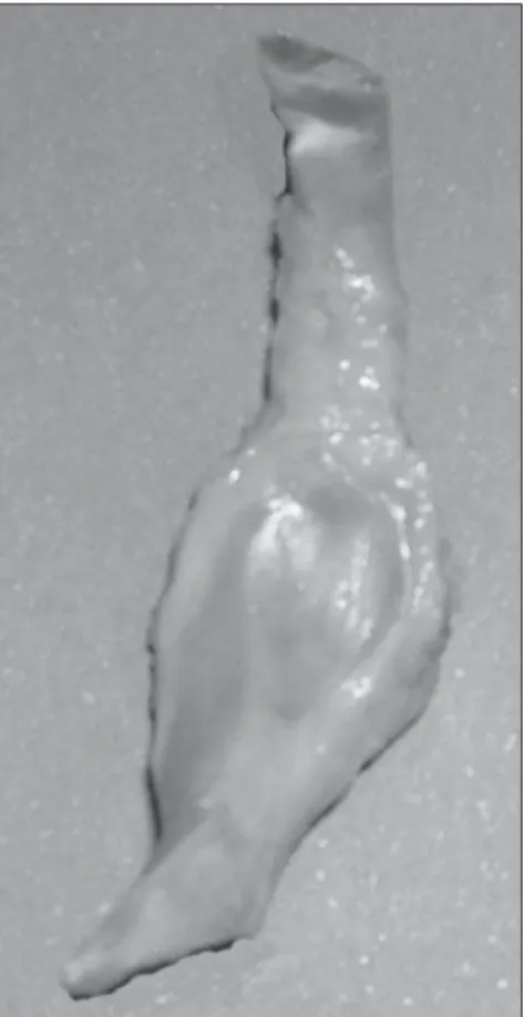
The harvested tendinous segments pre-sented a macroscopic flat, spindle-shaped morphology, with internally concave and externally convex surfaces. Another mor-phological characteristic observed in all of the specimens was a tendinous dilatation, exactly in the passage at the groove of the cuboid bone, which measured approxi-mately 3 cm in length, 2 cm in width and 1 cm in thickness (Figure 1). Radiographi-cally, the ossicle was demonstrated in 13 tendons (26% – group 1), absence of ossi-fication was observed in 29 (58% – group 2) and doubtful cases in 8 tendons (16% – group 3) (Figure 2). After macroscopic
lon-gitudinal section in all groups, the results of previous radiographic analysis were confirmed for groups 1 and 2. However, in group 3, the absence of ossification was demonstrated in five cases, and presence in only one case. The doubt remained in two tendons, whose microscopic analysis dem-onstrated absence of ossification. The final result of the study demonstrated the pres-ence of the fibular ossicle in 14 tendons (28%). In 36 tendons (72%) the accessory bone could not be visualized.
During the histological study of these two doubtful tendons and one segment with the accessory ossicle, the presence of fibrocartilaginous tissue was identified in the segment. Chondrocytes and matrix as-sociated with collagen fibers were also observed (Figure 4). The distal and proxi-mal tendon stumps showed microscopic characteristics typical of tendinous tissue, but on the internal surface of the tendon, hyaline cartilage was demonstrated at his-tology.
153
Radiomorphological study of the peroneus longus tendon
Radiol Bras. 2009 Mai/Jun;42(3):151–154 DISCUSSION
The peroneus longus tendon is exposed to trauma and degeneration, especially in the region where it bends around the cuboid bone(14,15). Anatomical adaptations,
such as the formation of a focal fibrocarti-laginous area(16) are important to reduce
friction and to resist compression and trac-tion forces(4). Compressive forces may be
responsible for this type of tissue differen-tiation, replacing the healthy tendinous tis-sue for fibrocartilage, with production of proteoglycans that retain water, increasing the resistance of the tendon against exter-nal forces.
Some studies have demonstrated that the tendon composition is variable in areas of change in direction, especially around bone structures, particularly in the pero-neus longus around the cuboid bone(4,5).
This fibrocartilaginous differentiation and mechanical forces may predispose to the development of ossification, and
conse-quently, to the formation of a fibular os-sicle(17).
Studies about the fibular ossicle are not recent, defining this condition in the pero-neus longus tendon as being a sesamoid-type bone(18). A morphological study has
demonstrated the presence of fibrocarti-laginous tissue in the fibular tendon adja-cent to the cuboid bone that may ossify and become an ossicle(19). Testut & Latarget(16)
have also mentioned this fibrocartilaginous structure susceptible to ossification. The reported incidence of fibular ossicle is vari-able, from 8.5% to 26% in anatomical re-search(8,20), and from 2.3% to 9% in
radio-logical studies(7,8). In fact, part of the
dif-ference between the rates of incidence of fibular ossicle at radiological and morpho-logical studies may be explained by sudden occurrence of calcification non-identifiable by radiography, or by less-than-optimal exposure, without inclusion of oblique view instead of dorsoplantar view (7).
Oyedele et al. have demonstrated the
pres-ence of this accessory ossicle in 90% of South African specimens, by means of ca-daver dissection, suggesting that environ-mental and ethnical factors may be associ-ated with the incidence of this ossicle(9).
Many authors agree that the presence of the fibular ossicle is actually not a mechanical advantage, and may indeed predispose the tendon to injury(11–13,21–24). The fibular os-sicle may appear as a consequence of an intense friction between the tendon and the cuboid bone, inducing the ossification of the fibrocartilaginous tissue. A comparative study has speculated that this ossicle is a structure that is disappearing in humans, being present in 100% of the primates to facilitate the peroneus longus in the abduc-tion of the hallux, important for the twee-zer-like movement of the fingers in these species(10). Hyaline cartilage was found on
the cuboid surface of the tendon in the cuboid groove, as reported by previous studies(4,6) and corroborated by the present
study. Ebraheim et al. have described the presence of a separate synovial joint be-tween the cuboid and the peroneus longus tendon, without communication with the tendinous sheath, called peroneal-cuboid joint(6). The present study evaluated only
qualitative histological aspects of the pero-neus longus tendon articular surface, and not the presence of such joint.
The present study faced some limita-tions; in situ radiographic study could not be performed to correlate the incidence of the fibular ossicle with the dissected speci-mens. Also, the radiographic study could have been performed with mammographic films and equipment, the ideal combination to investigate small calcifications. Besides that, only two doubtful specimens were submitted to histological study. Finally, considering that the present study was de-veloped with cadaver specimens, the loss of the previous clinical history as regards diseases on the lateral region of the feet may have affected the final results.
CONCLUSION
All the specimens included in the present study demonstrated focal tendon thicken-ing in the region of curvature under the cuboid. However, after quantitative analy-sis, of the two specimens submitted to
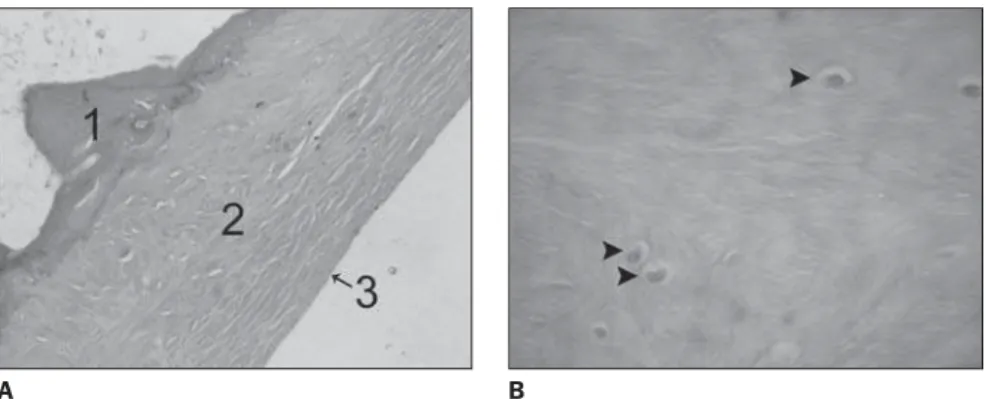
his-Figure 3. Histology of the tendinous segment. A: One specimen with fibular ossicle (1) demonstrating fibrocartilage (2) and hyaline cartilage (3) on the internal surface of peroneus longus tendon (hematoxy-lin-eosin staining, 63× magnification). B: Other specimen without the fibular ossicle, demonstrating fi-brocartilage, with chondroid cells (arrow heads) within the substance of the tendon (hematoxylin-eosin staining, 120× magnification).
A B
154
Grinbaum CEA et al.
Radiol Bras. 2009 Mai/Jun;42(3):151–154 tological study, it was observed that the
tendon was composed of fibrocartilage, and covered by hyaline cartilage on the c surface adjacent to the cuboid bone. Thus, the fibular ossicle was observed in 26% (13/50) of the specimens on the radiologi-cal study, and in 28% (14/50) of the speci-mens morphologically evaluated (macro-and microscopy). There was disagreement over one specimen that was doubtful on the radiological study and positive for the pres-ence of the ossicle at macroscopic evalua-tion.
REFERENCES
1. Warwick R, Williams PL. Gray’s Anatomy. 35th ed. Philadelphia: WB Saunders; 1973. 2. Brandes CB, Smith RW. Characterization of
pa-tients with primary peroneus longus tendinopa-thy: a review of twenty-two cases. Foot Ankle Int. 2000;21:462–8.
3. Sarrafian SK. Anatomy of the foot and ankle. Descriptive, topographic, functional. Philadel-phia: Lippincott; 1983.
4. Benjamin M, Qin S, Ralphs JR. Fibrocartilage associated with human tendons and their pulleys. J Anat. 1995;187(Pt 3):625–33.
5. Benjamin M, Ralphs JR. Fibrocartilage in tendons and ligaments – an adaptation to compressive load. J Anat. 1998;193(Pt 4):481–94.
6. Ebraheim NA, Lu J, Haman SP, et al. Cartilage and synovium of the peroneocuboid joint: an anatomic and histological study. Foot Ankle Int. 1999;20:108–11.
7. Mellado JM, Ramos A, Salvadó E, et al. Acces-sory ossicles and sesamoid bones of the ankle and foot: imaging findings, clinical significance and differential diagnosis. Eur Radiol. 2003;13 Suppl 4:L164–77.
8. Clanton TO, Schon LC. Athletic injuries to the soft tissues of the foot and ankle. In: Mann RA, Coughlin MJ, editors. Surgery of the foot and ankle. 6th ed. London: Mosby; 1993. p. 1125–6. 9. Oyedele O, Maseko C, Mkasi N, et al. High inci-dence of the os peroneum in a cadaver sample in Johannesburg, South Africa: possible clinical implications? Clin Anat. 2006;19:605–10. 10. Le Minor JM. Comparative anatomy and
signifi-cance of the sesamoid bone of the peroneus lon-gus muscle (os peroneum). J Anat. 1987;151:85– 99.
11. Brigido MK, Fessell DP, Jacobson JA, et al. Ra-diography and US of os peroneum fractures and associated peroneal tendon injuries: initial expe-rience. Radiology. 2005;237:235–41. 12. Peacock KC, Resnick EJ, Thoder JJ. Fracture of
the os peroneum with rupture of the peroneus longus tendon. A case report and review of the literature. Clin Orthop Relat Res. 1986;(202): 223–6.
13. Truong DT, Dussault RG, Kaplan PA. Fracture of the os peroneum and rupture of the peroneus lon-gus tendon as a complication of diabetic neuropa-thy. Skeletal Radiol. 1995;24:626–8.
14. O’Donnell P, Saifuddin A. Cuboid oedema due to peroneus longus tendinopathy: a report of four cases. Skeletal Radiol. 2005;34:381–8. 15. Sammarco GJ. Peroneus longus tendon tears: acute
and chronic. Foot Ankle Int. 1995;16:245–53. 16. Testut L, Latarjet A. Tratado de anatomía humana.
Barcelona: Salvat; 1959.
17. Sarin VK, Carter DR. Mechanobiology and joint conformity regulate endochondral ossification of sesamoids. J Orthop Res. 2000;18:706–12. 18. Anatomical Society. Collective investigations,
sesamoids in the gastrocnemius and peroneus longus. J Anat Physiol. 1897;32:182–6. 19. Schaeffer JP. Morris’ Human anatomy. 11th ed.
New York: The Blakiston Company; 1953. 20. Benninghoff G. Anatomia generale, anatomia
spe-ciale dell’apparato locomotore. In: Benninghoff G, Goerttler K, editors. Benninghoff-Goerttler Trattato di anatomia umana funzionale. Padova: Piccin Editore; 1978. p. 432–3.
21. Patterson MJ, Cox WK. Peroneus longus tendon rupture as a cause of chronic lateral ankle pain. Clin Orthop Relat Res. 1999;(365):163–6. 22. Rosenberg ZS, Beltran J, Bencardino JTM, et al.
From the RSNA Refresher Courses. Radiological Society of North America. MR imaging of the ankle and foot. Radiographics. 2000;20 Spec No: S153–79.
23. Peterson JJ, Bancroft LW. Os peroneal fracture with associated peroneus longus tendinopathy. AJR Am J Roentgenol. 2001;177:257–8. 24. Dombek MF, Lamm BM, Saltrick K, et al.