Antral Follicles Count and Anti-Müllerian
Hormone Levels after Gonadotoxic
Chemotherapy in Patients with Breast Cancer:
Cohort Study
Contagem de folículos antrais e níveis de hormônio
anti-Mülleriano após quimioterapia gonadotóxica em
pacientes com câncer de mama: estudo de coorte
Ângela Marcon D
’
Avila
1Edison Capp
1,2Helena von Eye Corleta
1,2,31Graduate Program in Medicine: Medical Sciences, Faculdade de
Medicina da Universidade Federal do Rio Grande do Sul (UFRGS), Porto Alegre, RS, Brazil
2Department of Obstetrics and Gynecology, Hospital de Clínicas de
Porto Alegre, Faculdade de Medicina da Universidade Federal do Rio Grande do Sul, Porto Alegre, RS, Brazil
3Generar - Reprodução Humana, Porto Alegre, Rio Grande do Sul,
Brazil
Rev Bras Ginecol Obstet 2017;39:162–168.
Address for correspondence Edison Capp, MD, Prof., Serviço de Ginecologia e Obstetrícia, Hospital de Clínicas de Porto Alegre -UFRGS, Rua Ramiro Barcelos, 2350/11° andar, Porto Alegre, RS, CEP 90035-903, Brazil (e-mail: [email protected]).
Keywords
►
anti-Müllerian
hormone
►
antral follicle count
►
ovarian reserve
►
chemotherapy-induced amenorrhea
►
anovulation
Abstract
Aim
To assess ovarian reserve (OVR) by means of follicle-stimulating hormone (FSH),
anti-Müllerian hormone (AMH), and antral follicle count (AFC) measurement in
eumenorrheic women with breast cancer, exposed to gonadotoxic chemotherapy.
Method
Fifty-two women (35.3
3.8 years old) with breast cancer and undergoing
cyclophosphamide-containing chemotherapy were enrolled. The assessment was
performed before chemotherapy (T1) and after 2 (T2) and 6 months (T3).
Results
Six months after chemotherapy, the prevalence of regular cycles was 60%.
Anti-Müllerian hormone decreased down to undetectable levels at T2 and T3 (T1: 2.53
[1.00–5.31]; T2
<
0.08; T3:
<
0.08 [
<
0.08–1.07] ng/mL), (
p
<
0.0001). Antral
folli-cle count was 11 [8.0–13.5] follifolli-cles at T1 and lower at T2 (5.50 [3.75–8.0] and T3 (5.0
[2.5–7.0]) (
p
<
0.0001). In patients who remained with regular cycles during
chemo-therapy or resumed normal menses, FSH and estradiol levels remained unchanged.
Conclusion
Anti-Müllerian hormone and AFC are useful as markers of OVR decline in
women exposed to chemotherapy. Follicle-stimulating hormone is only adequate in
women who become amenorrheic.
received
July 22, 2016
accepted after revision
January 16, 2017
DOI http://dx.doi.org/ 10.1055/s-0037-1601438.
ISSN 0100-7203.
Copyright © 2017 by Thieme-Revinter Publicações Ltda, Rio de Janeiro, Brazil
Introduction
Ovarian reserve (OVR) is the measure used to assess the capacity of the ovary to produce oocytes. It can be inferred from serum levels of follicle-stimulating hormone (FSH), inhibin B, and anti-Müllerian hormone (AMH), as well as by the antral follicle count (AFC), which is the number of follicles 2–10 mm in size as assessed by transvaginal ultrasonography during thefirst phase of the cycle. Low OVR reflects ovarian
“aging”and jeopardizes reproductive potential.1,2
A systematic review of the literature assessing several markers of OVR in the assisted reproductive technology setting was published in 2006.2Levels of estradiol, thefirst marker assessed, were not shown to be effective predictors of OVR-related outcomes, even when combined with other markers.3Follicle-stimulating hormone measurements are affected only when OVR is critically jeopardized.4,5In a 2002 study of potential predictors of poor response to ovarian stimulation for in vitro fertilization (IVF) cycles, Bancsi et al3 showed that AFC alone provided the best prognostic value as compared with FSH, inhibin B, and estradiol.
More recently, AMH has been considered the best avail-able marker of OVR, particularly as its levels do not vary over the course of the menstrual cycle, and become undetectable after menopause.6–9 Anti-Müllerian hormone is a dimeric glycoprotein produced by granulosa cells from the 3rd month of intrauterine life. Its production increases in puberty, and it is believed to exert autocrine and paracrine effects during follicle development. It modulates the primordial to primary follicle transition by inhibiting granulosa cell proliferation, aromatase activity, and luteinizing hormone (LH) receptor expression.7,10–13
In the 1950s, investigators found that women exposed to chemotherapy (CTX) developed premature ovarian failure.14 This effect was later attributed to the gonadotoxicity of chemotherapeutic agents and found even in patients who
resumed regular menses after CTX.15,16The main mecha-nism underlying CTX-induced anovulation, particularly in women treated with alkylating agents, are follicular deple-tion by apoptosis with loss of germ cells (oocytes), steroido-genic theca and granulosa cells.17
This study sought to assess OVR by means of AMH measurement in eumenorrheic women with breast cancer, exposed to gonadotoxic CTX. Other OVR markers (FSH and AFC) were compared with AMH in an attempt to ascertain which one is most sensitive for measurement of the decline in OVR after CTX.
Methods
Design
Cohort study.
Patients
The study sample comprised women with a diagnosis of breast cancer and indications for cyclophosphamide-con-taining CTX. From July of 2007 to November of 2009, 52 women aged 40 years or younger, with regular menses and no prior history of CTX (inclusion criteria), were recruited from six hospitals in Brazil. Previous ovarian surgery was an exclusion criterion. The study protocol (#07–061) was ap-proved by the corresponding Research Ethics Committees and was conducted in accordance with the Brazilian guide-lines and standards for human subject research.
Assessments were performed at baseline before the start of the CTX cycles (T1) and then at 2 (T2) and 6 months (T3) after completion of therapy. Each assessment consisted of an interview, blood sample collection for hormone measure-ment, and gynecologic ultrasonography for AFC quantifi ca-tion. The number of CTX cycles (4 or 6) and the dose of chemotherapeutic agent by total body surface area were also collected.
Palavras-chave
►
hormônio
anti-Mülleriano
►
contagem de folículos
antrais
►
reserva ovariana
►
amenorreia induzida
por quimioterapia
►
anovulação
Resumo
Objetivo
Avaliar a reserva ovariana (OVR) através da contagem de folículos antrais
(AFC), dosagem sérica de hormônio folículo estimulante (FSH) e hormônio
anti-Mülleriano (AMH) em mulheres com câncer de mama submetidas a quimioterapia
gonadotóxica.
Método
Foram incluídas na pesquisa 52 mulheres (35,3
3,8 anos) com câncer de
mama, em tratamento com quimioterapia com ciclofosfamida. As dosagens e medidas
foram realizadas antes do início da quimioterapia (T1) e após 2 (T2) e 6 meses (T3).
Resultados
Seis meses após quimioterapia, a prevalência de ciclos regulares foi de
60%. O AMH sérico diminuiu a níveis indetectáveis em T2 e T3 (T1: 2,53 [1,00–5,31] ];
T2
<
0,08; T3:
<
0,08 [
<
0,08–1,07] ng/mL) (
p
<
0,0001). A contagem de folículos
antrais foi de 11 [8,0–13,5] folículos em T1, e ainda menor em T2 (5,50 [3,75–8,0] e T3
(5,0 [2,5–7,0]), (
p
<
0,0001). Em pacientes que mantiveram ciclos regulares durante a
quimioterapia ou retomaram a menstruação normalmente, os níveis de FSH e estradiol
permaneceram inalterados.
Anti-Müllerian Hormone Measurement
Blood samples were centrifuged at 3500 rpm for 15 minutes and the supernatant serum stored at - 80°C for later analysis. Follicle-stimulating hormone and estradiol were measured by chemiluminescence performed with Siemens ADVIA Cen-taurXP Immunoassay System (Munich, Germany). Anti-Mül-lerian hormone was measured with a commercially available enzyme-linked immunosorbent assay (ELISA) kit made by Beckman Coulter - Immunotech (Marseille, France), as de-scribed by Long et al.18
Ultrasonography
All sonograms were performed by the same examiner using the Sonoline Adara system by Siemens (Munich, Germany) and a 5 MHz transvaginal transducer. In view of the urgency of instituting CTX, ultrasonography was performed on any day of the menstrual cycle. Determina-tion of the AFC took into account follicles with a mean diameter of 2–10 mm.19–21
Statistics
The sample size required to detect a 1.4 ng/mL difference in AMH levels from baseline until 6 months after CTX, with a significance level of 0.05, and a statistical power of 90%, was 44 patients. Calculation was based on thefindings of van Rooij et al.9
Data processing and analysis were performed in the SPSS 18 software (SPSS Inc., Chicago, IL, USA) environment. Data were asymmetrically distributed and are thus expressed as
medians. The following tests were used: Mann-Whitney U test and Kruskal-Wallis test for independent samples; Fried-man test for related samples, and Wilcoxon test for compar-isons. The significance level was set atp>0.05.
Results
The mean age of the patients was 35.33.8 years (range, 27–40 years). Ductal invasive carcinoma was the most com-mon histologic tumor type (51/98%) and there was one case of Paget disease (1/2%). Most cases were treated by breast-conserving surgery with adjuvant (31/60%) or neoadjuvant (21/41%) CTX and 75% of the patients received local radio-therapy. The mean length of follow-up was 143 months. Over the course of the study,five patients were lost to follow-up (three were unable to keep appointments due to disease recurrence, one discontinued treatment, and one died). Complete follow-up was obtained from 49 patients in T2 and 47 in T3.
During CTX, 40% of the women developed irregular cycles (amenorrhea or oligomenorrhea); 2 months after comple-tion of CTX (4 to 6 cycles of cyclophosphamide), 85% of the patients were irregular. At 6 months post-CTX, 60% of the patients remained with irregular menses or amenorrheic, whereas 40% had resumed normal menses.
►Table 1shows changes in the evaluated markers of OVR
before and after CTX. Baseline assessment of OVR before CTX yielded values within the expected range for women of reproductive age (►Table 2).
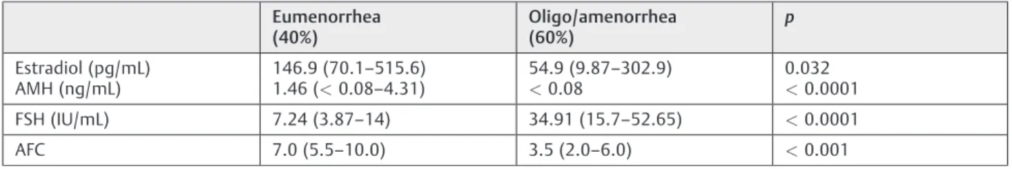
Table 2 Markers of OVR 6 months after chemotherapy (T3), stratified by menstrual status. Data expressed as median [interquartile range]
Eumenorrhea (40%)
Oligo/amenorrhea (60%)
p
Estradiol (pg/mL)
AMH (ng/mL) 146.9 (70.11.46 (<0.08–515.6)–4.31)
54.9 (9.87–302.9)
<0.08
0.032 <0.0001
FSH (IU/mL) 7.24 (3.87–14) 34.91 (15.7–52.65) <0.0001 AFC 7.0 (5.5–10.0) 3.5 (2.0–6.0) <0.001
Abbreviations: AFC, antral follicle count; AMH, anti-Müllerian hormone; FSH, follicle-stimulating hormone; OVR, ovarian reserve.
Table 1 Markers of OVR before chemotherapy (T1), at 2 (T2) and at 6 months (T3) after completion of chemotherapy. Data expressed as median [interquartile range]. The limit of detection for AMH was 0.08 ng/mL
Marker T1
n¼52
T2 n¼49
T3 n¼57
Estradiol (pg/mL) 79.42 (48.22–149) 27.55 (8.47–108.85) 95.50 (29.37–310.85)
FSH (IU/mL) 6.71 (3.87–8.64)a,b 47.67 (32.51
–88.01)a,c 16.31 (7.41
–41.16)b,c AMH (ng/mL) 2.53 (1.0–5.31)a,b <0.08a,d <0.08 (<0.08–1.07)b AFC 11.0 (8–13.5)a,b 5.50 (3.75–8.0)a 5.0 (2.5–7.0)b
aT1 versus T2;
p<0.05.
bT1 versus T3;
p<0.05.
cT2 versus T3;
p<0.05.
dThere is no variation because all patients had values below the detection limit.
There were no significant differences in estradiol levels at any of the time points of the assessment. Follicle-stimulating hormone was 6.71 [3.87–8.64] IU/mL at T1, significantly higher at T2 (47.67 [32.51–88.01] IU/mL), and significantly lower at T3 (16.31 [7.41–41.16] IU/mL), (p<0.001). Anti-Müllerian hormone levels declined significantly between T1 (2.53 [1–5.31] ng/mL) and T2 (undetectable), with p<0.0001. Six months after CTX (T3), AMH levels the same as in T2 (p¼0.128), even though some patients had resumed normal menses. The median AFC at T1 was 11 [8–
13.5] follicles, significantly higher than at T2 and T3 (p<0.0001). There were no significant differences between AFCs at T2 (5.5 [3.75–8]) and T3 (5 [2.5–7]) (►Table 1).
►Table 2shows the levels of OVR markers in
eumenor-rheic and oligo/amenoreumenor-rheic patients (40 and 60% of the sample respectively) at 6 months post-CTX (T3). There were statistically significant differences in estradiol, FSH, AMH levels, as well as in AFCs. Median estradiol in the eumenor-rhea group was 146.9 [70.1–515.6] pg/mL versus 54.9 [9.87–
302.9] pg/mL in the oligo/amenorrheic group (p¼0,032). Median FSH in the eumenorrhea group was 7.24 [3.87–14] IU/mL versus 34.91 [15.71–52.65] IU/mL in the anovulatory group (p<0.0001). Median AMH levels were 1.46 ng/mL (range, undetectable–4.31) in the eumenorrheic patients, whereas all oligo/amenorrheic women had levels below the threshold of detection (p<0.0001). Antral follicle counts also differed between the groups, with a median of 7 [5.5–10] follicles in patients who had resumed normal menses versus 3.5 [2–6] in oligo/anovulatory patients (p¼0.001) (►Table 2).
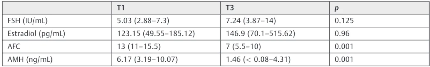
Assessment of patients who remained eumenorrheic at T3 and comparison by pre-CTX OVR markers showed that AMH and AFC declined significantly, despite normal ovulation. In these patients, FSH and estradiol remained unchanged from baseline (►Table 3).
At T1, there were significant negative correlations be-tween AMH and age (r¼-0.523,p<0.0001) and between AFC and age (r¼-0.469, p<0.001). Antral follicle count and AMH were positively correlated at T1 and T3 only (►Fig. 1).
Assessment of OVR markers was not influenced by the number of CTX cycles (4 or 6), nor by the dose of chemother-apeutic agent by total body surface area (mg/m2), (data not shown). There was no difference in OVR markers between women who received adjuvant radiation therapy and those who did not.
Discussion
This study assessed the impact of chemotherapeutic agents on OVR in 52 young women with breast cancer. From a clinical standpoint, CTX exerts rapid and major impact on ovarian function, as 60% of the patients had become oligo/ anovulatory by the end of the study. These findings are comparable to those of Stearns et al22, who reported amen-orrhea as a common event in women exposed to CTX, although it is transient in 50% of cases. Goodwin et al23 assessed women with breast cancer exposed to cyclophos-phamide-containing CTX before the age of 40 and found that 40% of them became permanently amenorrheic.24 Neverthe-less, even patients who continue to have normal menses are not spared the negative effects of CTX on ovarian function.25 In young patients, assessment of reproductive prognosis after cancer therapy required determination of OVR by reliable markers. In our study, higher AMH and AFC after CXT was associated with menstrual cyclicity. In a previous study, we have demonstrated that the age of 32 years pre-sented 96% of sensitivity and 39% of specificity to predict amenorrhea or oligomenorrhea with receiver operating characteristic area under the curve (ROC AUC) of 0.77.26
Partridge et al27 showed that CTX-induced damage to ovarian function becomes dose-dependent only after the 6th treatment cycle; this corroborates our findings, in which no significant differences in OVR or menstrual pat-tern were observed after four or six CTX cycles with cyclophosphamide.
According to Petrek et al,28 the time to resumption of menses in women with transient amenorrhea after CTX does not exceed 15 months. Therefore, assessment of OVR in our study, which was conducted on average 14 months after diagnosis and treatment of breast cancer, should accurately reflect OVR after CTX.
As shown elsewhere in the literature, estradiol was not a good marker of OVR.2,3Follicle-stimulating hormone levels followed the change in the menstrual pattern of the patients at T2 and T3, but did not represent the actual decline in reproductive capacity, as oocyte depletion occurs even with normal menses. A reliable marker of OVR should exhibit changes before amenorrhea is established. As FSH reflects these changes too late, as reported by other authors, it is not an adequate marker of OVR.4
Anti-Müllerian hormone remained stable at T2 and T3 despite resumption of menstrual cycles in some patients
Table 3 Comparative analysis of OVR markers in eumenorrheic patients before chemotherapy (T1) and 6 months after completion of chemotherapy (T3). Data expressed as median [interquartile range]
T1 T3 p
FSH (IU/mL) 5.03 (2.88–7.3) 7.24 (3.87–14) 0.125
Estradiol (pg/mL) 123.15 (49.55–185.12) 146.9 (70.1–515.62) 0.96
AFC 13 (11–15.5) 7 (5.5–10) 0.001
AMH (ng/mL) 6.17 (3.19–10.07) 1.46 (<0.08–4.31) 0.001
who were oligo/amenorrheic at T2, which demonstrates that changes in AMH reflect the true impact of CTX on OVR. As the AMH is secreted by developing follicles, their depletion after exposure to chemotherapeutic agents leads to a decline in
levels of the hormone, even in eumenorrheic patients and, according to some authors,29,30it seems to be of value in assessing ovarian function and advising patients before31 and after cancer treatment.
In 2006, Anderson et al32published the 1-year results of a cohort of 50 women with breast cancer treated with CTX. Anti-Müllerian hormone was the earliest marker of chemo-therapy-induced follicular depletion. Lie Fong et al33 com-pared AMH levels in a cohort of 185 women with regular menses who had been exposed to CTX before the age of 18 (at least 5 years before the study) to those of 42 control subjects, and found that survivors with lower levels of AMH were under greater risk of irregular menses and ovarian failure.
Antral follicle counts decrease after CTX and, despite resumption of menstrual cycles in some patients at T3, counts remained unchanged as compared with T2. These findings suggest that AFC and AMH reflect the gonadotox-icity of chemotherapeutic agents on OVR regardless of men-ses. These markers demonstrate substantial changes even before clinically apparent menstrual pattern alteration and hence, they can be used to identify significant OVR reduction (whether induced by a gonadotoxic insult or by physiological changes, such as age) in women that are still ovulatory.
Although CTX damages the ovarian stroma and affects oocytes at any stage, it appears to be particularly deleterious to primary follicles and spare resting follicles, which explains the transient anovulation exhibited by some patients at T2.34 Patients with a higher primordial follicle count (usually younger ones or those with a better pre-CTX ovarian reserve) still preserve a significant number of these follicles, which, upon development as part of the normal folliculogenesis process, lead to resumption of regular menses at T3. We believe the correlation between AMH and AFC is probably lost at T2 due to a decline in the number of AMH-producing follicles, which is expected to resume at T3, once the primor-dial follicles that remained unaffected at T2 reach the antral stage and start producing AMH.
As expected, on comparison between anovulatory (amen-orrheic or oligomen(amen-orrheic) and eumen(amen-orrheic patients at T3, all OVR-related parameters showed significant differ-ences. However, this finding does not appear relevant to determination of the optimal marker of OVR because clinical changes were already present.
Comparison of the AMH levels and AFCs of eumenorrheic patients at T3 with their T1 levels clearly showed the sensitivity of these parameters as markers of OVR, as both were decreased despite resumption of normal menses. Sim-ilarfindings were reported by de Vet et al35, who assessed ovarian aging over time among eumenorrheic patients not exposed to gonadotoxic agents. The authors found that in women whose cycles remained regular, only AMH decreased significantly over time, unlike the other markers assessed.35 In a similar study, van Rooij et al9,36concluded that AMH at any age and AFC, FSH and inhibin B in older women, accurately reflect changes in OVR among eumenorrheic women at 4-year follow-up.37
Markers that allow early identification of a decline in OVR before clinical changes in the menstrual cycle become
apparent could help to determine reproductive progno-sis.38,39This study shows that, in cases of overt decline in ovarian function induced by a proven gonadotoxic agent, AMH and AFC are efficient markers of OVR. Anti-Müllerian hormone is readily measured in serum, not investigator-dependent, and provides more reproducible results, whereas AFC can be assessed immediately on ultrasound by a gyne-cologist or sonographer.
Declaration of Interest Section
The authors report no conflict of interest.
References
1 Practice Committee of the American Society for Reproductive Medicine. Testing and interpreting measures of ovarian reserve: a committee opinion. Fertil Steril 2012;98(06):1407–1415
2 Broekmans FJ, Kwee J, Hendriks DJ, Mol BW, Lambalk CB. A systematic review of tests predicting ovarian reserve and IVF outcome. Hum Reprod Update 2006;12(06):685–718
3 Bancsi LF, Broekmans FJ, Eijkemans MJ, de Jong FH, Habbema JD, te Velde ER. Predictors of poor ovarian response in in vitro fertiliza-tion: a prospective study comparing basal markers of ovarian reserve. Fertil Steril 2002;77(02):328–336
4 Lambalk CB, van Disseldorp J, de Koning CH, Broekmans FJ. Testing ovarian reserve to predict age at menopause. Maturitas 2009; 63(04):280–291
5 Domingues TS, Rocha AM, Serafini PC. Tests for ovarian reserve: reliability and utility. Curr Opin Obstet Gynecol 2010;22(04): 271–276
6 La Marca A, Pati M, Orvieto R, Stabile G, Carducci Artenisio A, Volpe A. Serum anti-müllerian hormone levels in women with secondary amenorrhea. Fertil Steril 2006;85(05):1547–1549
7 La Marca A, Volpe A. Anti-Müllerian hormone (AMH) in female reproduction: is measurement of circulating AMH a useful tool? Clin Endocrinol (Oxf) 2006;64(06):603–610
8 D’Avila AM, Fracasso GL, Capp E, Brum IS, Corleta HE. Hormônio anti-mülleriano: um marcador maisfidedigno da reserva ovari-ana? Reprod Clim 2008;23:14–19
9 van Rooij IA, Tonkelaar Id, Broekmans FJ, et al. Anti-müllerian hormone is a promising predictor for the occurrence of the menopausal transition. Menopause 2004;11(6 Pt 1):601–606
10 van Rooij IAJ, Broekmans FJM, te Velde ER, et al. Serum anti-Müllerian hormone levels: a novel measure of ovarian reserve. Hum Reprod 2002;17(12):3065–3071
11 Durlinger AL, Gruijters MJ, Kramer P, et al. Anti-Müllerian hor-mone attenuates the effects of FSH on follicle development in the mouse ovary. Endocrinology 2001;142(11):4891–4899
12 Durlinger AL, Kramer P, Karels B, et al. Control of primordial follicle recruitment by anti-Müllerian hormone in the mouse ovary. Endocrinology 1999;140(12):5789–5796
13 La Marca A, De Leo V, Giulini S, et al. Anti-Mullerian hormone in premenopausal women and after spontaneous or surgically induced menopause. J Soc Gynecol Investig 2005;12(07): 545–548
14 Louis J, Limarzi LR, Best WR. Treatment of chronic granulocytic leukemia with myleran. AMA Arch Intern Med 1956;97(03): 299–308
15 Apperley JF, Reddy N. Mechanism and management of treatment-related gonadal failure in recipients of high dose chemoradiother-apy. Blood Rev 1995;9(02):93–116
16 Hudson MM. Reproductive outcomes for survivors of childhood cancer. Obstet Gynecol 2010;116(05):1171–1183
17 Blumenfeld Z. Ovarian rescue/protection from chemotherapeutic agents. J Soc Gynecol Investig 2001;8(1, Suppl Proceedings) S60–S64
18 Long WQ, Ranchin V, Pautier P, et al. Detection of minimal levels of serum anti-Müllerian hormone during follow-up of patients with ovarian granulosa cell tumor by means of a highly sensitive enzyme-linked immunosorbent assay. J Clin Endocrinol Metab 2000;85(02):540–544
19 Frattarelli JL, Levi AJ, Miller BT, Segars JH. A prospective assess-ment of the predictive value of basal antral follicles in in vitro fertilization cycles. Fertil Steril 2003;80(02):350–355
20 Fanchin R, Schonäuer LM, Righini C, Guibourdenche J, Frydman R, Taieb J. Serum anti-Müllerian hormone is more strongly related to ovarian follicular status than serum inhibin B, estradiol, FSH and LH on day 3. Hum Reprod 2003;18(02):323–327
21 Broekmans FJ, de Ziegler D, Howles CM, Gougeon A, Trew G, Olivennes F. The antral follicle count: practical recommendations for better standardization. Fertil Steril 2010;94(03):1044–1051
22 Stearns V, Schneider B, Henry NL, Hayes DF, Flockhart DA. Breast cancer treatment and ovarian failure: risk factors and emerging genetic determinants. Nat Rev Cancer 2006;6(11):886–893
23 Goodwin PJ, Ennis M, Pritchard KI, Trudeau M, Hood N. Risk of menopause during thefirst year after breast cancer diagnosis. J Clin Oncol 1999;17(08):2365–2370
24 Shapiro CL, Recht A. Side effects of adjuvant treatment of breast cancer. N Engl J Med 2001;344(26):1997–2008
25 Su HI, Sammel MD, Green J, et al. Antimullerian hormone and inhibin B are hormone measures of ovarian function in late reproductive-aged breast cancer survivors. Cancer 2010;116-(03):592–599
26 D’Avila AM, Biolchi V, Capp E, Corleta Hv. Age, anti-müllerian hormone, antral follicles count to predict amenorrhea or oligo-menorrhea after chemotherapy with cyclophosphamide. J Ovar-ian Res 2015;8:82
27 Partridge A, Gelber S, Gelber RD, Castiglione-Gertsch M, Gold-hirsch A, Winer E. Age of menopause among women who remain premenopausal following treatment for early breast cancer: long-term results from International Breast Cancer Study Group Trials V and VI. Eur J Cancer 2007;43(11): 1646–1653
28 Petrek JA, Naughton MJ, Case LD, et al. Incidence, time course, and determinants of menstrual bleeding after breast cancer treat-ment: a prospective study. J Clin Oncol 2006;24(07):1045–1051
29 Dunlop CE, Anderson RA. Uses of anti-Müllerian hormone (AMH) measurement before and after cancer treatment in women. Maturitas 2015;80(03):245–250
30 Bozza C, Puglisi F, Lambertini M, Osa EO, Manno M, Del Mastro L. Anti-Mullerian hormone: determination of ovarian reserve in early breast cancer patients. Endocr Relat Cancer 2014;21(01): R51–R65
31 Ruddy KJ, O’Neill A, Miller KD, et al. Biomarker prediction of chemotherapy-related amenorrhea in premenopausal women with breast cancer participating in E5103. Breast Cancer Res Treat 2014;144(03):591–597
32 Anderson RA, Themmen AP, Al-Qahtani A, Groome NP, Cameron DA. The effects of chemotherapy and long-term gonado-trophin suppression on the ovarian reserve in premenopausal women with breast cancer. Hum Reprod 2006;21(10): 2583–2592
33 Lie Fong S, Laven JS, Hakvoort-Cammel FG, et al. Assessment of ovarian reserve in adult childhood cancer survivors using anti-Müllerian hormone. Hum Reprod 2009;24(04):982–990
34 Ben-Aharon I, Shalgi R. What lies behind chemotherapy-induced ovarian toxicity? Reproduction 2012;144(02):153–163
36 van Rooij IAJ, Broekmans FJM, Scheffer GJ, et al. Serum antimul-lerian hormone levels best reflect the reproductive decline with age in normal women with proven fertility: a longitudinal study. Fertil Steril 2005;83(04):979–987
37 Di Cosimo S, Alimonti A, Ferretti G, et al. Incidence of chemother-apy-induced amenorrhea depending on the timing of treatment by menstrual cycle phase in women with early breast cancer. Ann Oncol 2004;15(07):1065–1071
38 Barnabei A, Strigari L, Marchetti P, et al. Predicting ovarian activity in women affected by early breast cancer: a meta-analysis-based nomogram. Oncologist 2015;20(10): 1111–1118