Electrophysiological Responses to Different
Follicle-Stimulating Hormone Isoforms on Human Cumulus
Oophorus Cells: Preliminary Results
Respostas eletro
fi
siológicas a diferentes isoformas de FSH em
células humanas do cumulus oophorus: resultados preliminares
Laura Silveira Ayres
1Adriana Bos-Mikich
1Nilo Frantz
2Letícia Schmidt Arruda
1Eloísa da Silveira Loss
11Department of Physiology, Instituto de Ci^encias Basicas da Saude (ICBS), Universidade Federal do Rio Grande do Sul, Porto Alegre, RS, Brazil
2Embriology Laboratory, Nilo Frantz Research and Human Reproduction Center, Porto Alegre, RS, Brazil
Rev Bras Ginecol Obstet 2018;40:763–770.
Address for correspondence Laura Silveira Ayres, PhD, Departamento de Fisiologia, Instituto de Ciências Básicas da Saúde (ICBS), Universidade Federal do Rio Grande do Sul, Rua Sarmento Leite 500, Sala 212, 90050-170, Porto Alegre, RS, Brasil (e-mail: [email protected]).
Keywords
►
ovarian stimulation
►
endocrinology
►
ovarian follicles
►
cumulus cells
►
FSH
Abstract
Objective
The aim of the present study was to provide a better understanding of the
speci
fi
c action of two follicle-stimulating hormone (FSH) isoforms (
β
-follitropin and
sheep FSH) on the membrane potential of human cumulus cells.
Methods
Electrophysiological data were associated with the characteristics of the
patient, such as age and cause of infertility. The membrane potential of cumulus cells
was recorded with borosilicate microelectrodes
fi
lled with KCl (3 M) with tip resistance
of 15 to 25 M
Ω
. Sheep FSH and
β
-follitropin were topically administered onto the cells
after stabilization of the resting potential for at least 5 minutes.
Results
In
cumulus
cells,
the
mean
resting
membrane
potential
was
-34.02
2.04 mV (
n
¼
14). The mean membrane resistance was 16.5
1.8 M
Ω
(
n
¼
14). Sheep FSH (4 mUI/mL) and
β
-follitropin (4 mUI/mL) produced depolarization
in the membrane potential 180 and 120 seconds after the administration of the
hormone, respectively.
Conclusion
Both FSH isoforms induced similar depolarization patterns, but
β
-folli-tropin presented a faster response. A better understanding of the differences of the
effects of FSH isoforms on cell membrane potential shall contribute to improve the use
of gonadotrophins in fertility treatments.
Resumo
Objetivo
O objetivo do presente estudo foi fornecer uma melhor compreensão da
ação especí
fi
ca de duas isoformas de hormônio folículo estimulante (FSH, sigla em
inglês) (
β
-folitropina e FSH ovino) no potencial de membrana de células do cumulus
oophorus humanas.
received June 6, 2018 accepted October 2, 2018
DOIhttps://doi.org/ 10.1055/s-0038-1676037. ISSN 0100-7203.
Introduction
The preovulatory follicle is surrounded by several granulosa cell layers. A specific type of granulosa cells, cumulus oophorus, arefirmly attached to each other and to the oocyte, surrounding it.1The highly specialized cumulus cells have transzonal cytoplasmic projections (TZPs).2 These projec-tions cross the zona pellucida and reach the oolemma. The TZPs present gap junctions at their endings, which allow the transfer of low-molecular weight molecules between the oocyte and the cumulus cells.2The communication between the cumulus cells and the oocyte is essential for the devel-opment of the follicle, for the maturation process of the oocyte, and for fertility.1Otherwise, the complete matura-tion of the follicle only occurs in the presence of follicle stimulating hormone (FSH).1In females, FSH has only one well known target: the follicle granulosa cells (which include cumulus cells), in which the gonadotrophin initiates and mediates multiple functions required for the maturation of the oocyte.1
Before it is released into the circulation, the FSH molecule is glycosylated by the addition of oligosaccharides in two N-linked glycosylation sites in each FSH subunit.3Each carbo-hydrate branch added to the molecule may end in a nega-tively charged sialic acid residue, conferring different isoforms of the FSH, with different isoelectric points.3It is already known that the effect of FSH on in vitro follicle culture depends on the degree of purity of the commercial preparations.4Besides, not only the hormone concentration, but also its quality, isoform type, and purity have different effects in the early phase of follicular development.4
Electrophysiological studies may provide an additional understanding of the mechanism of hormonal action. The action of FSH on the granulosa cells of swine was associated with a raise of intracellular Ca2þ
.5 Other studies using immature Sertoli cells from rats have shown that FSH causes
depolarization in the membrane potential, which is associ-ated with L-type voltage-gassoci-ated Ca2þ
channels (L-VDCC).6,7 However, to date, no studies evaluating the action of FSH on ionic channels in human cumulus oophorus cells have been found. Based on these previous studies, the aim of the present study was to standardize the intracellular electrophysiological register technique to human cumulus cells and to evaluate the effects of two FSH isoforms (β-follitropin and sheep FSH) on the membrane potential of cumulus cells. Sheep FSH was previously tested in Sertoli cells6,7 and presents a different isoelectric point from β-follitropin, which is used in human ovarian stimulation protocols. In addition, the electrophysiological data obtained in the present study were associated with some character-istics of the patient, such as age and cause of infertility.
Methods
Study Design
This is an experimental study.
Setting
The cumulus oophorus cells were obtained from an assisted reproduction center (Nilo Frantz Research, Porto Alegre, RS, Brazil). The present study was approved by the ethics com-mittee of the Universidade Federal do Rio Grande do Sul (UFRGS, in the Portuguese acronym), with the process num-ber 20173.
Participants
The criteria of eligibility for the present study were: con-senting patients, assigned to intracytoplasmic sperm injec-tion (ICSI). All of the patients who participated in the study signed the informed consent form, approved by the Ethics Committee of the UFRGS (process number 20173), before the beginning of the procedures.
Métodos
Dados eletro
fi
siológicos foram associados às características da paciente,
como idade e causa da infertilidade. O potencial de membrana das células do cumulus
foi registrado com microeletrodos de borossilicato preenchidos com KCl (3 M) com
uma resistência de 15 a 25 M
Ω
. O FSH ovino e a
β
-folitropina foram administrados
topicamente nas células após a estabilização do potencial de repouso durante pelo
menos 5 minutos.
Resultados
Nas células do cumulus, o potencial médio de membrana em repouso foi
de -34,02
2,04 mV (
n
¼
14). A resistência média da membrana foi de 16,5
1,8
M
Ω
(
n
¼
14). O FSH ovino (4 mUI/mL) e a
β
-folitropina (4 mUI/mL) produziram
despolarização no potencial de membrana 180 e 120 segundos após a aplicação do
hormônio, respectivamente.
Conclusão
Ambas as isoformas de FSH induzem padrões de despolarização
seme-lhantes, mas a
β
-folitropina apresentou uma resposta mais rápida. Uma melhor
compreensão das diferenças dos efeitos das isoformas do FSH no potencial da
membrana celular contribuirá para aprimorar o uso das gonadotro
fi
nas no estímulo
ovariano controlado e em protocolos de maturação oocitária in vitro.
Palavras-chave
Variables
A data bank containing information on age, cause of infertility, number of mature oocytes (MII), number of normal fertilized oocytes (2-pronuclei), number of embryos graded from 1 to 5, obtained from the medical history of the patients, and mean membrane potential of cumulus cells at rest was organized.
Study Size
For the intracellular registration experiments, the treat-ments were repeated at least 4 times (n¼4). The sample calculation was performed with WINPEPI software version 9 (Abramson JH and Peritz E, Hebrew University and Hadassah faculty of Medicine, Jerusalem, Israel), using a sample power of 80% and a confidence interval (CI) of 95%.
Cumulus Oophorus Cells
The collection of the oocytes was performed between 10 and 14 days after ovarian stimulation. Pituitary suppression was achieved using a gonadotropin-releasing hormone (GnRH) antagonist, and ovarian stimulation was achieved using recombinant FSH (rFSH). When at least one follicle reached 18 mm in diameter, the patients received a single dose of human chorionic gonadotropin (hCG) (10,000 IU). The col-lection of the oocyte was performed 36 hours after the administration of hCG, and the insemination was performed by ICSI. After denudation, the cumulus cells were placed in a culture dish in human tubalfluid (HTF) medium (Life Global, Guilford, CT, USA) with 10% synthetic serum substitute (SSS) (Life Global, Guilford, CT USA) and left to attach to the bottom of the dish for between 24 and 48 hours, as shown in►Fig. 1.
Solutions and Hormones
Sheep FSH (50 UI) (Sigma, St. Louis, MO, USA), andβ-follitropin (625 UI/mL) (Puregon, Merck/Schering-Plough, North Wales, PA, USA) were used at a final concentration of 4 mUI/mL. Hank’s Balanced Salt Solution (HBSS) contained: CaCl2· 2H2O, MgSO4(anhyd), KCl, KH2PO4(anhyd), NaHCO3, NaCl, Na2HPO4 (anhydrous), D-Glucose and Phenol Red · Na (H9269-1L, Sig-ma, St. Louis, MO, USA). Sodium hydroxide (NaOH [1N]) was added to this solution to reach a pH of 7.4.
Electrophysiological Experiments
The dish containing the cumulus cells was positioned in a Nikon Diaphot-TMD inverted microscope (Nikon Corporation, Tokyo, Japan) and connected to a perfusion pump tubing. The dish was then perfused with 1 mL/min of HBSS with HEPES and main-tained at 37° C in water bath (DeLeo & Cia Ltda., Porto Alegre, RS Brazil). Borosilicate microelectrodes werefilled with KCl (3 M) with a tip resistance of 15 to 25 MΩ. The intracellular recording of each cell was amplified using an Intra 767 WPI intracellular amplifier (World Precision Instruments Inc., Sarasota, FL, USA). Square current pulses of 0.5 nA, 0.5 Hz, and 250 milliseconds were applied by the microelectrode to estimate membrane resistance using the S48 stimulator (Grass Instrument, West Warwick, RI, USA). A Tektronix TDS 210 2-Channel Digital Oscilloscope (Tektronix, Beaverton, OR, USA) and the Wavestar Lite software, Version 1.0.10 (Tektronix, Beaverton, OR, USA) were used to record the variations in the membrane potential. Sheep FSH andβ-follitropin were topically administered onto the cells after the resting potential was stabilized for at least 5 minutes. Each treatment was repeated at least four times with different cells from different patients, and the variations in the membrane potential were recorded. Each cell was tested with one FSH isoform. The results are presented as meanstandard error of the mean (SEM).
Statistical Analysis
Statistical analyses were performed by one-way analysis of variance (ANOVA) with the Bonferroni posttest or with the Fischer exact test. The analyses were performed using GraphPad InStat version 3.01, 32 bits for Windows 95/NT (GraphPad Software, San Diego, CA, USA). Differences were considered significant ifp<0.05.
Results
Participants
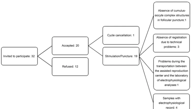
The resting membrane potential of the cumulus cells from 14 patients was recorded (►Fig. 2 presents a flowchart of
patient selection and electrophysiological data). Of these patients, six presented a male cause of infertility and eight presented female infertility. The age of the patients, the number of oocytes collected, the number of mature and fertilized oocytes, as well as the number of embryos grades 1 and 2 or grades 3, 4, and 5, human chorionic gonadotrophin (hCG) test results, and the mean cellular membrane potential recorded are shown in►Table 1.
Clinical Variables
The analysis of the resting membrane potential of the cumulus cells revealed one group of patients presenting less negative membrane potential (-6 to -16 mV), and the other group presenting a more negative membrane potential (-16 to -60 mV). A comparison between some of the characteristics of the patients from the two groups was made. It was observed that in patients with male infertility factor, most of the cells have less negative membrane potentials, whereas in the cases of poly-cystic ovary syndrome (POS), most of the cells have more negative membrane potentials (►Table 2). Comparing the Fig. 1 Cumulus cells attached to the bottom of a culture dish for
membrane potential with the age of the patients, a slight difference was observed. Women aged between 20 and 35 years old showed a tendency to present cells with less negative membrane potential when compared with older women (►Table 3). The comparison between the number of immature
and mature oocytes (MII) from patients with male factor of
infertility and from patients with POS presented no significant difference (p¼0.0941, odds ratio [OR] ¼1.792; 95%: CI: 0.9110–3.525). There was also no difference in oocyte maturity status between patients with male factor of infertility and with female factor of infertility (except POS) (p¼0.1018; OR ¼2.133; 95% CI: 0.8962–5.078). In addition, the number of
Fig. 2 Flowchart of patient selection and electrophysiological data.
Table 1 Descriptive data and follow-up of patients and samples
Patient Age (years old)
Infertility cause
Number of oocytes
MII 2PN Embryo grades 1 and 2
Embryo grades 3, 4 and 5
hCG mlU/ml Mean membrane potential (mV)
1 28 PCOS 33 21 17 3 15 No ET 35.83
2 39 OI 7 7 7 3 4 No ET 54.63
3 37 MF 15 12 7 2 7 <5 15.3
4 42 MFþOI 8 8 6 0 2 113 23.22
5 31 CR 21 8 0 0 0 No ET 14.5
6 39 PCOS 19 18 12 5 7 No ET 17.71
7 25 MF 7 6 5 4 1 No ET 15.02
8 39 PCOS 12 9 8 5 3 No ET 14.08
9 41 E 6 4 2 1 1 No ET 17.84
10 31 TF 6 5 5 1 4 No ET 11.97
11 34 MF 8 7 6 1 5 347,74 17.04
12 32 TFþUN 7 6 2 1 2 150 8.95
13 30 MFþPCOS 8 8 3 2 0 99 36.29
14 35 MF 21 17 14 10 6 No ET 6.65
fertilized oocytes (2-pronuclei) and unfertilized oocytes from patients with male factor of infertility and with POS (p¼0.6914; OR¼0.6250; 95% CI: 0.1075–3.634) or with female factor of infertility (except POS) (Fischer exact test:
p¼1.0000; OR¼1.375; 95% CI: 0.1133–16.121) presented no significant difference. For the number of embryos with better viability (grades 1 and 2) and less viability (grades 3, 4 and 5), there was also no statistical difference between the male factor, the POS or the non-POS groups (p¼0.4866, OR¼1.491; 95% CI: 0.5969–3.725).
Basal Electrophysiological Values of the Membrane of Human Cumulus Cells
In our experimental conditions, the basal electrical charac-teristics of the membrane of the cumulus cells were: resting membrane potential of 34.022.04 mV (n¼14); and resting membrane resistance of 16.54.03 MΩ(n¼14). These values remained steady for at least 5 minutes before the administration of the hormone (►Fig. 3).
Effect of Sheep FSH in the Membrane Potential of Human Cumulus Cells
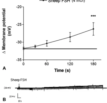
Sheep FSH (4 mUI/mL) induced depolarization in the mem-brane potential of cumulus cells. This response was signifi -cantly different from the resting value described above, after
180 seconds of FSH administration (►Fig. 4A and B). The
resistance of the membrane of the cumulus cells was not significantly affected by the experimental conditions.
Effect ofβ-follitropin in the Membrane Potential of the Cumulus Cells
Beta follitropin (4 mUI/mL) induced membrane depolariza-tion in cumulus cells. This effect was significantly different from the resting value after 120 seconds of β-follitropin administration (►Fig. 5A and B). The resistance of the
membrane of the cumulus cells was not significantly differ-ent under the experimdiffer-ental conditions.
Discussion
The standardization of the electrophysiological register tech-nique for the cumulus cells was successfully achieved and the mean resting membrane potential obtained was 34.02 2.04 mV (SEM). The mean resistance of the membrane to the ionflow was 16.51.8 MΩ(SEM). Sheep FSH application (4 mUI/mL) led to a statistically significant slow depolarization 180 seconds after the administration of the hormone (p<0.01). The administration of β-follitropin (4 mUI/mL) Table 2 Comparison between membrane potential and infertility
factors
Membrane potential (mV)
Male Factor
Female PCOS
Factor Non-PCOS
Total
6.0 to 16.0 4 (40%) 1 (11%) 2 (20%) 7 (50%)
16.1 to 60.0 1 (10%) 3 (33%) 3 (30%) 7 (50%)
Total 5 (50%) 4 (44%) 5 (50%) 14 (100%)
Abbreviation: PCOS, polycystic ovary syndrome.
Table 3 Comparison between membrane potential and patient age range
Membrane potential (mV)
20–35
years old
>35–40 years old
Total
6.0 to 16.0 5 (36%) 2 (14%) 7 (50%)
16.1 to 60.0 3 (21%) 4 (29%) 7 (50%)
Total 8 (57%) 6 (43%) 14 (100%)
Fig. 3 Recording of the membrane resting potential of a typical cumulus cell with 47.7 mV. The vertical lines provide the membrane input resistance values achieved by the application of pulses of 0.5 nA.
Fig. 4 Effect of sheep FSH on the membrane potential of cumulus cells. (A) Depolarizing effect of sheep FSH at 4 mUI/mL on the membrane potential of cumulus cells compared with the resting potential (
led to a statistically significant slow depolarization 120 and 180 seconds after the application of the hormone (p<0.001). The depolarization pattern was similar between both isoforms. Beta follitropin had a more immediate effect than sheep FSH. The limitations of the present study were the small sample size (further investigations using a large cohort are needed), the inclusion of participants with different clinical variables that may have interfered with the results (age, and cause of infertility), and that the data were obtained from samples collected after controlled ovarian stimulation, which may not necessarily be extrapolated to natural cycles. The standardization of the intracellular electrophysiolog-ical register technique to human cumulus oophorus cells was achieved based on previous studies using immature Sertoli cells from rats.6,7The pretreatment of the culture dishes was not necessary, since the cells adhered to the bottom of the dish by themselves (►Fig. 1). Gilula et al (1978)8used rat
cumulus-oocyte complexes pretreated culture dishes with poly(L-lysine). Their report is, to our best knowledge, the only previous study using the intracellular register technique in cumulus cells. However, there are several differences between their study and the present one. The most effective electrode tip resistance value was found to range between 15 and 25 MΩ, while Gilula et al (1978)8used electrodes with resistances ranging between 50 and 70 MΩ. In human cumulus cells, the average membrane potential obtained was 34.022.04 mV (►Fig. 2). This resting membrane
potential was different from that observed in rat cumulus-oocyte complexes, which was 50 to 60 mV.8However, it has to be taken into account that the present experiments were performed using isolated human cumulus cells, while
Gilula et al (1978)8used rat cumulus-oocyte complex, ob-serving an ionic coupling between cumulus cells and the oocyte. The average membrane resistance of human cumulus cells in the present study was 16.51.8 MΩ.
The isoforms of FSH induced a rapid depolarizing effect on the membrane of human cumulus cells (►Fig. 3and►Fig. 4).
Even though the responses were similar to both isoforms, the action ofβ-follitropin was apparently faster than that of sheep FSH. The depolarizing effect of both FSH isoforms achieved their maximum at 180 seconds and returned to the resting potential at300 seconds. The action of FSH on the membrane potential has previously been studied in Sertoli cells from immature rats.6In these cells, FSH induces biphasic membrane potential changes. Very short hyperpolarization, with the duration of seconds, occurs followed by a prolonged depolarization (>6 minutes).6,9The hyperpolarization was blocked by tolbu-tamide, an inhibitor of ATP-sensitive Kþ
channels (Kþ
ATP).7The FSH-induced depolarization in the membrane of Sertoli cells was nullified by verapamil, a voltage-dependent calcium chan-nel blocker.9Therefore, FSH-induced depolarization in imma-ture Sertoli cells is related to the uptake of Ca2þ
through voltage-dependent calcium channels.6,9The same mechanism may be involved in FSH-induced depolarization in cumulus cells, which may be evaluated in future studies.
Using swine granulosa cells, Flores et al (1990)5found that FSH raises the intracellular calcium concentration, and that this effect was completely abolished by verapamil. The expression of a variety of Ca2þ
-sensitive Kþ
channels was observed in human granulosa cells.10 These channels are associated to the production of sex hormones, which is stimulated by gonadotrophins.10In addition, another study observed the presence of Kþ
ATP in human granulosa cells.10 All those previous studies assessed ionic channels without relating theirfindings with the different FSH isoforms, which was the main objective of the present study.
Comparing the depolarization induced by sheep FSH and the one induced byβ-follitropin, one can observe a similarity in the pattern of depolarization between the two isoforms, whereas β-follitropin had a faster effect. Nevertheless, there was no statistical difference in the depolarization effect between the 2 isoforms at 120 and 180 seconds. Sheep FSH is a less purified mixture of FSH isoforms. On the other hand,β-follitropin is a recombinant human FSH (rhFSH) produced by a Chinese hamster ovary cell lineage transfected with two plasmids containing genes forα andβ FSH chains. Beta follitropin is composed by two times less acidic isoforms and a proportion two times higher of less acidic isoforms than FSH from the urine of postmenopausal women (urofollitropin).11This may explain the differences in the pattern of depolarization be-tween the isoforms.
A previous study using rat and mouse ovarian follicles showed that naturally occurring FSH isoforms can have differ-ent, and even opposite effects in target cells.12It was shown that less acidic isoforms (pH 6.6–4.6) were able to induce higher cyclic adenosine monophosphate (cAMP) release, higher estro-gen production, and higher activity of citochrome P450 aroma-tase than more acidic isoforms (pH>7.10). On the other hand, more acidic isoforms induced a higher expression ofα-inhibin
Fig. 5 Effect ofβ-follitropin on the membrane potential of cumulus cells. (A) Depolarizing effect ofβ-follitropin at 1 µM on the membrane potential of cumulus cells compared with the resting potential (
p<0.01;
RNA messenger. Concerning in vivo effects, less acidic isoforms were as effective as or more effective than more acidic isoforms in sustaining rat granulosa cell proliferation when administered immediately after hypophysectomy.12A higher activity of less acidic FSH isoforms, compared with more acidic isoforms, was also observed related to other parameters of hormonal actions.13–15The present report also observed a tendency to
an earlier effect of the less acidic FSH isoform (β-follitropin). Cruz et al analyzed the gene expression profile in cumulus cells according to the type of gonadotropin received during ovarian stimulation and revealed greater differences between the urinary FSH (uFSH) and the human menopausal gonadotropin (hMG) groups compared with the rest of the pairwise compar-isons; rFSH versus hMG and uFSH versus rFSH.16Their results suggest that controlled ovarian stimulation induces specific gene expression profiles in human cumulus cells depending on the type of gonadotropin used.16The choice of different iso-forms to modulate the activity of cumulus cells may be a useful tool for both in vivo and in vitro oocyte maturation. More studies on the FSH electrophysiology of human cumulus cells are necessary to clarify the different action mechanisms of FSH isoforms.
A tendency toward a higher percentage of cells with a more negative mean resting membrane potential was ob-served in patients with female factor of infertility, a result that might be further explored, with the inclusion of a greater number of patients and the evaluation of the differences in the membrane channels between fertile and subfertile patients. Also, older women (36–50 years old) seem to have a higher percentage of cells with more negative mean resting membrane potential than younger women (20–35 years old). A previous report that assessed the capacity of FSH to affect the expression and the internalization of gap junctions in hypophysectomized rat granulosa cells ob-served that FSH and luteinizing hormone (LH) may have antagonistic effects in gap junctions.17The authors conclud-ed that during the initial follicular growth, FSH stimulates the expression of gap junctions in the cell surface, while gap junction renewal occurs during the later stages of follicular growth.17 Older patients and those affected by ovarian ill-nesses generally have an increased FSH production to trigger and improve folliculogenesis through a greater ovarian stim-ulation.18Similarly, cells obtained from older patients may present an altered expression of other molecules, as well as of ionic channels, leading to changes on the resting mem-brane potential.
Several studies using FSH isoforms demonstrated that the development of normal follicles and of healthy oocytes depends on the balanced distribution of isoforms in specific moments of the follicular maturation.3,19–21 In addition,
although uFSH isoforms have been used successfully for years, recombinant human FSH (r-hFSH) have presented better results and safer use.22,23On the other hand, another study, which included only women>37 years old, observed that the patients treated with uFSH had significantly higher rates of 2PN zygotes, of gradeІembryos, and of endometrial thickness on the day of hCG application, and a lower rate of no transferable embryos (1.2 versus 5.3%, p¼0.019) than
women treated with recombinant follicle stimulating hor-mone (rFSH).24 In agreement with this study, Colacurci et al,25 in a study with women between 35 and 40 years old, performed a standard downregulation with a GnRH-analogue and assigned 115 women to stimulation with uFSH for 6 days and then shifting to rFSH (group A).25Other 115 women underwent a stimulation protocol with only rFSH (group B).25In this study, the number of days of stimulation was lower in group A than in group B, there was a higher proportion of MII oocytes and of grade 1 embryos, higher implantation and pregnancy rates in group A versus group B, concluding that a sequential protocol using uFSH in the early days of stimulation and, subsequently, rFSH, may improve the in vitro fertilization (IVF) outcome in patients of ad-vanced reproductive age.25In the present study, the tenden-cy of a more negative resting membrane potential in older women is indicative of the possible different responses to FSH according to the age.
Wang et al16 compared the glycosylation of urinary human FSH (uhFSH), obtained from human urine with that of rhFSH. They showed that highly sialylated, branched, and macro-heterogeneity glycans are predominant in the uhFSH, compared with rhFSH, as well as a high degree of heteroge-neity in the N-glycopeptides of both human FSH isoforms.16 The earlier depolarization of β-follitropin in this study indicates a difference in action between FSH isoforms. Future studies may further explore the specific responses to FSH isoforms according to age ranges and for which age and infertility causes each isoform is recommended.
In the present study, there were no differences in membrane potential, number of immature and mature oocytes, number of fertilized oocytes, and number of embryos with better viability (grades 1 and 2) between patients with male and female infertility causes. This may be due to the small sample size, but in terms of electrophysiology, it is a good aspect, indicating homogeneity between the patients evaluated.
Although the function of ionic currents in oocyte matura-tion is still unclear, the changes in the electrical characteristics of the plasma membrane seem to be involved in oocyte growth, in meiosis progression, and in the preparation for fertilization.26 A better knowledge of electrical properties during follicle growth may help to develop new culture systems for in vitro oocyte maturation protocols and improved ovarian stimulation regimens.26It has also been demonstrated that FSH intersects with the follicular epidermal growth factor network to activate the phosphatidylinositol 3-phosphate/ AKT cascade in the oocyte to control translation and develop-mental competence, providing a molecular rationale for the use of FSH to improve egg quality in vitro.27
Conclusion
Contributors
Ayres L. S., Bos-Mikich A., Frantz N., Arruda L. S. and Loss E. S. declare to have contributed to the project conception, to the data analysis and interpretation, to the writing of the manuscript, to the relevant critical review of the intellec-tual content, and to thefinal approval of the version to be published.
Conflicts of Interest
The authors have no conflicts of interest to declare.
References
1 Sutton ML, Gilchrist RB, Thompson JG. Effects of in-vivo and in-vitro environments on the metabolism of the cumulus-oocyte complex and its influence on oocyte developmental capacity. Hum Reprod Update 2003;9(01):35–48 Doi: 10.1093/humupd/dmg009 2 Eppig JJ. Intercommunication between mammalian oocytes and
companion somatic cells. BioEssays 1991;13(11):569–574 Doi: 10.1002/bies.950131105
3 Yding Andersen C. Effect of FSH and its different isoforms on maturation of oocytes from pre-ovulatory follicles. Reprod Biomed Online 2002;5(03):232–239 Doi: 10.1016/S1472-6483(10)61826-3 4 Magalhães DM, Araújo VR, Lima-Verde IB, et al. Impact of pituitary FSH purification on in vitro early folliculogenesis in goats. Biocell 2009;33(02):91–97
5 Flores JA, Veldhuis JD, Leong DA. Follicle-stimulating hormone evokes an increase in intracellular free calcium ion concentra-tions in single ovarian (granulosa) cells. Endocrinology 1990;127 (06):3172–3179 Doi: 10.1210/endo-127-6-3172
6 Loss ES, Jacobus AP, Wassermann GF. Rapid signaling responses in Sertoli cell membranes induced by follicle stimulating hormone and testosterone: calcium inflow and electrophysiological changes. Life Sci 2011;89(15-16):577–583 Doi: 10.1016/j.lfs.2011.05.017 7 Jacobus AP, Loss ES, Wassermann GF. Pertussis toxin nullifies the
depolarization of the membrane potential and the stimulation of the rapid phase of Ca entry through L-type calcium channels that are produced by follicle stimulating hormone in 10- to 12-day-old rat Sertoli cells. Front Physiol 2010;1:138 Doi: 10.3389/fphys.2010.00138 8 Gilula NB, Epstein ML, Beers WH. Cell-to-cell communication and ovulation. A study of the cumulus-oocyte complex. J Cell Biol 1978;78(01):58–75 Doi: 10.1083/jcb.78.1.58
9 Wassermann GF, Monti Bloch L, Grillo ML, Silva FRMB, Loss ES, McConnell LL. Electrophysiological changes of Sertoli cells pro-duced by the acute administration of amino acid and FSH. Horm Metab Res 1992;24(07):326–328 Doi: 10.1055/s-2007-1003324 10 Traut MH, Berg D, Berg U, Mayerhofer A, Kunz L. Identification and characterization of Ca2þ-activated Kþchannels in granulosa cells of the human ovary. Reprod Biol Endocrinol 2009;7:28 Doi: 10.1186/1477-7827-7-28
11 European Medicines Agency.Scientific Discussion2005http://www. ema.europa.eu/docs/en_GB/document_library/EPAR_-_Scientific_ Discussion/human/000086/WC500045613.pdf. Accessed April 29, 2018.
12 Barrios-De-Tomasi J, Timossi C, Merchant H, et al. Assessment of the in vitro and in vivo biological activities of the human
follicle-stimulating isohormones. Mol Cell Endocrinol 2002;186(02): 189–198 Doi: 10.1016/S0303-7207(01)00657-8
13 Creus S, Chaia Z, Pellizzari EH, Cigorraga SB, Ulloa-Aguirre A, Campo S. Human FSH isoforms: carbohydrate complexity as determinant of in-vitro bioactivity. Mol Cell Endocrinol 2001; 174(1-2):41–49 Doi: 10.1016/S0303-7207(00)00453-6 14 Timossi CM, Barrios-de-Tomasi J, González-Suárez R, et al.
Differ-ential effects of the charge variants of human follicle-stimulating hormone. J Endocrinol 2000;165(02):193–205
15 Zambrano E, Zariñán T, Olivares A, Barrios-de-Tomasi J, Ulloa-Aguirre A. Receptor binding activity and in vitro biological activity of the human FSH charge isoforms as disclosed by heterologous and homologous assay systems: implications for the structure-function relationship of the FSH variants. Endocrine 1999;10(02):113–121 Doi: 10.1385/ENDO:10:2:113
16 Wang H, Chen X, Zhang X, et al. Comparative Assessment of glycosylation of a recombinant human FSH and a highly purified FSH extracted from human urine. J Proteome Res 2016;15(03): 923–932 Doi: 10.1021/acs.jproteome.5b00921
17 Burghardt RC, Matheson RL. Gap junction amplification in rat ovarian granulosa cells. I. A direct response to follicle-stimulating hormone. Dev Biol 1982;94(01):206–215 Doi: 10.1016/0012-1606(82)90084-7
18 Aires MM. Fisiologia. Rio de Janeiro, RJ: Guanabara Koogan; 2012 19 Ulloa-Aguirre A, Timossi C, Barrios-de-Tomasi J, Maldonado A, Nayudu P. Impact of carbohydrate heterogeneity in function of follicle-stimulating hormone: studies derived from in vitro and in vivo models. Biol Reprod 2003;69(02):379–389 Doi: 10.1095/ biolreprod.103.016915
20 Nayudu PL, Vitt UA, Barrios De Tomasi J, Pancharatna K, Ulloa-Aguirre A. Intact follicle culture: what it can tell us about the roles of FSH glycoforms during follicle development. Reprod Biomed Online 2002;5(03):240–253 Doi: 10.1016/S1472-6483(10)61827-5 21 D’Antonio M, Borrelli F, Datola A, et al. Biological characterization
of recombinant human follicle stimulating hormone isoforms. Hum Reprod 1999;14(05):1160–1167
22 Andersen CY, Westergaard LG, van Wely M. FSH isoform composi-tion of commercial gonadotrophin preparacomposi-tions: a neglected aspect? Reprod Biomed Online 2004;9(02):231–236 Doi: 10.1016/S1472-6483(10)62135-9
23 Hugues JN. Recombinant human follicle-stimulating hormone: a scientific step to clinical improvement. Reprod Biomed Online 2001;2(01):54–64 Doi: 10.1016/S1472-6483(10)62188-8 24 Liu X, Hao C, Wang J. Efficacy of highly purified urinary FSH versus
recombinant FSH in Chinese women over 37 years undergoing assisted reproductive techniques. Int J Fertil Steril 2015;8(04): 385–392
25 Colacurci N, Caprio F, La Verde E, et al. Sequential protocol with urinary-FSH/recombinant-FSH versus standard protocol with recom-binant-FSH in women of advanced age undergoing IVF. Gynecol Endocrinol 2014;30(10):730–733 Doi: 10.3109/09513590.2014.92 7856
26 Tosti E, Boni R, Gallo A, Silvestre F. Ion currents modulating oocyte maturation in animals. Syst Biol Reprod Med 2013;59(02):61–68 Doi: 10.3109/19396368.2012.758790