134
Revista da Sociedade Brasileira de Medicina Tropical 45(1):134-137, jan-fev, 2012
1.Laboratório de Imunologia Clínica, Instituto Oswaldo Cruz, Fundação Oswaldo Cruz, Rio de Janeiro, RJ. 2. Laboratory of Clinical Infectious Diseases, National Institutes of Health, Bethesda, MD, USA. 3. Division of Infectious Diseases, Department of Medicine, Vanderbilt University School of Medicine, Nashville, TN, USA. 4. Center for Health Services Research, Department of Medicine, Vanderbilt University School of Medicine, Nashville, TN, USA.
Address to: Dr. Paulo Antas. Lab. Imunologia Clínica/IOC/FIOCRUZ. Av. Brasil 4365, 21045-900 Rio de Janeiro, RJ, Brasil.
Fax: 55 21 2290-0479 e-mail: [email protected] Received in 26/11/2009 Accepted in 17/01/2011
Case Report/Relato de Caso
INTRODUCTION
CASE REPORT
Abnormal spontaneous interleukin 8 receptor expression: a brief
report of two cases
Expressão espontânea anômala do receptor de interleucina 8: um breve relato de dois casos
Paulo Antas
1,2,3, Steven Holland
2and Timothy Sterling
3,4ABSTACT
Interleukin 8 (CXCL8) is an autocrine chemokine specific for the chemoattraction and activation of granulocytes, NKT cells and T lymphocytes. Patients with tuberculosis and latent Mycobacterium tuberculosis
infection were assessed for the spontaneous expression of CXCR1 (CD128) and CXCR2 on lymphocytes and monocytes. Compared with ex vivo proiles, increased spontaneous CXCR2 expression and normal CXCR1 expression were found on lymphocytes in two out of 59 individuals. Monocytes showed normal ex vivo proiles for both receptors. Ater stimulation with puriied protein derivative, the in vitro levels of CXCL8 were below the median levels of all patients with prior tuberculosis. Spontaneous CXCR2 modulation did not cause notable variation in the in vitro levels of CXCL8.
Keywords: Interleukin 8 receptor. Lymphocytes. Cytokines.
RESUMO
Interleucina 8 (CXCL8) é quimiocina autócrina especíica para atração e ativação de granulócitos, assim como NKT e linfócitos T. Pacientes com infecção por Mycobacterium tuberculosis e latentes foram recrutados para comparar expressão espontânea dos receptores CXCR1 (CD128) e CXCR2 nos mononucleares. Comparado com peris ex vivo dos linfócitos, observou-se aumento em CXCR2; porém, expressão normal de CXCR1 em dois dos 59 indivíduos. Monócitos mostraram peris ex vivo normais; após estimulação especíica in vitro das citocinas estudadas com extrato bruto total, não se encontrou correspondência na anomalia observada ex vivo. Modulação espontânea de CXCR2 não causou grande variação in vitro nos níveis de CXCL8.
Palavras-chaves: Receptor de interleucina 8. Linfócitos. Citocinas.
Interleukin 8 (CXCL8), an important autocrine chemokine, belongs to the glutamic acid-leucine-arginine-CXC chemokine family that specifically chemoattracts and activates human neutrophilic granulocytes (as previously reviewed1). To date, mice
do not appear to produce or respond to this putative chemokine. Two CXCL8 receptor subtypes have been identiied on human leukocytes: receptor A, or CXCR1 (later designated CD128), and receptor B,
or CXCR22. Both receptors share 77% amino acid homology but
difer in their binding properties. CXCR1 is speciic for CXCL6 and CXCL8 with high binding ainity, whereas CXCR2 binds to CXCL8 and other CXC chemokine family members with equally high ainity. Binding to CXCR2 usually promotes angiogenesis and endothelial cell chemotaxis. Normal granulocytes not only produce a large amount of CXCL8 but also promptly respond to CXCL8 stimulation via these two receptors (as previously reviewed1). In a pioneering
study, staining of T cells for induced CXCR1 and CXCR2 expression showed strongly elevated levels of expression exclusively in NKT cells and CD8+ T cells3. In addition, CD8+ T lymphocytes, but not
B cells or CD4+ T cells, expressed CXCL8 receptors. Lymphocytes
also expressed higher levels of CXCR2 than CXCR13.
In this study, the speciic and spontaneous ex vivo CXCR1 and CXCR2 expression levels were compared by low cytometry. In parallel, cytokine levels were also measured in stimulated primary cell cultures. We compared 2 cohorts of individuals and determined the presence of any abnormality in either CXCL8 receptors or spontaneous expression.
Twenty-nine prior tuberculosis (TB) patients and 30 control patients with latent Mycobacterium tuberculosis infection were identiied through the Baltimore City Health Department Eastern Chest Clinic and the Nashville Metropolitan Health Department Tuberculosis Clinic (USA). he eligibility and exclusion criteria have been described elsewhere4. his study was approved by the
institutional review boards (IRB) of both sites. All study participants provided writen informed consent. Peripheral blood mononuclear cells (PBMC) were purified (to > 92% purity) within 24h of obtaining the specimens from study participants using a previously described protocol4, and the low cytometric staining was assayed
and analyzed as described before with slight modiications5. Cytokine
135
Antas P et al - Abnormal IL-8R expression
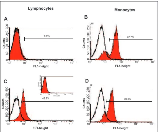
TABLE 1 -Speciic staining for CXCR1 and CXCR2 expression (%) and the 96h puriied protein derivative-stimulated CXCL8 levels (pg/ml) on both lymphocyte and monocyte populations (106 cells/ml) performed in prior tuberculosis (TB) patients and controls with latent M. tuberculosis infection (LTBI). Likewise, the 2 discrepant individuals for CXCR2 staining (please see text) are also shown for comparison.
Parameters
CXCR1* CXCR2*
lymphocytes monocytes lymphocytes monocytes CXCL8** TB 5.7 (+ 0.5) 69.7 (+ 2.2) 2.4 (+ 0.2) 67.4 (+ 2.6) 374.9 (106.6-1,010.8) LTBI 2.6 (+ 0.3) 59.2 (+ 3.5) 2.1 (+ 0.3) 60.2 (+ 3.6) 344.3 (157.0-1,296.8)
ID #125 1.0 50.6 53.1 84.8 242.6
ID #162 5.0 47.7 42.9 98.3 316.1
*mean (+SEM), **median (min-max), PPD at 10mg/ml.
TB: tuberculosis, LTBI: latent tuberculosis infection, ID: identiication. The levels of ex vivo CXCR1 and CXCR2
expression at thescreening and the single-cell levels were compared among persons who had had been previously treated for TB and among control patients with latent M. tuberculosis infections. Table 1
depicts the spontaneous levels found for each patient group. Due to the homogeneous patern, all data were further combined for receptor expression (below). As an internal control, negative responses to isotype control antibodies were detected in all individuals tested (Figure 1). All individuals assayed demonstrated standard staining for CXCR1, showing means (+ SEM) values of 3.7% (+ 0.3) and 63.6% (+ 2.1) in the lymphocyte and monocyte (R1) gated populations, respectively (Figures 1A and B).
Likewise, 57 examined individuals showed a similar, regular patern for CXCR2 expression on both lymphocytes (Figure 1C) and monocytes (2.2% + 0.1 and 65.2% + 2.3, respectively). In contrast, two clinically characterized prior extrapulmonary TB patients showed spontaneously high CXCR2 expression ex vivo. he irst patient was a 47-year-old male (#ID: 125) who was culture-positive
for M. tuberculosis with a PPD reaction of 18 mm, who responded
well to the standard treatment and who had had pleural efusion plus extensive infection (Table 1). he second subject was a 52-year-old male (#ID: 162) who was culture-positive for M. tuberculosis with no reaction to PPD, who responded well to standard treatment and who exhibited bone/joint lesions (Table 1; Figure 1C). During
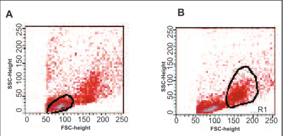
sample handling, the later patient showed an increased number of erythrocytes when the PBMCs were puriied. However, a normal blood patern was exhibited when the monocytes of those cases were also compared, indicating that the abnormality did not extend to the other mononuclear populations, making it unlikely that an artifact was observed during the cell puriication and staining procedures (Figure 1D). In addition, the remote possibility of cross-staining of the granulocyte population was ruled out because all studied patients showed similar light scatering proiles (Figure 2). Remarkably, the cytokine proile did not change signiicantly (data not shown), even though the CXCL8 level was slightly decreased at 96h when PBMCs (106 cells/ml) were cultured after PPD
98.3% R1
Lymphocytes
A
C
5.0%
42.9%
D B
42.7% R1
3.8%
Monocytes
0 60 120 180 240 300
Counts Count
s
Counts Count
s
Counts
100 101 102 103 104
100 101 102 103 104
100 101 102 103 104
100 101 102 103 100
FL1-height
FL1-height FL1-height
FL1-Height
FL1-height
0 50 100 150 200 250
0 100 200 300 400 500 0 50 100 150 200 250
100 101 102 103 104
0 60 120 180 240 300
FIGURE 1 -Two ex vivo proiles of CXCR1 (A and B) and CXCR2 (C and D) expression on lymphocytes (A and C) and monocytes (B and D) obtained from peripheral blood mononuclear cells of a prior extrapulmonary tuberculosis patient.
136
Rev Soc Bras Med Trop 45(1):134-137, jan-fev, 2012
DISCUSSION
FIGURE 2 -Two ex vivo proiles of lymphocytes (A) and monocytes (B) from peripheral blood mononuclear cells derived from a prior extrapulmonary tuberculosis patient.
As denoted in the dot plots, the two distinct populations, lymphocytes (A) and monocytes (B - R1), were gated on the basis of their light scatering properties.
(10mg/ml) stimulation (Table 1). To beter explore this abnormality, staining for HLA was also performed in parallel for one patient (#ID: 162). As expected, monocytes exhibited a positive modulation for HLA when LPS (200ng/ml) and IFNg (1,000U/ml) were used alone or in combination: 19.0-fold, 92.6-fold and 67.4-fold for LPS, IFNg and LPS+IFNg, respectively. his result ruled out possible non-responsiveness during the cell culture procedures. Unfortunately, because of IRB conlicts at the time that this study was performed, no additional analysis could be carried out.
0 50 100 150 200 250 0 50 100 150 200 250
SSC-Height
SSC-Height
0 50 100 150 200 250 0 50 100 150 200 250 FSC-height FSC-height
R1
A B
It is hypothesized that increased serum CXCL8 regulates its own receptors on granulocytes by inducing receptor internalization and proteolytic cleavage(as previously reviewed1). he same hypothesis
could be extended to other cell populations, such as T lymphocytes. In addition, blocking experiments with neutralizing anti-CXCL8 antibodies and pretreatment with bacterial LPS, which rapidly downmodulates CXCL8 receptor levels, showed that the inducing activity was due to the presence of CXCL8 in the conditioned medium6.
Nevertheless, the almost consistent in vitro CXCL8 levels that were found in the present study cannot explain the disparities in ex vivo CXCR2 expression. A recent study showed that CXCR2 expression is afected by medications (as previously reviewed1). In another seting,
increased gene expression of CXCL8 and the corresponding receptors was detected in PBMCs, mainly during chronic heart failure, resulting in the selective recruitment of leukocyte subpopulations into inlamed tissues7. However, marked abnormalities in genes from those patients
were signiicantly modulated in a normal direction during speciic therapy. hree single-nucleotide polymorphisms (SNP) have been reported in the CXCR2gene. he +1208 T/C SNP, located in the non-coding region of the CXCR2gene, might provide valuable information for the pathogenesis of and susceptibility to chronic inlammatory disease8. In addition, a highly signiicant association has been found
between the homozygous +1208 T genotype and an aggressive phenotype of breast carcinoma8. Furthermore, the +1440 G/A SNP
and some haplotypes are associated with periodontitis in Brazilian individuals. In contrast, the +785 C/T SNP, located in exon 11 and resulting in a silent codon change (no amino-acid substitution), may be important in protecting against pulmonary inlammation, despite exhibiting no association with Kawasaki disease. hus, CXCR2is
an important and interesting new candidate gene that is potentially involved in regulating the inlammatory process in the airways9. It
has been postulated previously that the CXCR2 +785 T allele may be associated with protection against decreased expiratory low rates and anomalous gas exchange in chronic obstructive pulmonary disease (COPD) and with a number of quantitative spirometric abnormalities, such as reduced lung function and respiratory symptoms10. Conversely,
this SNP is associated with angiogenesis in systemic sclerosis in individuals homozygous for the CXCR2 +785 C allele8. Although
diferential expression in both PBMC populations ruled out a primary deficiency, any association of the CXCR2gene SNPs and the 2 individuals described here deserve further investigation, as does the analysis of the intrinsic regulation of monocytes and lymphocytes by CXCR2 because chemokine receptors are selectively and diferentially expressed on diferent leukocyte subsets in individuals with arthritis.
In the current study, 2 patients showed stimulated-CXCL8 production that fell within the range of all patients tested and the normal range of CXCR1 expression, but those 2 patients showed abnormal CXCR2 expression when compared with the other analyzed individuals. A probable pharmacological explanation seems unlikely because those 2 individuals were medication-free (except for TB treatment) for more than two years. Unfortunately, for logistical reasons, neither patient could be followed ater the completion of this study. Detailed clinical histories were not available. However, previous complete blood count analyses did not show any discrepancies between the 2 cases and the rest of the cohort. Unlike CXCR1, CXCR2 can bind other CXC chemokines with high ainity. Accordingly, CXCR2 plays more a pleiotropic role in inlammation than does CXCR1 (as previously reviewed1). his diference may be detected in the 2
patients found here. It is worth noting that the regular cytokine levels found in the 2 patients do not seem to explain the disparities found in this study. In addition, the CXCR2 observed on granulocytes in patients sufering from active systemic lupus erythematosus is more important than CXCR1 in CXCL8-mediated inlammatory responses. he real mechanism of the increased spontaneous CXCR2 expression on lymphocytes from the 2 patients remains to be elucidated.
137
ACKNOWLEDGMENTSREFERENCES FINANCIAL SUPPORT
he authors are grateful to Mrs. Liza Krassner and Dr. Stuart Krassner (UCI, Irvine, USA) for editing the text of this manuscript, the staf for assistance with laboratory facilities, and the nursing staf of the TB ward for helping with patient recruitment.
his study was partly inanced by Coordenação de Aperfeiçoamento de Pessoal de Nível Superior (CAPES) (Brazil). P.A. is currently under a
Conselho Nacional de Desenvolvimento Cientíico e Tecnológico (CNPq)
research fellowship program (PQ-2, Brazil).
1. Baggiolini M, Dewald B, Moser B. Interleukin-8 and related chemotactic cytokines-CXC and CC chemokines. Adv Immunol 1994; 55:97-179. 2. Holmes WE, Lee EJ, Kuang WJ, Rice GC, Wood WI. Structure and functional
expression of a human interleukin-8 receptor. Science 1991; 253:1278-1280. 3. Chuntharapai A, Lee J, Hébert CA, Kim KJ. Monoclonal antibodies detect
diferent distribution paterns of IL-8 receptor A and IL-8 receptor B on human peripheral blood leukocytes. J Immunol 1994; 153:5682-5688.
4. Antas PR, Ding L, Hackman J, Reeves-Hammock L, Shintani AK, Schifer J, et al. Decreased CD4+ lymphocytes and innate immune responses in adults with previous extrapulmonary tuberculosis. J Allergy Clin Immunol 2006; 117:916-923.
5. Antas PRZ, Oliveira EB, Milagres AS, Franken LMC, Otenhof THM, Klatser P, et al. Kinetics of T cell-activation molecules in response to Mycobacterium
tuberculosis antigens. Mem Inst Oswaldo Cruz 2002; 97:1097-1099.
6. Khandaker MH, Xu L, Rahimpour R, Mitchell G, DeVries ME, Pickering JG, et al. CXCR1 and CXCR2 are rapidly down-modulated by bacterial endotoxin through a unique agonist-independent, tyrosine kinase-dependent mechanism. J Immunol 1998; 161:1930-1938.
7. Damås JK, Gullestad L, Aass H, Simonsen S, Fjeld JG, Wikeby L, et al. Enhanced gene expression of chemokines and their corresponding receptors in mononuclear blood cells in chronic heart failure--modulatory effect of intravenous immunoglobulin. J Am Coll Cardiol 2001; 38:187-193.
8. Snoussi K, Mahfoudh W, Bouaouina N, Fekih M, Khairi H, Helal AN, et al. Combined efects of IL-8 and CXCR2 gene polymorphisms on breast cancer susceptibility and aggressiveness. BMC Cancer 2010; 10:283.
9. Breunis WB, Biezeveld MH, Geissler J, Kuipers IM, Lam J, Otenkamp J, et al. Polymorphisms in chemokine receptor genes and susceptibility to Kawasaki disease. Clin Exp Immunol 2007; 150:83-90.
10. DeMeo DL, Celedon JC, Lange C, Reilly JJ, Chapman HA, Sylvia JS, et al. Genomewide linkage of forced mid-expiratory low in chronic obstructive pulmonary disease. Am J Respir Crit Care Med 2004; 170:1294-1301.