INTRODUCTION
NK cells
NK cells are a subtype of mononuclear cells morphologically distinct of lymphocytes B and T due to its increased granular density. NK cells participate in immunoregulation, haematopoiesis, reproduction and neuroendocrine interactions. Also play signiicant role in host defense against certain microorganisms, in part through their ability to secrete cytokines, such as IFN-γ, TNF, and GM-CSF,1 among others.2 They are capable of producing cytokines, pro and anti-inlammatory, and lead to death of target cells by apoptosis.
NK cells in pregnant patients with
SLE: a preliminary study
Alessandra Cardoso Pereira1, Mônica Cristina Brandão dos Santos Lima2, Nilson Ramires de Jesús3, Marcus Montello França4, Homero da Silva Nahum Junior5, Roger Abramino Levy6
Received on 06/26/08. Approved on 04/28/09. Financial support: FAPERJ E-26/170.034/06, 2006.
Discipline of Rheumatology, Pedro Ernesto University Hospital (UERJ) and Discipline of Obstetrics, Pedro Ernesto University Hospital (UERJ) 1. Assistant Professor, Discipline of Rheumatology, School of Medicine (UNIGRANRIO) and master of Medical Sciences (PGCM-UERJ) 2. Biologist Professor, School of Medical Sciences, Department of General Pathology, Laboratory of Immunopathology (UERJ) 3. Assistant Professor, Discipline of Obstetrics, School of Medical Sciences (UERJ)
4. Resident of the Discipline of Ophthalmology, Pedro Ernesto University Hospital (UERJ) 5. Professor, Discipline of Biostatistics (Universidade Estácio de Sá)
6. Associate Professor, Discipline of Rheumatology, School of Medical Sciences (UERJ)
Correspondence to: Alessandra Cardoso Pereira. Rua Marechal Bitencourt, 184, apt. 601, Bl. 1, Riachuelo, Rio de Janeiro - RJ. CEP: 20950-200. Tel: (11) 9948-0820 FAX: (11) 2567-7530. Email: [email protected]
ABSTRACT
The innate immune system plays an important role in reproduction, with marked involvement of NK cells. These cells behavior during pregnancy may clarify crucial points in the pathogenesis of complications that may occur in pregnant women with SLE. Objective: To measure the amount of circulating NK cells and their viability in pregnant SLE patients. Materials and methods: Blood samples from four groups of ten patients each were evaluated:1. GLES: Pregnant SLE patients; 2. PLES: Non-pregnant SLE patients; 3. Gcontrols: Pregnant controls; 4. Controls: Healthy non-pregnant
wo-men. In all patients the amount and viability of NK cells was measured by low cytometry, as well as the total apoptosis
by annexin V and propidium iodite staining. Results: Due to the great variability, median of each group was used for evaluation: CD56+ count [GLES (0.10), PLES (0.12), Gcontrols (0.15), Controls (0.08)]; total apoptosis (addition of
initial and late apoptosis to total number of dead cells) [GLES (0.06), PLES (0.04), Gcontrols (0.11), Controls (0.11)]. The results for live cells count had low variability, so the averages and standard deviations were used for comparisons: [GLES (0.91±0.06), PLES (0.95±0.03), Gcontrols (0.86±0.11), Controls (0.88+0.08)]. Conclusion: Although not
sta-tistically signiicant, the total apoptosis in the SLE groups was lower than in the control groups, and the live cell count
was higher. This suggests that in SLE patients, pregnant or not, the NK cells have a prolonged life cycle (or have a lower/different turnover), and that there may be a higher immune stimulus making the NK cells take longer to activate the apoptosis process.
Keywords: pregnancy; systemic lupus erythematosus; NK cells.
Thus, have implications in the pathogenesis of several human diseases. By the nature of cytokine production, NK cells takes part in the production regulation of antibodies dependent on T cells in autoimmune diseases. A reduction in NK cell activity is seen in SLE. Thus, the modulation of NK cell activity may be important in the pathogenesis and control of autoimmune diseases, infectious, and neoplastic.3
NK cells in pregnancy
of illness or pre-existing changes. From the immunological standpoint, it has been a great challenge trying to understand what mechanisms are involved in no rejection of the placenta by the maternal immune system, since the placenta, being of embryonic origin, contains genetic material, both maternal and paternal.4 The innate immune system is active in pregnancy and, due to the relative suppression of adaptive immunity, may play an important role in maternal immune defense.5 Furthermore, the innate immune system has a dominant inluence on reproduction. Analyses of cell types, which are present at the egg implantation site, show that T and B cells, typical of the adaptive immune system, are rare, while the predominant population consists of NK cells.6 The main types of immune cells in secretory endometrium are T cells, NK cells, and macrophages. T cells comprise approximately 45% of leukocytes in the proliferative phase endometrium, and this number remains constant throughout the secretory phase, and during pregnancy. However, their number apparently decreases due to the increase of NK cells during the secretory phase and early pregnancy.7 Several reports suggest that uterine NK cells have an important role in reproduction. They are hormonally regulated, increasing in number during the luteal phase of the menstrual cycle, when the implantation occurs; are present in early pregnancy when trophoblastic cells invade the spiral arterioles. They are particularly abundant around the fetal iniltration derived from extravillous trophoblastic cells. NK cells (CD56+) proliferate actively in the decidua during the luteal phase. In these phases, other mucosal elements (epithelial glands and stromal cells) cease proliferation and begin differentiation, except for endothelial cells, which continue to proliferate at this stage. However, the stimulus for the proliferation of NK cells in vivo is still unknown.8 NK cells in decidua are in close contact with the trophoblast fetal-maternal interface. However, decidual NK do not exert cytolytic activity against trophoblastic cells. Several studies have shown that the general cytotoxicity of decidual NK is reduced, when compared with peripheral blood NK cells.9
Pregnancy in SLE
For many years, SLE patients were warned not to become pregnant, but with a greater knowledge of the disease pathophysiology mechanisms and clinical manifestations during pregnancy, associated with medical care, pregnancy is a normal event in women with SLE. Normal but not monotonous. The pregnancy of SLE patients remains a high risk pregnancy, although most women do not present major complications.10 The autoimmune disease is primarily a disorder of immune
reactivity, which changes tolerance against various substances that starts behaving as autoantigen. Similarly, the success of pregnancy depends on maternal tolerance or non-immune reactivity against paternal antigens. Maternal tolerance seems to be associated with the development of several speciic mechanisms that protect the fetus from maternal immune cytotoxic attacks.11
NK cells in SLE
Since 1980, many evidences have been collected suggesting an association between the decrease in the number and activity of NK cells and autoimmune diseases. In a study of 71 patients with SLE, it was found that the number of NK cells (CD16+ CD56+) in the peripheral blood of these patients was only one third of the levels of controls - a highly signiicant difference. The proportion of NK cells in peripheral blood was signiicantly lower in patients with moderate or severe disease, compared with patients with inactive disease, and more depressed in patients with severe lupus nephritis.1 Green et al. found low levels of NK cell activity in irst-degree relatives of patients with SLE, suggesting that the correlation of NK cell deiciency with patient activity is important in the pathogenesis of the disease and not secondary to the disease process or to treatment with drugs. This deiciency was both in cell number and function.12
SLE NK cells seem to change on quantity and cytotoxicity, which is related to its pathogenesis, and yet there is no investigation about these cells presentation during pregnancy in SLE. Perhaps, a better control of immune response is the key to prevent the majority of SLE patients to develop disease activity during pregnancy. This pilot study objective was to evaluate the quantity and the life cycle of NK cells in peripheral blood and try to correlate with disease activity.
MATERIAL AND METHODS
This study was reviewed and approved by the Research Ethics Committee of Pedro Ernesto University Hospital and the Research Ethics Committee of the Maternity School of UFRJ, and was registered in SISNEP.
non-pregnant women. Each group was composed by 10 child-bearing age women, except the Control group with 11 individuals. Inclusion criteria were: pregnant women with SLE who began monitoring the pregnancy latest by twenty weeks of pregnancy, pregnant control groups, patients with SLE and controls matched for age and skin color. Exclusion criteria were: 1) more than twenty weeks of pregnancy at the time of material collection (blood and urine) for the study; 2) non-pregnant women over 40 (not in child-bearing age); 3) SLE carrier associated with another autoimmune disease, except antiphospholipid antibody syndrome; 4) infectious processes that require treatment with systemic antibiotics up to seven days before study entry. In the PLES group, were accepted only patients treating SLE with prednisone (Pred), chloroquine diphosfate (CQN), hydroxychloroquine (HCQ) and azathioprine (AZA), as these are the most commonly used medications for SLE treatment during pregnancy. At the day of urine and blood sample collection, it was applied a questionnaire to patients to assess factors that could interfere with the immune response: fever, lu, allergies, use of medication, physical activity, exposure to sun and sea, smoking, use of alcohol, and mood state.
To determine the total number of NK cells, 50 μL of blood (containing up to 0.5 x 10⁶ cells/mL per tube) were stained with 3 μL of anti-CD56-PE (BD 347747 California, USA) and incubated for twenty minutes at 4 ºC. Then the cells were lysed with Quicklise (Cytognos, Salamanca, Spain) for 15 minutes, and the remaining cells were washed twice with buffer saline pH 7.2 frozen. Soon after that, the cells were centrifuged for three minutes at 400 x g. To quantify the percentage of apoptosis in NK cells (CD56+), it was used the kit for detection of apoptosis from BD Pharmingen™ (California, USA), stained with annexin V and propidium iodide. In this test, the apoptosis is quantiied by the detection of fosfatidilserina exposed on the surface of apoptotic cells using annexing V that has afinity for it,16 and PI detects cells alive or dead. The cells are resuspended in 300 μL of saline buffer, 2.5 μL of annexin-V-FITC and 2.5 μL of PI (FL-3) were added, followed by homogenization, and incubated for 15 minutes in a darkroom. After incubation, it was added 300 μL of buffered saline to cells, which were processed and acquired in the FACSCalibur cytometer (BD Biosciences, CA, USA) at the maximum of one hour. 10 thousand events were acquired in channels FL-1, FL-2 and FL-3. The analysis was performed with multiparametric approach, to visualize the movement of migration living cells to those in apoptosis (initial and late) and dead. It was used the Paint’A’Gate program for this analysis and quantiication of these population.
Statistical evaluation was divided into descriptive and inferential analysis, the irst aimed to characterize the groups investigated, while to obtain inferences, we tried to compare them to identify possible statistical differences, always considering α = 0.05.
The descriptive analysis led to the estimates measures of location (mean and median) and dispersion (standard deviation and coeficient of variation) for quantitative variables (age, CD56%+, CD56+ absolute/mm3, living cell, initial apoptosis, late apoptosis, dead cells, total apoptosis, gestational age), as advocated by Costa Neto.17 This reference was also the base for frequency tables development of qualitative variables (skin color, humor, questionnaire, Pred, CQN / HCQ, AZA, SLEPDAI and SLEDAI14,15).
Also in the description ield, Pearson’s linear correlation and covariance were estimated, both estimated by quantitative variables.18 Cramer’s V coeficient correlation was estimated for the questionnaire variable against CD56+% CD56+ abs/mm3 and total apoptosis, since it involves qualitative variable.
Comparison among groups demanded that the proximity to the normal distribution were evaluated for quantitative variables, which was conducted by the Shapiro-Wilk test, given the low number of individuals/group.17
The normality enabled a comparison of four groups to be performed by two-way ANOVA.19 When proximity to the normal distribution was not observed, it was applied the Kruskal-Wallis test20 (CD56+%, CD56+ abs, initial apoptosis, late apoptosis, dead cell).
PLES and GLES groups were compared for some variables that were present only in those groups, then the statistical difference investigation was designed by Mann-Whitney test20 (gestational age).
For qualitative variables, the comparison of frequencies was done by the Chi-Square test19 (skin color, humor, questioning, Pred, CQN / HCQ, AZA). Statistical analysis were made based on the coeficient of variation (CV).17
CV < 20.00% - low variability, the individuals had uniform results, and the characterization of the variable was performed by mean and standard deviation.
CV > 20.00% - high variability, the individuals had discrepant results, and the characterization of the variable was performed by median and coeficient of variation.
RESULTS
estimated in days, and the GLES group was 105.00 days ± 23.55%, and Gcontrols group 100.45 days ± 16.73%. The age of women studied in the four groups ranged from 17 years and 11 months to 40 years old, the GLES group was 25 years old ± 24.66%, while in PLES, Gcontrols and Controls were respectively 32.40 ± 6.20 years, 25.80 ± 4.85 years and 30.91 ± 5.01 years, which a priori indicated that the groups were uniform with regard to age. Therefore, this variable should have no impact on other variables, except in GLES. Counting of CD56+ cells was obtained (percentage and absolute value). Percentage showed high variability, and the medians were: GLES group, 0.10 ± 91.01%; PLES, 0.12 ± 27.65%; Gcontrols, 0.15 ± 45.70% and Controls, 0.08 ± 47.47%. Individual values are shown in Table 1.
After determining the total amount of NK cells, these were analyzed for their viability, and differentiated in the stages of: live cells, cells at initial apoptosis, cells at late apoptosis and dead cells. Value of total apoptosis was considered as the sum of initial apoptosis, late apoptosis and dead cell stages. This determination was based on the consideration that, once the cells enter into apoptosis, its natural course is death. The results of each stage were calculated as percentages and analyzed in relation to its average, standard deviation, median and the coeficient of variation (Table 2). Living cells showed low variability, with means: GLES group, 0.91 ± 0.06%; PLES, 0.95 ± 0.03%; Gcontrols, 0.86 ± 0.11%; and Controls, 0.88 ± 0.08. The groups with SLE patients had a higher number of live NK, compared with patients without the disease; and the GLES group had lower value than PLES group, as well as Gcontrols group when compared to Control group (Table 2).
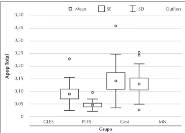
The initial apoptosis, late apoptosis and dead cell stages showed high variability, as well as total apoptosis stage. The median total apoptosis, GLES 0.06 ± 73.45%, 0.04 ± 54.86% PLES, Gcontrols 0.11 ± 75.72%, and Controls 0.11 ± 62.30%, was lower in groups of SLE patients when compared with the groups of patients without disease (Table 2) (Figure 1).
Some patients were receiving medications during the study period, except treatment with systemic antibiotics. We analyzed the frequency of the use of these medications (Table 3).
The Shapiro-Wilk test was applied for variables of age, CD56+, living cell, initial apoptosis, late apoptosis, dead cell and total apoptosis (Table 4). Then, the ANOVA test was applied for the variables of age (P = 0.02), living cell (P = 0.06), total apoptosis (P = 0.03), and we also applied the Fisher’s test for age and total apoptosis (Table 5). There was signiicant difference in total apoptosis of Gcontrols and Controls groups
in relation to PLES group, but it was not possible to identify differences in GLES group.
Results of the activity indices of SLE, SLEPDAI13 and SELENA/SLEDAI14,15 were analyzed in relation to CD56+%, CD56+ abs, living cell and total apoptosis (Table 6), and was observed a dependence with CD56+ abs, but reverse, i.e., when the variability of CD56+ increased, SLEPDAI13 and SELENA/ SLEDAI14,15 decreased. There was a relationship, but no a correlation between CD56+% and CD56+ abs: the dependence only occurred in CD56+ abs. The correlations of live cells and total apoptosis were not signiicant (P > 0.05). There was no dependency between the variables. Due to the small number of individuals in each group and/or a variable that interfered in the results, it was not possible to check values statistically signiicant.
DISCUSSION
NK cells are the predominant population of leukocytes in the uterine mucosa, and much has been studied to examine their role at the time of implantation and maintenance of pregnancy. Changes in regulation of NK cells have been associated with reproductive changes, such as spontaneous abortion, implantation failure and infertility, and preeclampsia.21 Miscarriage is associated with an increase in NK CD56dimCD16+cells and decrease in CD56brightCD16- in the endometrium during the luteal phase.22 A study that assessed the levels of NK cells in peripheral blood in the pre-conception and post-conception
Table 1
Percentage of CD56+ per person for each group Group
Patient GLES PLES Gcontrols Controls
1 10.0% 15.7% 13.6% 5.7%
2 8.0% 6.3% 15.7% 4.6%
3 11.3% 14.3% 22.4% 9.4%
4 10.6% 12.4% 16.3% 8.3%
5 46.6% 13.4% 18.1% 6.7%
6 5.9% 9.2% 2.9% 7.9%
7 10.8% 9.4% 7.5% 8.4%
8 8.4% 11.1% 15.8% 17.1%
9 3.8% 8.6% 10.9% 12.0%
10 26.6% 15.7% 6.7% 19.8%
11 - - - 15.7%
Median 10.3% 11.7% 14.6% 8.4%
found no signiicant changes of these levels when women with a history of recurrence miscarriages were studied and compared to normal controls. However, when subpopulations of NK cells were assessed, the CD56+CD16+ cells were moderately reduced in pregnant compared with non-pregnant women.21 It is important to remember that the values of NK cells count does not speciically relect the immune response of pregnancy, and that the quantity and activity of these cells can luctuate according to different variables, such as hormonal effects, Table 2
Descriptive results of the NK feasibility stage
Group
Variable GLES PLES Gcontrols Controls
Living Cell
Average 0.91 0.95 0.86 0.88
Standard
deviation 0.06 0.03 0.11 0.08
Median 0.94 0.96 0.89 0.90
Coefficient
of Variation 7.15 2.71 12.41 8.82
Initial Apop.
Average 0.05 0.03 0.11 0.07
Standard
deviation 0.02 0.02 0.09 0.05
Median 0.04 0.02 0.08 0.04
Coefficient
of Variation 53.61 74.81 79.37 80.10
Late Apop.
Average 0.02 0.01 0.01 0.04
Standard
deviation 0.03 0.01 0.02 0.04
Median 0.01 0.00 0.01 0.03
Coefficient
of Variation 188.18 148.12 109.55 93.57
Dead Cell
Average 0.03 0.01 0.02 0.04
Standard
deviation 0.03 0.01 0.01 0.04
Median 0.01 0.01 0.02 0.03
Coefficient
of Variation 115.31 81.58 57.61 93.57
Total Apop.
Average 0.09 0.04 0.14 0.13
Standard
deviation 0.07 0.02 0.11 0.08
Median 0.06 0.04 0.11 0.11
Coefficient
of Variation 73.45 54.86 75.72 62.30
Figure 1. Descriptive results of the percentage of total apoptosis (Total Apop) in the groups studied. Mean = average; ± SE = standard error, ± SD = standard deviation ; outliers = anomalous.
Apop
Total
0,40 0,35 0,30 0,25 0,20 0,15 0,10 0,05 0
GLES PLES Gest MN
Grupo
Mean SE SD Outliers
Table 3
Frequency of the use of medication
Variable
Medication GLES PLES Gcontrols Controls
AZA 0 2
CQN/HCQ 4 7
Prednisone 4 6
Acetylsalicylic acid 3 1
Folic Acid 2 1
Amitriptyline 1
Contraception 7
Cabergoline 1
Calcium + Vitamin D 3
Candesartan A 1
Chlordiazepoxide 1
Inhaled corticosteroids 2
Acid gamma-aminobutírico + del-lysine monohydrochloride
1
Heparin 1
ACE inhibitor 5
Levothyroxine 1
Clonazepam 1
Sertraline 1
Sibutramine 1
Ferrous sulfate 1 1
Tioconazole + tinidazole (vaginal cream)
1
Warfarin 1 1
physical activity, time of day, and sympathetic response to stress. Moreover, the number of NK cells of the peripheral blood is not necessarily correlated with their cytotoxicity.21 In our study, we used a questionnaire containing some of these variables, which could interfere in the values of NK cells, to try to minimize errors in the interpretation of the results. There was no correlation between the values of NK cells and the variables studied (fever, lu, allergies, use of medication, physical activity, exposure to sun and sea-bathing, smoking, use of alcohol and humor).
Although still controversial, some studies show an increase in levels of SLE activity adapted for pregnancy. The increased in SLE activity has profound implications on the fetal outcome, being associated with the lower number of live births and prematurity. However, the presence of lupus anticoagulant increases the risk of fetal loss.23 There are reports of reduction in the number of NK cells in the peripheral blood of patients with SLE, and this decrease was more pronounced in patients with active disease.1 This study found no reduction in the number of NK cells in pregnant patients and/or controls with or without SLE, but the small number of individuals in each group may have hindered the evaluation. The NK cell activity was not evaluated, only the cell cycle. There were no reports found on the evaluation of cell cycle of the NK cells in SLE patients.
Schepis et al. found an increase of NK cells in patients with SLE, regardless of the activity of the disease. This increase was related to the increase of INF type I (irst-response cytokine produced mainly by dendritic cells; appears to be involved in the pathogenesis of SLE), an abundant cytokine in SLE that can lead to an increase of NK cells in vitro. Serum levels of INF-α were increased in patients with disease activity, but not in those without disease activity.24 In this study, we found no signiicant differences in NK cell count between the groups, there was a high variability in the results. However, when related to the indices of activity of SLE (SLEPDAI13 and SELENA / SLEDAI14,15), we veriied an inverse relationship with the absolute number of CD56+, but this relationship was not maintained in the percentage of CD56+cells, so there was no correlation between the percentage and the absolute number. These results were not statistically signiicant.
There is a discussion whether the blood tests with increased levels of NK cells can be reproduced, or the values of patients with pregnancy loss are within the rate of normality, and if high levels in the blood may represent a mobilization in response to stress. The increase of the levels of NK cells may derive, in part, from a response to the stress of the venipuncture (elevation of NK count was found in the initial sample but not twenty minutes later). However, high counts of NK cells in the peripheral blood could be important for events in the maternal-fetal interface, the increase of stress in the activity of NK cells and their mobilization, suggesting an important role in the pathophysiology of fetal loss.25 These differences in literature on the values found for NK cells in SLE patients may be related to the dificulty of determining a relationship between the values of NK cells and its correlation with the disease activity, due to several factors that can interfere with the immune response.
Table 6
Correlation with activity index of SLE
Correlation* Covariance
Variable
SLEPDAI (GLES)
SLEDAI (PLES)
SLEPDAI (GLES)
SLEDAI (PLES)
CD56+ % 0.13 -0.27 0.06 -0.04
CD56+ abs -0.4 -0.28 -392.3 -80.52
Célula viva 0.51 -0.49 0.13 -0.06
Apop. total -0.5 0.58 -0.13 0.06
*Value-P > 0.05. Table 5
Results of Fisher’s test (α = 0.05*)
Variable GLES PLES Controls
Age (years)
PLES 0.02
Gcontrols 0.94 0.01
Controls 0.05 0.55 0.05
Apop. Total
PLES 0.20
Gcontrols 0.14 0.01
Controls 0.24 0.01 0.72
*Value-P < 0.05, variable not close to normal distribution. PLES age group different of GLES and Gcontrols groups. Apoptosis total of PLES group different of GLES and Controls. Table 4
Results of Shapiro-Wilk’s test (n < 50; α = 0.05*)
Variable
GLES PLES Gcontrols Controls
CD56+(%) 0.00 0.62 0.86 0.20
Living cell 0.06 0.30 0.21 0.18
Initial apop. 0.87 0.00 0.30 0.04
Late apop. 0.00 0.00 0.02 0.11
Dead cell 0.01 0.60 0.59 0.12
Apop. total 0.06 0.11 0.21 0.32
In this study, it was evaluated in addition to the count of NK cells its viability and quantiied the number of living cells in the initial and late stages of apoptosis, dead cells and total apoptosis. The results of living cells showed low variability and the groups of SLE patients (GLES and PLES) had a higher number of NK living cells, compared with patients without the disease.The group of pregnant women (GLES) showed a lower value group than the non-pregnant women group (PLES), as well as the group of women without disease (Gcontrols) when compared to the Control group. Perhaps, this shows that in the peripheral blood there are a smaller number of live NK cells in early pregnancy. In general, apoptotic cells are rapidly removed by phagocytosis, due to the induction of changes in the cell surface by the process of apoptosis. This prevents the release of physiological intracellular constituents, including nucleosomes, which are formed during the process of apoptosis through cleavage of chromatin by nucleases. Therefore, disorders in apoptosis or phagocytosis of apoptotic cells have been proposed in the development of autoimmune diseases, especially SLE.26 Several studies using mice with molecule deiciencies (e.g., C1q, and IgM) or receptors involved in phagocytosis of apoptotic cells have shown that the change in the removal of apoptotic cells leads to autoimmunity against nucleosomes and glomerulonephritis. Moreover, defects in the clearance of apoptotic cells have been described in mice and patients with lupus. Nucleosomes are not only important for the induction of the disease, but also may inluence the clearance of apoptotic cells. Furthermore, autoantibodies formed in SLE may also modulate this process. Therefore, during disease progression when nucleosomes autoantibodies begin to move, they may amplify the disease by inhibiting the clearance of apoptotic cells.26 In this study, it was found that total apoptosis was decreased in the groups of patients with SLE, when compared with groups without disease.
It was not possible to determine the predominant proile of NK cells, but perhaps this increase in the amount of living cells and decrease in total apoptosis may suggest that NK cells in SLE patients, pregnant or not, remain alive and maybe active for a greater period.
CONCLUSION
In this pilot study, it was not possible to demonstrate diffe-rences in the NK cells values of patients with or without the disease. As for the cell cycle of NK cells, the living cells in the groups of SLE patients were in greater number than in the controls without the disease; pregnant women had lower
values than non-pregnant women in groups with SLE and in groups of patients without the disease. The total apoptosis was decreased in all groups of SLE patients when compared with groups without the disease, but less in non-pregnant patients than in pregnant women.
These differences between living cells counts and total apoptosis may show that in SLE patients, pregnant or not, the NK cells have a long life (or a lower/different turnover), suggesting that there may be a greater immune stimulation, making NK cells take longer to activate the process of apoptosis. Due to the small number of patients in each group, the results may have been just a coincidence. We will continue collecting samples of new cases within the same protocol.
ACKNOWLEDGMENTS
To the doctors and residents of Rheumatology and Obstetrics at the State University of Rio de Janeiro and the Obstetrics sec-tionof UFRJ, where we have performed part of our study with pregnant women; to the employees of Bronstein Laboratory, RJ (Diagnostics of America SA), where the determinations were processed.
REFERENCES
1. Baxter AG, Smyth MJ. The Role of NK Cells in Autoimmune Disease. Autoimmunity 2002;35:1-14.
2. Ruddy S, Harris Jr ED, Sledge CB. Kelley’s Textbook of Rheumatology, 6. ed, Philadelphia: Saunders, 2001.
3. Sigal LH. Natural Killer Cell Receptors and Activation Mechanisms. J Clin Rheumatol 2003;9:55-9.
4. Zhou Y, Fisher SJ, Janatpour M, Genbacev O, Dejana E, Wheelock M
et al. Human Cytotrophoblasts Adopt a Vascular Phenotype as They
Differentiate. A Strategy for Successful Endovascular Invasion? J Clin Invest 1997;99:2139-51.
5. Sacks G, Sargent I, Redman C. An Innate View of Human Pregnancy. Immunol Today 1999;20:114-8.
6. Moffett-King A. Natural Killer and Pregnancy. Nat Rev Immunol 2002;2:656-63.
7. Lobo SC, Huang ST, Germeyer A, Dosiou C, Vo KC, Tulac S et al. The Immune Environment in Human Endometrium during the
Window of Implantation. Am J Reprod Immunol 2004;52:244-51. 8. Verma S, Hiby SE, LokeYW, King A. Human Decidual Natural
Killer Cells Express the Receptor for and Respond to the Cytokine Interleukin 15. Biol Reprod 2000;62:959-68.
9. Manaster I, Mandelboim O. The Unique Properties of Human NK Cells in the Uterine Mucosa. Placenta 2008;29:S60-6.
10. Ruiz-Irastorza G, Khamashta MA. Evaluation of Systemic Lupus Erythematous Activity during Pregnancy. Lupus 2004;13:679-82. 11. Wilder RL. Hormones, Pregnancy, and Autoimmune Diseases. Ann
12. Green MRJ, Kennell ASM, Larche MJ, Seifert MH, Isemberg DA, Salaman MR. Natural Killer Cell Activity in Families of Patients with Systemic Lupus Erythematosus: Demonstration of a Killing Defect in Patients. Clin Exp Immunol 2005;141:165-73.
13. Buyon JP, Kalunian KC, Ramsey-Goldman R, Petri MA, Lockshin MD, Ruiz-Irastorza G et al. Assessing Disease Activity in SLE
Patients during Pregnancy. Lupus 1999;8:677-84.
14. Bombardier C, Gladman DD, Urowitz MB, Caron D, Chang CH. Derivation of the SLEDAI. A Disease Activity Index for Lupus Patients. The Committee on Prognosis Studies in SLE. Arthritis Rheum 1992;35:630-40.
15. Buyon JP, Petri MA, Kim MY, Kalunian KC, Grossman J, Hahn BH et al. The Effect of Combined Estrogen and Progesterone
Hormone Replacement Therapy on Disease Activity in Systemic Lupus Erythematosus: a Randomized Trial. Ann Intern Med 2005;142:953-62.
16. Vermes I, Haanen C, Steffens-Nakken H, Reutelingsperger C. A Novel Assay for Apoptosis. Flow Cytometric Detection of Phosphatidylserine Expression on Early Apoptotic Cells Using Fluorescein Labelled. Annexin V. J Immunol Meth 1995;184:39-51.
17. Costa Neto, PLO. Estatística, 2 ed. São Paulo: Edgard Blucher, 2002.
18. Bunchaft G, Kellner SRO. Estatística sem mistério. Petrópolis: Vozes, 1999, pp. 350-899.
19. Beiguelman B. Curso prático de bioestatística. 5 ed. Ribeirão Preto: FUNPEC 73-212, 2002.
20. Siegel S, Castellan Jr NJ. Estatística não-paramétrica para ciências do comportamento. 6 ed. Porto Alegre: Artmed 2006, pp. 134-178. 21. Kwak-Kim J, Gilman-Sachs A. Clinical Implication of Natural Killer
Cells and Reproduction. Am J Reprod Immunol 2008;59:388-400. 22. Kalkunte S, Chichester CO, Gotsch F, Sentman CL, Romero R,
Sharma S. Evolution of Non-Cytotoxic Uterine Natural Killer Cells. Am J Reprod Immunol 2008;59:425-32.
23. Petri M. Prospective Study of Systemic Lupus Erythematosus Pregnancies. Lupus 2004;13:688-9.
24. Schepis D, Gunnarsson I, Eloranta ML, Lampa J, Jacobson SH, Kärre K et al. Increased Proportion of CD56(bright) Natural Killer Cells
in Active and Inactive Systemic Lupus Erythematosus. Immunology 2008 Jun 18 - in press.
25. Clark DA. Immunological Factors in Pregnancy Wastage: Fact or Fiction. Am J Reprod Immunol 2008;59:277–300.