Cop
yright
© ABE&M t
odos os dir
eit
os r
eser
vados
.
New insights into steroidogenesis
in normo- and hyperandrogenic
polycystic ovary syndrome patients
Novos aspectos na esteroidogênese em pacientes normo ou hiperandrogênicas com síndrome de ovários policísticos
Sebastião Freitas de Medeiros1,2,3, Ângelo Barrionuevo Gil-Junior2,
Jacklyne Silva Barbosa3, Érico Duarte Isaías2, Márcia Marly Winck Yamamoto3
ABSTRACT
Objective: This study sought to examine corticosteroidogenic enzyme activities in normo- and hype-randrogenic polycystic ovary syndrome (PCOS) patients. Subjects and methods: This cohort study included 81 patients with biochemical hyperandrogenism and 41 patients with normal androgen levels. Enzyme activities were assessed according to the serum steroid product/precursor ratios at baseline and after adrenal stimulation. Results: At baseline, in the delta 4 (Δ4) pathway, hyperandro-genic patients showed greater 17-hydroxylase and 17,20 lyase activities in converting progesterone (P4) into 17-hydroxyprogesterone (17-OHP4) and 17-hydroxypregnenolone (17-OHPE) into androste-nedione (A) (p = 0.0005 and p = 0.047, respectively) compared to normoandrogenic patients. In the delta 5 (Δ5) pathway, the 17-hydroxylase and 17,20 lyase enzymes showed similar activities in both groups. Hyperandrogenic patients presented lower 21-hydroxylase, lower 11β-hydroxylase (p = 0.0001), and statistically signiicant increases in 3β-hydroxysteroid dehydrogenase II (3β-HSDII) ac-tivities (p < 0.0001). Following tetracosactrin stimulation, only the 17,20 lyase activity remained up--regulated in the Δ4 pathway (p < 0.0001). Conclusion: Hyperandrogenic patients had higher 17,20 lyase activity, both at baseline and after adrenal stimulation. Greater conversion of dehydroepian-drosterone (DHEA) into A with normal conversion of 17-OHPE to 17-OHP4 in hyperandrogenic PCOS patients indicated different levels of 3β-HSDII activity in adrenal cells, and hyperandrogenic patients had lower 11β-hydroxylase and 21-hydroxylase activities. Arq Bras Endocrinol Metab. 2013;57(6):437-44 Keywords
Polycystic ovary syndrome; steroidogenesis; hyperandrogenism; enzyme activity; adrenal stimulation
RESUMO
Objetivo: O objetivo deste estudo foi examinar a atividade de enzimas responsáveis pela pro-dução de corticosteroides em pacientes normo e hiperandrogênicas com síndrome de ovários policísticos (SOP). Sujeitos e métodos: A coorte estudada incluiu 81 pacientes com hiperandro-genismo bioquímico e 41 pacientes com níveis normais de androgênio. A atividade enzimática foi avaliada de acordo com as proporções de produto/precursor do esteroide sérico, no momen-to inicial do estudo e depois de estimulação adrenal. Resultados: No momento inicial, na via delta 4 (Δ4), as pacientes hiperandrogênicas mostraram maior atividade da 17-hidroxilase e 17,20 liase na conversão da progesterona (P4) em 17-hidroxiprogesterona (17-OHP4) e na conversão da 17-hidroxipregnenolona (17-OHPE) em androstenediona (A) (p = 0,0005 e p = 0,047, respectiva-mente) em comparação com pacientes normoandrogênicas. Na via delta 5 (Δ5), a 17-hidroxilase e a 17,20 liase mostraram atividades similares nos dois grupos. As pacientes hiperandrogênicas mostraram menor atividade da 21-hidroxilase, menor atividade da 11β-hidroxilase (p = 0,0001) e aumento estatisticamente signiicativo na atividade da 3β-hidroxiesteroide desidrogenase II (3β-HSDII) (p < 0.0001). Após a estimulação com tetracosactrin, apenas a atividade da 17,20 liase permaneceu regulada para cima na via Δ4 (p < 0.0001). Conclusão: As pacientes hiperandrogêni-cas apresentaram atividade mais alta da 17,20 liase, tanto no momento inicial quanto depois da estimulação adrenal. Maior conversão da desidroepiandrosterona (DHEA) em A com conversão normal da 17-OHPE em 17-OHP4 em pacientes hiperandrogênicas com SOP indica níveis diferen-tes de atividade da 3β-HSDII em células da adrenal, e pacientes hiperandrogênicas apresentaram menores atividades da 11β-hidroxilase e da 21-hidroxilase. Arq Bras Endocrinol Metab. 2013;57(6):437-44 Descritores
Síndrome dos ovários policísticos; esteroidogênese; hiperandrogenismo; atividade enzimática; estimulação adrenal
1 Department of Gynecology
and Obstetrics at Medical Science School, Universidade Federal de Mato Grosso (UFMT), Cuiabá, MT, Brazil
2 Julio Muller University
Hospital, Cuiabá, MT, Brazil
3 Tropical Institute of Reproductive
Medicine and Menopause, Cuiabá, MT, Brazil
Correspondence to:
Sebastião Freitas de Medeiros Rua Almirante Henrique Pinheiro Guedes, 195
78043-306 – Cuiabá, MT, Brazil [email protected]
Cop
yright
© ABE&M t
odos os dir
eit
os r
eser
vados
.
INTRODUCTION
P
atients with polycystic ovary syndrome (PCOS)have an increased luteinizing hormone (LH) pul-se frequency, higher LH pulpul-se amplitude, and greater secretion of ovarian testosterone (T) and androstene-dione (A). As the action of follicle-stimulating hormo-ne (FSH) is not increased, there is a relatively lower aromatization of these androgens in the granulosa cells
of PCOS patients(1). PCOS patients also seem to have
generalized adrenal hyperactivity and higher adrenal androgen secretion as a consequence of greater
hor-mone adrenocorticotrophic (ACTH) action(2) or
ac-celeration in cortisol (F) catabolism(3). Following the
most recent standardization of diagnostic criteria (4-6), biochemical hyperandrogenism was found in 75%-82%
of PCOS patients(7,8). Short- and long-term
meta-bolic consequences of biochemical hyperandrogenism require that the androgen levels and the source of such androgens be determined to provide a rationale for prognosis and the establishment of optimal therapy.
Androgen synthesis requires the action of the cho-lesterol side-chain cleavage (P450 ssc, CYP11A) and
cytochrome P450c17α (CYP17) enzymes. The CYP17
enzyme mediates both 17-hydroxylase and 17,20 lyase activities, with greater 17-hydroxylase activity in the ad-renal fasciculate layer and equivalent activities in the re-ticular layer. The 17-hydroxylase enzyme promotes rapid conversion of pregnenolone (PE) and progesterone (P4) into 17-hydroxypregnenolone (17-OHPE) and 17-hy-droxyprogesterone (17-OHP4), respectively; in tan-dem, and more slowly, 17,20 lyase converts 17-OHPE and 17-OHP4 into dehydroepiandrosterone (DHEA)
and A, respectively(9). Two other adrenal enzymes,
3β-hydroxysteroid dehydrogenase II (3β-HSDII) and
sulfotransferase (SULT2A1), drive these precursors to-wards the synthesis of A and dehydroepiandrosterone sulfate (DHEAS), respectively (9,10).
After excluding classic 21-hydroxylase, 3β-HSDII,
and 11β-hydroxylase deiciencies, thyroid
dysfunc-tions, hyperprolactinemia, small defects, or imbal-anced enzyme activities may account for the bio-chemical hyperandrogenism seen in PCOS patients (9,11,12). Currently, few studies have investigated the activity of corticosteroidogenic enzymes in this syndrome (13), and these studies have not compared their results to those obtained in studies performed prior to the introduction of the NIH/Rotterdam di-agnostic criteria. Considering the current exclusion
criteria, differences in steroidogenic enzyme activities may exist between PCOS patients for whom diagnosis was performed before (11) or after the (14) NIH/ Rotterdam consensus. The present study sought to examine the activities of these enzymes in both nor-mo- and hyperandrogenic PCOS patients in order to compare these new indings with those obtained prior to the National Institute of Health (NIH) meeting and the Rotterdam consensus.
SUBJECTS AND METHODS
One hundred and forty two PCOS patients diagnosed with PCOS were prospectively enrolled at the Júlio Muller University Hospital and Tropical Institute of Reproductive Medicine and Menopause in Cuiabá, MT, Brazil from January 2003 to August 2012. Each patient signed an informed consent form that was ap-proved by the Federal University of Mato Grosso Committee for Ethics in Research. Patients who had used sex steroids or insulin sensitizing drugs over the last 6 months or who did not fulill the NIH/Rotter-dam criteria were excluded. Classic 21-hydroxylase,
3β-HSDII, and 11β-hydroxylase deiciencies were
ex-cluded in the following cases: 17-OHP4 levels ≤ 5 ng/ mL (≤ 15 nmol/L), 17-OHPE < 0.42 ng/mL (< 13.5 nmol/L), and 11-DOC < 8 ng/mL (< 230 nmol/L) (12,15). Eighty-one patients had biochemical hyperan-drogenism, and 41 had normal androgen levels. De-spite normal baseline concentrations of 17-OHP4, 2 of these patients (one from each group) were subse-quently excluded because their 17-OHP4 levels were > 30 nmol/L at 60 min after adrenal stimulation.
On average, included patients were 27.2 ± 5.5 years old. Anthropometric parameters were measured as
pre-viously described(8). Hyperandrogenism was deined
as total T ≥ 2.4 nmol/L, A ≥ 8.7 nmol/L, DHEAS ≥
6.7 nmol/L, and free androgen index (FAI) ≥ 7(8,16).
The FAI was estimated as the total T (nmol/L)/sex hormone-binding globulin (SHBG; nmol/L) x 100. The comparison between normo- and hyperandro-genic PCOS patients, as opposed to comparing PCOS subjects to healthy women, was consistent with recent
recommendations(17).
Cop
yright
© ABE&M t
odos os dir
eit
os r
eser
vados
.
Data distribution was assessed by Shapiro-Wilkins test and those with nonparametric distribution were transformed into a logarithmic scale prior to analysis, and subsequently retransformed into the original units. Anthropometric, endocrine, and enzymatic activity re-sults are presented as the mean ( ) and standard de-viation (SD) and were analyzed using the Welch test.
Clinical data were compared using a proportion test; p
values ≤ 0.05% were considered statistically signiicant.
RESULTS
Infertility (20% vs. 50%) and secondary amenorrhea
(37.5% vs. 60%) tended to be more frequent in
hype-randrogenic patients, although these differences were
not statistically signiicant (p = 0.064 and p = 0.117,
respectively). The rates of hirsutism, acne, acanthosis nigricans, and oligomenorrhea were also similar between normo- and hyperandrogenic patients, and no differen-ces in age, BMI, waist/hip ratio, or body surface area were observed (Table 1).
Hyperandrogenic patients had higher basal levels of LH, T, A, DHEAS, 17-OHP4, 11-DOC, and insulin compared with normoandrogenic patients. Baseline concentrations of F, PRL, P4, E2, and thyroid hor-mones were similar in both groups (Table 2).
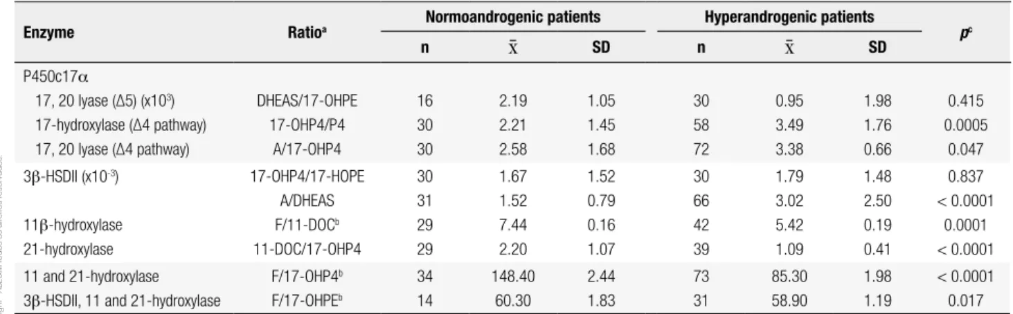
At the baseline condition, 17,20 lyase activity was not different between normoandrogenic and hyper-androgenic PCOS patients for the conversion of 17-OHPE into DHEA (Δ5 pathway; Table 3). Hyperan-drogenic patients demonstrated higher 17-hydroxylase
(17-OHP4/P4 of 3.49 vs. 2.21; p = 0.0005) and 17,20
lyase (A/17-OHP4 of 3.38 vs.2.58; p = 0.047) activities
for the conversion of P4 to 17-OHP4 and 17-OHP4 to
A, respectively (Δ4 pathway). The 3β-HSDII enzyme
demonstrated greater conversion of DHEA to A in
hy-perandrogenic patients (A/DHEAS of 3.02 vs. 1.52; p
< 0.0001). In contrast, 17-OHPE was converted into 17-OHP4 at the same rate in both groups (17-OHP4
∕17-OHPE of 1.79 vs. 1.67; p = 0.837). The activities
of 21-hydroxylase (11-DOC/17-OHP4 of 1.0 vs. 2.2;
p < 0.0001) and 11β-hydroxylase (F/11-DOC of 5.42
vs. 7.44; p = 0.0001) were lower in hyperandrogenic
patients (Table 3).
Following adrenal stimulation, the maximal lev-els of F, A, 17-OHP4, and P4 were not different
be-tween normo- and hyperandrogenic patients (p > 0.05).
However, the areas under the response curves of 17-OHP4 and A were greater in hyperandrogenic patients stimulation test was performed between 8:00 and 9:00
AM after a 12-hour fast. At baseline and 30 and 60 min after the injection of 0.25 mg of tetracosactrin
(Synac-then®, Novartis Pharmaceuticals, NJ, USA), blood
sam-ples were collected and assayed for 17-OHP4, F, A, and P4. The adrenal response was evaluated by measuring the difference between the basal value and the highest value, divided by the basal value (maximum increment, Δ) and the area under the curve (AUC).
Cop
yright
© ABE&M t
odos os dir
eit
os r
eser
vados
.
Table 1. Anthropometric characteristics of normo-and hyperandrogenic PCOS patients
Variable Normoandrogenic patients Hyperandrogenic patients pa
n SD n SD
Age (years) 39 26.6 5.8 79 27.0 5.4 0.719
Weight (kg) 38 74.6 25.6 75 75.4 16.3 0.861
Height (m) 37 1.58 0.06 64 1.57 0.07 0.450
BMI 37 29.0 10.0 64 30.6 6.5 0.386
Waist (cm) 32 87.9 18.6 59 90.9 13.1 0.422
Hip (cm) 32 106.6 16.9 59 107.7 12.8 0.749
W:H 32 0.82 0.06 59 0.84 0.08 0.182
Body area (m2) 37 1.76 0.3 64 1.81 0.2 0.369
a Welch Test.
Table 2. Comparison of demographic and endocrine characteristics in patients with PCOS with and without hyperandrogenemia
Hormone
Normoandrogenic patients Hyperandrogenic patients
pb
n SD n SD
TSH (µUI/mL) 36 2.50 1.31 72 2.19 1.16 0.233
Free thyroxine (pmol/L)a 35 14.56 1.16 62 14.23 1.46 0.224
Prolactin (nmol/L)* 37 0.48 0.25 75 0.43 0.21 0.268
LH (mUI/mL)a 38 6.66 1.91 74 9.32 1.69 < 0.0001
FSH (mUI/mL)a 38 6.10 1.49 74 5.64 1.39 0.118
LH:FSH ratioa 38 2.57 1.54 74 2.85 1.41 0.352
Total testosterone (nmol/L)* 35 1.50 0.50 74 2.20 1.04 < 0.0001
SHBG (nmol/L)a 35 39.35 1.67 43 25.38 1.70 < 0.0001
FAI (%) 31 4.16 1.77 63 10.17 5.86 < 0.0001
Cortisol (nmol/L)* 35 347.80 139.40 78 358.90 172.10 0.717
17-hydroxypregnenolone (nmol/L)* 17 4.98 2.80 34 6.07 3.55 0.239
17-hydroxyprogesterone (nmol/L)a* 35 3.36 1.60 76 4.74 1.66 < 0.0001
11-deoxycortisol (nmol/L)a* 32 61.3 17.3 44 72.9 17.1 0.004
DHEAS (µmol/L)* 36 3.74 1.54 71 5.31 2.70 0.0002
Androstenedione (nmol/L)* 34 5.15 1.51 75 11.49 4.96 < 0.001
Estradiol (nmol/L)* 31 162.51 64.80 50 191.97 97.0 0.105
Progesterone (nmol/L)* 31 2.23 1.46 61 1.86 1.15 0.224
Insulin (pmol/L) 32 61.29 1.92 71 82.14 1.96 0.0001
a The data underwent logarithmic transformation to meet the Gaussian distribution precepts. b Welch unpaired test.
*To convert gravimetric international unit (SI) values to mass units, we divided by 0.0347 for total T, 0.0271 for DHEAS, 0.0301 for 17-OHPE, 0.0303 for 17-OHP4, 0.0349 for A, 0.02886 for
11-DOC, 27.59 for cortisol, 3.671 for estradiol, 3.18 for P4, 0.04348 for PRL, 12.87 for free thyroxine, and 6.945 for insulin.
Table 3. Corticosteroidogenic enzyme activity in normo-and hyperandrogenic patients with PCOS
Enzyme Ratioa Normoandrogenic patients Hyperandrogenic patients pc
n SD n SD
P450c17α
17, 20 lyase (∆5) (x103)
17-hydroxylase (∆4 pathway) 17, 20 lyase (∆4 pathway)
DHEAS/17-OHPE 17-OHP4/P4
A/17-OHP4
16 30 30
2.19 2.21 2.58
1.05 1.45 1.68
30 58 72
0.95 3.49 3.38
1.98 1.76 0.66
0.415 0.0005
0.047
3β-HSDII (x10-3)
11β-hydroxylase 21-hydroxylase
17-OHP4/17-HOPE A/DHEAS F/11-DOCb
11-DOC/17-OHP4
30 31 29 29
1.67 1.52 7.44 2.20
1.52 0.79 0.16 1.07
30 66 42 39
1.79 3.02 5.42 1.09
1.48 2.50 0.19 0.41
0.837 < 0.0001
0.0001 < 0.0001
11 and 21-hydroxylase 3β-HSDII, 11 and 21-hydroxylase
F/17-OHP4b
F/17-OHPEb
34 14
148.40 60.30
2.44 1.83
73 31
85.30 58.90
1.98 1.19
< 0.0001 0.017
Cop
yright
© ABE&M t
odos os dir
eit
os r
eser
vados
.
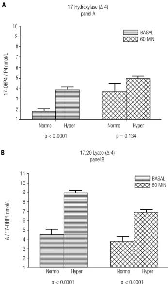
(p < 0.0001 and p = 0.049, respectively). Although they
presented different activities under basal conditions af-ter tetracosactrin stimulation, normoandrogenic and hyperandrogenic patients did not differ in their 17-hy-droxylase activity for converting P4 into 17-OHP4 in
the Δ4 pathway (p = 0.134; Figure 1). On the other
hand, 17,20 lyase activity for converting 17-OHP4 into A was greater in this pathway among hyperandrogenic
women (p < 0.0001; Figure 1). Sixteen out of 40 (40%)
normoandrogenic patients and 58 out of 80 hyperan-drogenic patients showed baseline levels of 17-OHP4 ≥ 1.0 and < 5.0 ng/mL. All of these patients were sub-jected to adrenal stimulation, and after tetracosactrin in-jection, 2 out of 16 (12.5%) normoandrogenic patients had 17-OHP4 levels between 15 and 30 nmol/L; in the hyperandrogenic PCOS group, 11/58 (18.9%) patients
presented 17-OHP4 levels in this range (p = 0.551).
Figure 1. 17-hydroxylase ∆4 (17-OHP4/P4; panel A) and 17,20 lyase ∆4 (A/17-OHP4; panel B) activities before (basal) and after (60 min) tetracosactrin injection in normo- and hyperandrogenic PCOS patients.
10
17 Hydroxylase (∆ 4) panel A
17-OHP4 / P4 nmoI/L
9
8
7
6
5
4
3
2
1
Normo Hyper
p < 0.0001 p = 0.134
Normo
BASAL 60 MIN
Hyper
A / 17-OHP4 nmoI/L
17,20 Lyase (∆ 4) panel B
11
10 9
8
7
6
5
4
3
2
1
BASAL
Normo Hyper
p < 0.0001 p < 0.0001
Normo Hyper
60 MIN
A
B
DISCUSSION
The present study compared the activities of diffe-rent steroidogenic enzymes between normoandroge-nic and hyperandrogenormoandroge-nic PCOS patients, rather than between PCOS and healthy subjects, as recently
sug-gested(17), both before and after adrenal stimulation.
To the best of our knowledge, this is the irst study comparing corticosteroidogenic enzyme activities be-tween normo- and hyperandrogenic PCOS patients. Previous studies have shown that a single early morning basal serum androgen measurement, as used in the cur-rent study, is an extremely accurate screening method (18). In previous studies, using several androgens as markers, biochemical hyperandrogenism was found in up to 80% of PCOS patients in different populations, as diagnosed using the NIH/Rotterdam criteria (7,8). In addition, adrenal hyperandrogenism was found in up to
62% of PCOS patients(19), and ovarian
hyperandroge-nism was reported to be even more frequent(7). No
consistent corticosteroidogenic enzyme abnormality has been reported in PCOS patients. After the intro-duction of the NIH/Rotterdam criteria in the clinical setting, only a few studies have examined the activities
of these enzymes(14), so this was the principal goal of
the current study.
A detailed understanding the Δ4 and Δ5 cortico-steroidogenic pathways is illustrated in igure 2 (9).
Dysregulation of the P450c17α enzymatic complex
in PCOS was reported previously, both before(11,20)
and after(21) dissemination of the NIH/Rotterdam
criteria, at least in the presence of biochemical hyper-androgenism. In the current study, 17,20 lyase activity was higher for the conversion of 17-OHP4 into A in hy-perandrogenic patients, both at baseline and after adre-nal tetracosactrin stimulation. Both decreases in
17-hy-droxylase activity with increases in 17,20 lyase(18) and
increases in 17-hydroxylase in relation to 17,20 lyase (12) activity have been detected in PCOS patients, even after the adoption of the new exclusion criteria. An early study demonstrated that in PCOS patients, estrogen may activate 17,20 lyase for the conversion of 17-OHP4 into A without affecting the conversion
of 17-OHPE into DHEA(13). The increase in 17,20
lyase activity was also seen in PCOS patients, even af-ter diagnostic standardization, and it was attributed to the phosphorylation of Ser/Thr residues, cytochrome b5, and cytochrome c allosteric actions (22-24). In
Cop
yright
© ABE&M t
odos os dir
eit
os r
eser
vados
.
the modulatory effects of cytochrome b5, insulin, and
estradiol(13,23). In the current study, estradiol levels
were not increased in the hyperandrogenic group, and this inding does not support previous observations in-dicating that estrogen may increase 17,20 lyase activity in the conversion of 17-OHP4 to A in PCOS patients (13,14). Although hyperandrogenic patients presented higher baseline insulin levels, reports of the role of
in-sulin in P450c17α enzyme activity have been
inconsis-tent (13,23,25).
The higher baseline 17-OHP4 and A levels found in the hyperandrogenic patients in the present study indicate that PCOS patients with biochemical hyperan-drogenism have greater 17-hydroxylase and 17,20 lyase activities in the Δ4 pathway, which results in greater conversion of P4 to 17-OHP4 and 17-OHP4 to A (Figure 2). Higher baseline levels of DHEAS and 17-OHPE in hyperandrogenic patients also indicate higher
P45017α complex activity in the Δ5 pathway. These
observations are relevant because, in normal
condi-tions, the human P450c17α enzyme possesses normal
17,20 lyase activity for the conversion of 17-OHPE to DHEA (Δ5 pathway) but minimal, if any, activity for the conversion of 17-OHP4 to A (Δ4 pathway) (26). Compared to healthy individuals, greater activity of 17,20 lyase was previously demonstrated in PCOS patients when they were diagnosed according to the
Rotterdam criteria and taken as the only population group (14). On the other hand, 17-hydroxylase activity was shown to be the same in both PCOS and healthy
women(14). There is currently no clear explanation for
this inconsistency, but it is possible that 17-hydroxylase activity may be maximally driven, precluding its further increase in the conversion of P4 to 17-OHP4.
Hyperandrogenic patients may present decreased
3β-HSDII activity with a lower conversion rate of
Δ5-3β-hydroxysteroids to Δ4-3β-hydroxysteroids(10,11),
which results in a higher amount of substrates for
an-drogen synthesis(26,27). Prior to the NIH/Rotterdam
criteria, a non-classic clinical form of 3β-HSD
deicien-cy was reported in hyperandrogenic patients with
hir-sutism and abnormal menstrual cycles(27,28),and
dif-ferent 3β-HSD activities have been reported in PCOS
patients both before (27) and after the implementation
of the Rotterdam criteria(26). In the current study,
normo- and hyperandrogenic PCOS patients demon-strated equal ratios of progesterones
(17-OHP4/17-OHPE p = 0.837), suggesting equal 3β-HSDII activity
in these groups for the conversion of OHPE to
17-OHP4. Equivalent 3β-HSDII activity was previously
reported for the comparison betweenhealthy women
and PCOS patients (13). Using DHEAS as a surrogate for DHEA, the higher A/DHEAS ratio in hyperan-drogenic patients found in the current study suggests
Cop
yright
© ABE&M t
odos os dir
eit
os r
eser
vados
.
higher 3β-HSDII activity speciically for the conversion
of DHEA to A. This discrepancy and inconsistency in
the 3β-HSDII activity indicates that this enzyme may
not be similarly expressed in all adrenal cells (29). Even prior to diagnostic standardization, lower 21-hydroxylase activity was found in 1%-19% of PCOS patients, depending on their ethnicity (30). In the present study, this lower activity was found only in hy-perandrogenic patients for whom the 11-DOC/17-OHP4 ratio was signiicantly lower in comparison to normoandrogenic patients. Thus, the present study supports the tendency for lower 21-hydroxylase activ-ity in some PCOS patients, which was previously dem-onstrated both before (30) and after (31) the NIH/
Rotterdam standardization. A minor 11β-hydroxylase
deiciency was found in up to 8.4% of PCOS patients prior to NIH/Rotterdam standardization (13). In the present study, 10% (4/40) of the hyperandrogenic PCOS patients presented 11-DOC concentrations of 18.2 nmol/L, a threefold increase compared with the third quartile of the normoandrogenic patients,
in-dicating some degree of 11β-hydroxylase deiciency.
Similar results were previously observed when hyper-androgenic PCOS patients were compared to normal women (12). In addition, in the current study, hyper-androgenic patients had lower C/11-DOC ratios with 11-DOC accumulation, and these indings support
ear-lier observations of lower 11β-hydroxylase activity in a
group of clomiphene-resistant PCOS patients(11). A
signiicant decrease in the conversion of 17-OHP4 to C, representing the combined activities of 11-hydroxy-lase and 21-hydroxy11-hydroxy-lase, was present only in hyperan-drogenic patients in the present study. After the NIH/ Rotterdam standardization, the combined activities of these enzymes, as examined by the C/17-OHP4 ra-tio, were reported to be the same when PCOS patients as a whole were compared to healthy, normal cycling women (32). These discrepancies indicate that the de-creased combined activities of 11-hydrolase and 21-hy-droxylase may occur only in hyperandrogenic PCOS patients and reinforce the notion that PCOS patients with hyper- and normoandrogenic phenotypes should be examined separately.
Most anthropometric and clinical aspects are not thought to differentiate normoandrogenic from hy-perandrogenic PCOS patients. Dysregulation of the
P450c17α enzymatic complex with up-regulated
17,20 lyase activity was found only in the hyperandro-genic PCOS group, both at baseline and after adrenal
stimulation. The greater conversion of DHEA into A that was observed in hyperandrogenic PCOS patients without an increased conversion rate of 17-OHPE to 17-OHP4 supports previous reports of different
3β-HSDII activities in different adrenal cells.
Hyper-androgenic PCOS patients have lower 21-hydroxylase
and 11β-hydroxylase activities than normoandrogenic
patients, and the results of the current study conirm indings reported prior to the NIH/Rotterdam di-agnostic criteria. Taken together, the indings of this study and those of other publications with a similar de-sign suggest the existence of different pathophysiolo-gies in normo- and hyperandrogenic PCOS patients.
Although the present study clearly deined PCOS and biochemical hyperandrogenism, it may have suf-fered from some limitations. First, the criteria used to
exclude classic 21-hydroxylase, 11β-hydroxylase, and
3β-HSD deiciencies are not yet standardized, and
those used in this study may be not universally adopted. Second, the use of the A/DHEAS ratio as a surrogate
for the A/DHEA ratio to evaluate 3β-HSDII activity
may not be appropriate. However, DHEAS and DHEA
levels have been shown to be closely related(9,28), and
an advantage of measuring DHEAS is that it is stable throughout the day. Third, the assays used to measure androgens may not be very accurate; however, recent comparisons between early radioimmunoassays for T, 17-OHPE, 17OHP4, 11-DOC, and liquid chroma-tography tandem mass spectrometry have shown good
agreement between the methods (33). Fourth, the
comparison of enzyme activities between normo- and hyperandrogenic PCOS patients, as opposed to com-paring PCOS patients to healthy women, was selected due to recent recommendations that provided new information on the phenotypic heterogeneity seen in PCOS patients.
Acknowledgements: the authors would like to thank the Ameri-can Journal Experts for English review of the manuscript.
Disclosure: no potential conlict of interest relevant to this article was reported.
REFERENCES
1. Gilling-Smith C, Story H, Rogers V, Franks S. Evidence for a pri-mary abnormality of thecal cell steroidogenesis in the polycystic ovary syndrome. Clin Endocrinol. 1997;47:93-9.
Cop
yright
© ABE&M t
odos os dir
eit
os r
eser
vados
.
3. Gambineri A, Forlani G, Munarini A, Tomassoni F, Cognigni GE, Ciampaglia W, et al. Increased clearance of cortisol by 5beta-re-ductase in a subgroup of women with adrenal hyperandrogenism in polycystic ovary syndrome. J Endocrinol Invest. 2009;32:210-8. 4. The Rotterdam ESHRE/ASRM-Sponsored PCOS Consensus
Workshop Group. Revised 2003 consensus on diagnostic criteria and long-term health risks related to polycystic ovary syndrome. Fertil Steril. 2004;81:19-25.
5. Zawadski JK, Dunaif A. Diagnostic criteria for polycystic ovary syndrome: towards a rational approach. In: Dunaif AGJ, Haseltine F (eds). Polycystic ovary syndrome. Baston: Blackwall Scientiic; 1992. p. 377-84.
6. Azziz R, Carmina E, Dewailly D, Diamanti-Kandarakis E, Escobar--Morreale HF, Futterweit W, et al. Position statement: criteria for deining polycystic ovary syndrome as a predominantly hyperan-drogenic syndrome: an androgen excess society guideline. J Clin Endocrinol Metab. 2006;91(11):4237-45.
7. Azziz R, Woods KS, Reyna R, Key TJ, Knochenhauer ES, Yildiz BO. The prevalence and features of the polycystic ovary syn-drome in an unselected population. J Clin Endocrinol Metab. 2004;89:2745-9.
8. Gil-Junior AB, Rezende APR, Carmo AV, Duarte EI, Medei-ros MMWY, MedeiMedei-ros SF. Adrenal androgen participation in the polycystic ovary syndrome. Rev Bras Gynecol Obstet. 2010;32:541-8.
9. Payne AH, Hales DB. Overview of steroidogenic enzymes in the pathway from cholesterol to active steroid hormones. Endocr Rev. 2004; 25:947-70.
10. Carmina E, Gonzalez F, Chang L, Lobo RA. Reassessment of adre-nal androgen secretion in women with polycystic ovary syndro-me. Obstet Gynecol. 1995;85:971-6.
11. Rosenield RL, Barnes RB, Cara JF, Lucky AW. Dysregulation of cytochrome P450c 17 alpha as a cause of polycystic ovary syn-drome. Fertil Steril. 1990;53:785-91.
12. Sahin Y, Kelestimur F. The frequency of late-onset 21-hydroxylase and 11β-hydroxilase deiciency in women with polycystic ovary syndrome. Hum Reprod. 1997;137:670-4.
13. Ditkoff EC, Fruzzetti F, Chang L, Stancyzk FZ, Lobo RA. The im-pact of estrogen on adrenal androgen sensitivity and secre-tion in polycystic ovary syndrome. J Clin Endocrinol Metab. 1995;80:603-7.
14. Bayoumy HA, Alothman AN. Adrenal contribution to polycystic ovary syndrome. Med Princ Pract. 2001; 10:151-5.
15. New ML, Lorenzen F, Lerner AJ, Kohn B, Oberield SE, Pollack MS, et al. Genotyping steroid 21-hydroxylase deiciency: hormonal re-ference data. J Clin Endocrinol Metab. 1983;57:320-6.
16. Willenberg HS, Bahlo M, Schott M, Wertenbruch T, Feldkamp J, Scherbaum WA. Helpful diagnostic markers of steroidogenesis for deining hyperandrogenemia in hirsute women. Steroids. 2008;73:41-6.
17. Bloom MS, Schisterman EF, Hediger ML. Selecting controls is not selecting “normals”: design and analysis issues for studying the etiology of polycystic ovary syndrome. Fertil Steril. 2006;86:1-12. 18. Rothman MS, Carlson NE, Xu M, Wang C, Swerdloff R, Lee P, et al. Reexamination of testosterone, dihydrotestosterone, estradiol and estrone levels across the menstrual cycle and in postme-nopausal women measured by liquid chromatography-tandem mass spectrometry. Steroids. 2011;76:177-82.
19. Escobar-Morreale HF, Sanchón R, San Millán JL. A prospective study of the prevalence of nonclassical congenital adrenal hyper-plasia among women presenting with hyperandrogenic symp-toms and signs. J Clin Endocrinol Metab. 2008;93:527-33. 20. Barnes RB, Rosenield RL, Burnstein S, Ehrmann DA.
Pituitary--ovarian responses to nafarelin testing in the polycystic ovary syndrome. N Engl J Med. 1989;320:559-65.
21. Çolak R, Kelestimur F, Unluhizarci K, Bayram F, Sahin Y, Tutus A. A comparison between the effects of low dose (1 µg) and stan-dard dose (250 µg) ACTH stimulation tests on adrenal P450c17α
enzyme activity in women with polycystic ovary syndrome. Eur J Endocrinol. 2002;147:473-7.
22. Auchus RJ, Lee TC, Miller WL. Cytochrome b5 augments the 17,20-lyase activity of human P450c17 without direct electron transfer. J Biol Chem. 1998;273:3158-65.
23. Moghetti P, Castello R, Negri C, Tosi F, Spiazzi GG, Brun E, et al. Insulin infusion ampliies 17 alpha-hydroxycorticosteroid inter-mediates response to adrenocorticotropin in hyperandrogenic women: apparent relative impairment of 17,20 lyase activity. J Clin Endocrinol Metab. 1996;81:881-6.
24. Lin D, Black SM, Nagahama Y, Miller WL. Steroid 17 alpha-hidro-xylase and 17,20-lyase activities of P450c 17: contributions of seri-ne 106 and P450 reductase. Endocrinology. 1993;132:6:2498-506. 25. Nestler JE, Whitield JB, Williams TY, Zhu G, Condon J, Kirk KM,
et al. Genetics of serum dehydroepiandrosterone sulfate and its relationship to insulin in a population-based cohort of twin sub-jects. J Clin Endocrinol Metab. 2002;87:682-6.
26. Doi SAR, Al-Zaid M, Towers PA, Scott CJ, Al-Shoumer KA. Steroi-dogenic alterations and adrenal androgen excess in PCOS. Ste-roids. 2006;71:751-9.
27. Pang S, Lerner A, Stoner E, Levine LS, Oberield SE, Engel I, et al. Late-onset adrenal steroid 3 beta-hydroxysteroid dehydrogenase deiciency a cause of hirsutism in pubertal ans postpubertal wo-men. J Clin Endocrinol Metab. 1985;60:428-38.
28. Cobin RH, Futterweit W, Fiedler RP, Thornton JC. Adrenocorticotro-phic hormone testing in idiopathic hirsutism and polycystic ova-rian disease: a test of limited usefulness. Fertil Steril. 1985;44:224-6. 29. Lutfallah C, Wang W, Mason JI, Chang YT, Haider A, Rich B, et
al. Newly proposed hormonal criteria via genotypic proof for type II 3beta-hydroxylase deiciency. J Clin Endocrinol Metab. 2002;87:6:2611-22.
30. Pall M, Azziz R, Beires J, Pignatelli D. The phenotype of hirsu-te women: a comparison of polycystic ovary syndrome and 21-hydroxylase-deicient nonclassic adrenal hyperplasia. Fertil Steril. 2010;94: 684-9.
31. Trakakis E, Rizos D, Loghis C, Chryssikopoulos A, Spyropoulou M, Salamalekis E, et al. The prevalence of non-classical congeni-tal adrenal hyperplasia due to 21-hydroxylase deiciency in greek women with hirsutism and polycystic ovary syndrome. Endocr J. 2008;55:33-9.
32. Azziz R, Bradley Jr EL, Potter D, Boots LR. Adrenal androgen ex-cess in women: lack of a role for 17-hydroxylase and 17, 20-lyase dysregulation. J Clin Endocrinol Metab. 1995;80:400-5.