Calcium renal lithiasis: metabolic diagnosis and
medical treatment
Litiase renal cálcica: diagnóstico metabólico e tratamento médico
Miguel Angel Arrabal-Polo
I, Miguel Arrabal-Martin
II, Juan Garrido-Gomez
IIISan Cecilio University Hospital, Granada, Spain
ABSTRACT
Calcium renal lithiasis is a frequent condition that afects the worldwide population and has a high recur-rence rate. Diferent metabolic changes may trigger the onset of calcium stone disorders, such as hy-percalciuria, hyperoxaluria, hyperuricosuria, hypocitraturia and others. There are also other very prevalent disorders that are associated with calcium calculi, such as arterial hypertension, obesity and loss of bone mineral density. A correct diagnosis needs to be obtained through examining the serum and urinary pa-rameters of mineral metabolism in order to carry out adequate prevention and treatment of this condition. Once the metabolic diagnosis is known, it is possible to establish dietary and pharmacological treatment that may enable monitoring of the disease and prevent recurrence of stone formation. Some advances in treating this pathological condition have been made, and these include use of sodium alendronate in patients with calcium renal lithiasis and osteopenia/osteoporosis, or use of a combination of a thiazide with a bisphosphonate. In summary, calcium renal lithiasis often requires multidrug treatment with strict control and follow-up of patients.
RESUMO
Litíase renal cálcica é uma doença comum que afeta a população no mundo todo e tem alta taxa de recor-rência. Diferentes alterações metabólicas podem desencadear o aparecimento de distúrbios de pedras de cálcio, como hipercalciúria, hiperoxalúria, hiperuricosúria, hipocitratúria e outros. Existem também doen-ças altamente prevalentes associadas à doença de cálculo de cálcio, como hipertensão, obesidade e perda de densidade óssea mineral. Para realizar prevenção e tratamento adequados, é necessário diagnóstico correto, examinando o metabolismo mineral sérico e urinário. Depois de conhecer o diagnóstico meta-bólico, é possível estabelecer um tratamento dietético e farmacológico que permita controlar a doença e prevenir a recorrência de cálculos biliares. Há alguns avanços no tratamento dessa doença e incluem o uso de alendronato de sódio em pacientes com nefrolitíase de cálcio e osteopenia/osteoporose, ou a combinação de um tiazídico com um bifosfonato. Em resumo, litíase renal cálcica exige, muitas vezes, um tratamento multidroga com rigorosos controle e acompanhamento de pacientes.
IMD. Resident in training, Urology Department,
San Cecilio University Hospital, Granada, Spain.
IIMD, PhD. Attending Physician, Urology
Department, San Cecilio University Hospital, Granada, Spain.
IIIMD, PhD. Attending Physician, Traumatology
Department, San Cecilio University Hospital, Granada, Spain.
KEY WORDS:
Kidney calculi. Therapeutics. Metabolism. Diagnosis. Pharmacy. Calcium.
PALAVRAS-CHAVE:
INTRODUCTION
Calcium kidney stones are an important urological and nephro-logical pathonephro-logical condition that afects a high percentage of the population during life. he information available on their eti-ology, clinical presentation, diagnosis and treatment (medical and non-medical) is very extensive and therefore, in this review article, we will address their metabolic diagnosis and medi-cal treatment, particularly by reviewing published papers in the most important databases.
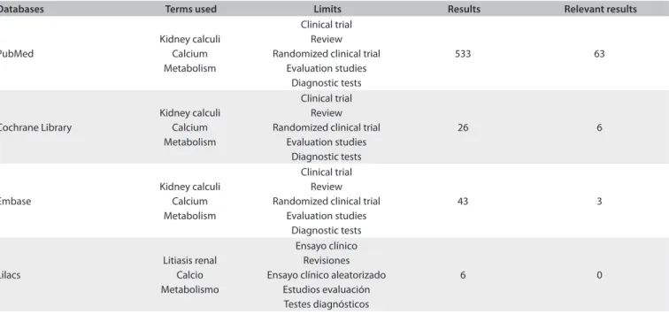
PubMed, Lilacs, Embase and Cochrane Library were searched for systematic reviews and practice guidelines, using the MeSH terms Kidney calculi AND Calcium AND Metabolism with the following limits: review, clinical trial, randomized clini-cal trial, diagnostic tests and evaluation studies. We found a total of 533 articles in PubMed, 43 articles in Embase, 26 articles in Cochrane and 6 in Lilacs. Finally, we selected a total of 63 articles from PubMed, 6 from Cochrane and 3 from Embase. he 6 arti-cles and 3 artiarti-cles from Cochrane and Embase were also among the 63 articles used from PubMed (Table 1).
EPIDEMIOLOGY
Renal lithiasis is a very frequent pathological condition among the worldwide population, with varying incidence according to distinct geographical regions. Calcium-based stones, both in oxalate and in phosphate form, prevail in developed countries, while infectious lithiasis remains the main cause of this condi-tion in developing countries.1 Renal lithiasis occurs at least once
in life in 15% of Caucasian men and in 6% of women, with recur-rence of around 50%.2
In most cases (around 75%), stones have a calcium composi-tion, and they generally present in the form of calcium oxalate, whereas other types of lithiasis are less frequent.2 Increased
fre-quency of calcium phosphate stones has been observed over the last few decades, and above all among women, with metabolic and bone-derived diseases.3
he geographical variability in the prevalence of lithiasis is also noteworthy, since in some zones it is very high, while in oth-ers it is nonexistent.4 In general, urinary lithiasis has a mean
inci-dence of 0.5-1%, and an annual prevalence of 4-5%.5
CLINICAL AND METABOLIC DIAGNOSIS Clinical diagnosis
Renal colic is the most common clinical presentation of calcium renal lithiasis and leads to a high admission rate in the emergency rooms of hospitals. It is very important to objectively describe the type of pain, quantify it, determine the origin of irradiation and collect any personal and family histories relating to calcium renal lithiasis. Patients should be asked about their dietary patterns, luid intake, use of medication and type of physical activity that they perform.6 Simple abdominal radiography and
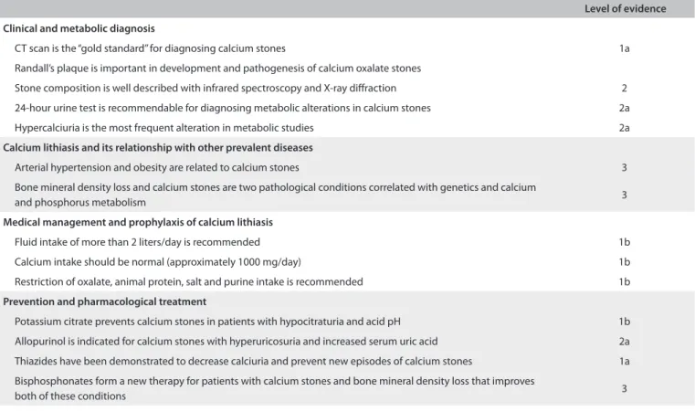
ultrasonogra-phy are the imaging techniques usually ordered at the beginning of the study, although abdominal computed tomography (CT) is the gold standard imaging technique (Table 2) for diagnosing and classifying renal lithiasis. Today, and thanks to new imaging techniques such as dual-energy computed tomography, it is pos-sible to obtain a suiciently accurate diagnosis to be able to dif-ferentiate calcium stones from uric acid, or from other types with less frequent chemical compositions.7
Databases Terms used Limits Results Relevant results
PubMed
Kidney calculi Calcium Metabolism
Clinical trial Review Randomized clinical trial
Evaluation studies Diagnostic tests
533 63
Cochrane Library
Kidney calculi Calcium Metabolism
Clinical trial Review Randomized clinical trial
Evaluation studies Diagnostic tests
26 6
Embase
Kidney calculi Calcium Metabolism
Clinical trial Review Randomized clinical trial
Evaluation studies Diagnostic tests
43 3
Lilacs
Litiasis renal Calcio Metabolismo
Ensayo clínico Revisiones Ensayo clínico aleatorizado
Estudios evaluación Testes diagnósticos
6 0
Metabolic diagnosis
Lithiasis composed of calcium oxalate is the most frequent type, followed by calcium phosphate. Numerous studies have highlighted the signiicance that Randall’s plaque has in rela-tion to development and pathogenesis of calcium oxalate lithi-asis (Table 2), thanks to the use of intraoperative biopsies.8,9 It
has even been possible to demonstrate that the singularity of the renal papilla depends on the type of renal calcium stone former, i.e. whether it is oxalate or phosphate.10 Microscopic lesions on
the renal papilla are the main cause of formation of monohydrate calcium oxalate stones, while the location of such lesions and their characteristics will in many instances determine the inal morphology of the renal calculus.11 Even the presence of lesions
in kidney tubular cells may favor aggregation and growth of crys-tals, which eventually leads to formation of calcium stones.12
Calcium stone formation is a complex process that involves numerous metabolic, anatomical and physiopathological mech-anisms. Occurrences of supersaturated states of crystallization with the capacity to precipitate in urine are a key factor in cal-culus formation. he saturation state of a substance is expressed as the ratio between a given substance and its solubility variable. he supersaturated state of the calcium oxalate is unrelated to the urinary pH; however, calcium phosphate saturation increases signiicantly when the urinary pH changes from 6 to 7.13,14
Metabolic disorders are, beyond any doubt, key factors in the formation and persistence of calcium lithiasis. It is therefore advisable to conduct a complete metabolic study on patients with histories of calcium lithiasis, multiple calcium lithiasis or hard-to-treat calcium lithiasis; and on calcium lithiasis-forming chil-dren, single kidney patients and occurrences of intestinal dis-orders, bone disorders or nephrocalcinosis.15 Metabolic studies
include analysis on the calculi, since each occurrence of lithia-sis comprises in itself a certain clinical form of lithialithia-sis disease. Firstly, the macroscopic structure of the calculi, before and ater fragmentation, should be examined in order to identify their exact composition through infrared spectroscopy and X-ray dif-fraction along with other electronic scanning microscopy tech-niques. On the other hand, chemical analysis of calculi is an inex-act method that has now become obsolete.16
Regarding blood and urine results, tests should be ordered when it is necessary to know the blood levels of calcium, phos-phorus, sodium, potassium, chlorine, magnesium, urea, creati-nine and uric acid. In 24-hour urine tests, it is recommendable to determine the urea, creatinine, uric acid, oxalate, citrate and magnesium levels, and to establish the diuresis volume, pH and urinary density. A fasting urine test to assess the levels of calcium, creatinine, oxalate and citrate is also recommendable.5,7-19 he
analysis should be extended in order to determine the levels of
Level of evidence
Clinical and metabolic diagnosis
CT scan is the “gold standard” for diagnosing calcium stones 1a
Randall’s plaque is important in development and pathogenesis of calcium oxalate stones
Stone composition is well described with infrared spectroscopy and X-ray difraction 2
24-hour urine test is recommendable for diagnosing metabolic alterations in calcium stones 2a
Hypercalciuria is the most frequent alteration in metabolic studies 2a
Calcium lithiasis and its relationship with other prevalent diseases
Arterial hypertension and obesity are related to calcium stones 3
Bone mineral density loss and calcium stones are two pathological conditions correlated with genetics and calcium
and phosphorus metabolism 3
Medical management and prophylaxis of calcium lithiasis
Fluid intake of more than 2 liters/day is recommended 1b
Calcium intake should be normal (approximately 1000 mg/day) 1b
Restriction of oxalate, animal protein, salt and purine intake is recommended 1b
Prevention and pharmacological treatment
Potassium citrate prevents calcium stones in patients with hypocitraturia and acid pH 1b
Allopurinol is indicated for calcium stones with hyperuricosuria and increased serum uric acid 2a
Thiazides have been demonstrated to decrease calciuria and prevent new episodes of calcium stones 1a
Bisphosphonates form a new therapy for patients with calcium stones and bone mineral density loss that improves
both of these conditions 3
bone resorption markers in cases of nephrocalcinosis, suspected hyperparathyroidism, fasting hypercalciuria or previous treat-ment for loss of bone mineral density. Blood tests should detect markers for intact parathyroid hormone (iPTH), vitamin D, alka-line phosphatase, osteocalcin and beta-crosslaps. Furthermore, any metabolic study conducted on patients with recurrent cal-cium renal lithiasis and altered bone remodeling markers can be complemented by performing bone densitometry to objectively assess bone mineral density.20
Metabolic studies reveal a series of alterations that can afect the calcium stone composition, and which can be summarized as follows:
Hypercalciuria
Hypercalciuria is, for many reasons, the most important meta-bolic risk factor for formation of calcium lithiasis. Hypercalciuria is the most frequently detected metabolic abnormality, in 35-65% of patients with stones; the relation between urinary calcium lev-els and occurrences of Randall’s plaques is another factor;19,21 also,
a genetic and molecular relationship with the presence of calcium in urine could be regarded as a further factor.22,23 Hypercalciuria
is deined, according to diferent studies, as excretion in urine of more than 260 mg of calcium in 24 hours (or 4 mg of cal-cium per kg/day). In clinical practice, it is also valuable to regard hypercalciuria as excretion of more than 250 mg/day in women and 300 mg/day in men.5,9,24 Although the term idiopathic
hyper-calciuria has been used for years to refer to increased urinary calcium levels, it is more accurate to talk about distinct types of hypercalciuria, on the basis that the calcium transport defect may be localized in the gastrointestinal tract, bones or kidneys. hus, hypercalciuria can be classiied into three groups: absorp-tive hypercalciuria; excretory, or renal, hypercalciuria; and reab-sorptive hypercalciuria.5,19-25
• Absorptive hypercalciuria: hree types of hypercalciuria have
been deined and are characterized by having a calcium/cre-atinine ratio in urine of less than 0.11 during fasting, and less than 0.22 when there is an overload of calcium in the diet. hey difer from each other in that type 1 has high calcium excretion in urine even with a restricted calcium diet; in type 2, lower calcium levels in urine are detected following restric-tion of calcium in the diet; and a deicit of serum phosphate is detected in type 3.26,27
• Excretory hypercalciuria: his occurs as a result of failure of
renal tubular reabsorption of calcium. hese patients have calcium/creatinine ratios greater than 0.11 while fasting, and higher than 0.22 ater calcium intake. Furthermore, iPTH is found to be within the normal range.19
• Reabsorptive hypercalciuria: his results from increased
bone resorption, basically in relation to primary hyperpara-thyroidism. Calcium/creatinine ratios are found to be high
while fasting and ater calcium overloads greater than 0.11 and 0.22, respectively, as well as the iPTH levels.
Hyperoxaluria
Hyperoxaluria is deined as urinary excretion of oxalate higher than 40 mg/day. Increased oxalate excretion in urine seems to be associ-ated with formation of monohydrate calcium oxalate stones, which is a metabolic risk factor in this type of lithiasis disorder. As with hypercalciuria, various types of hyperoxaluria can be distin-guished. Primary hyperoxaluria (type 1 and type 2) can be found in patients with enzyme deicits resulting from mutations of a variety of genes, and it involves anomalous and incomplete degradation of oxalates that accumulate in urine before their excretion. However, this condition is usually infrequent.28
Enteric hyperoxaluria is the most frequent disorder, and results from malabsorption of fats. Since these fats cannot be absorbed, they bind to calcium and diminish the formation of intestinal calcium-oxalate complexes, and thus increase the intes-tinal absorption of oxalates. Dietetic hyperoxaluria is caused by an increase in the intake of oxalate-rich food, or excessive intake of vitamin C.29
Moreover, the bacterium Oxalobacter formigenes has recently been described as the causative agent of oxalate absorption at intestinal level. It seems that low levels of this microorganism lead to increased oxalate levels, since oxalate metabolism dimin-ishes because of this bacterium.30
In the past, bariatric surgery played a signiicant role in the formation of oxalate calculi, since jejunum-ileum shunts would lead to high levels of hyperoxaluria through increased abdominal absorption of oxalates. his has now been replaced by other tech-niques with fewer side efects on metabolism.31
Hypocitraturia
Urinary citrate is a recognized inhibitor of oxalate and calcium phosphate stones, through avoiding the formation of the nucleus of stone, its growth and its aggregation.32-34 Hypocitraturia can
be deined as urinary excretion of citrate less than 320 mg/day. Citrate has a triple protective efect, since it can bind to calcium to prevent formation of complexes, alkalinize urine and have a direct inhibitory efect. Urinary citrate is reabsorbed under states of acidosis, with the result that its urinary excretion decreases. In addition, hypocitraturia has been correlated with chronic diar-rhea, extenuating physical exercise and excessive intake of ani-mal proteins.19,35
Hyperuricosuria
oxalate-calcium calculi. In general, increased urinary excretion of uric acid results from high intake of purines, although it can also be associated with other pathological conditions, such as gout, myeloproliferative diseases, myeloma etc.5,36,37
Hypomagnesuria
he disorder of hypomagnesuria is an infrequent cause of forma-tion of calcium oxalate stones. It can be detected in around 1% of patients and is associated with intestinal inlammatory disease.5,36
CALCIUM LITHIASIS AND ITS RELATIONSHIP WITH OTHER PREVALENT DISEASES
Arterial hypertension and calcium lithiasis
Numerous studies have analyzed the relationship between lithi-asis and arterial hypertension.38,39 It seems that increases in
cir-culatory volume and arterial pressure lead to decreased sodium reabsorption at the level of the proximal tubules, which results in diminished calcium reabsorption and subsequently, increased calciuria (Table 2). High sodium intake in these patients’ diet is another factor to bear in mind, since this increases the urinary excretion of calcium.40,41
Obesity and calcium lithiasis
A variety of studies in the literature have shown that there is a relationship between body mass index and kidney stones. It has been increasingly recognized that obesity is associated with uric acid-derived lithiasis, although the prevalence of calcium lithia-sis can also be high in these patients as a result of the process of hyperuricosuria, which is, as noted earlier, a metabolic risk fac-tor for calcium lithiasis.42 It has been observed that obese patients
who underwent bariatric surgery have higher levels of oxaluria, which may result in higher incidence of calcium lithiasis.43
Loss of bone mineral density and calcium lithiasis
he involvement of the kidneys in regulating phosphorus-cium metabolism through excretion and reabsorption of cal-cium and phosphorus, and in mediating 1.25 OH vitamin D and iPTH, is well known. It has been shown that the incidence of calcium renal lithiasis is higher in patients with losses of bone mineral density, while calciuria levels are also found to be high.44
Some patients present genetic predisposition towards both cal-cium renal lithiasis and loss of bone mineral density.45 Higher
incidence of pathological fractures is also seen in osteoporosis patients with calcium renal lithiasis.46,47 With regard to clinical
data, our group recently found that patients with recurrent cal-cium renal lithiasis have higher levels of bone remodeling mark-ers, caused by greater loss of bone mass (revealed though bone densitometry) and by higher levels of calciuria in 24-hour tests.20
It is worth bearing in mind the relationship between calcium renal lithiasis and osteopenia/osteoporosis (Table 2), especially
in patients with recurrent hypercalciuria and elevated bone marker levels, given that these patients could beneit from treat-ments aimed at improving bone mineral density and reducing the recurrence of stone formation.47-50
Diabetes mellitus and calcium lithiasis
So far, there is no clear evidence of higher prevalence of calcium lithiasis in patients with diabetes mellitus. Only higher incidence of uric acid lithiasis has been detected in these patients, along with increased urinary excretion of uric acid.51,52
MEDICAL MANAGEMENT AND PROPHYLAXIS OF CALCIUM LITHIASIS
Diet
Fluid intake
In general, luid intake of more than two liters per day is rec-ommended for all patients, since this decreases saturation states by making urine more diluted and lowering the concen-trations of crystallizable substances. Low-mineral water, espe-cially with low sodium and calcium levels, is recommended for consumption.53
Calcium intake in the diet
Some years ago, a calcium-restricted diet was recommended, especially among patients presenting both calcium lithiasis and hypercalciuria. However, it has been noticed that restricted-calcium diets result in higher absorption of enteric oxalate and increased loss of bone mineral density, thereby leading to osteo-penia/osteoporosis. A calcium-restricted diet is only recom-mendable for patients without any risk of loss of bone mineral density or of type 2 absorptive hypercalciuria. For other patients, a normal intake of 1000 mg of calcium per day is recommended in order to prevent both bone demineralization and increased intestinal absorption of oxalate.54,55
Fiber-rich food
Fiber intake leads to a change in bowel transit that diminishes the absorption of both calcium and oxalate and thus reduces the incidence of calcium lithiasis. herefore, moderate iber intake is recommended for patients with recurrent lithiasis. However, there is not enough scientiic evidence to corroborate the beneits of this measure.53,55
Oxalate restriction
Restriction of the intake of oxalate-rich food is recommended for patients with hyperoxaluria levels greater than 0.45 mmol/24 h, as a way of diminishing oxalate absorption in the bowel. his restriction includes rhubarb, chocolate, spinach, walnuts, black tea, etc.53 Moderate consumption of vitamin C is also
oxalate, which increases the levels of this substance, and hence those of oxaluria.55
Restriction of protein intake
It is advisable to avoid high protein intake, since this raises cal-ciuria and oxaluria levels. his is therefore harmful for patients with recurrent calcium lithiasis. High protein intake also dimin-ishes urinary citrate levels and lowers urinary pH.53-57
Restriction of salt and purine-rich food
In general, it is advisable to restrict the intake of salt and purine-rich foods among patients with hypercalciuria, or in cases of supersaturation of urinary calcium, since this it increases natri-uresis and consequently, calciuria.55 Intake of purine-rich
food-stufs (oily ish, meat ofal etc.) should also be restricted, since they increase uricosuria, which may be damaging for patients with calcium oxalate lithiasis and hyperuricosuria, as it favors heterogeneous crystallization.55,57
PREVENTION AND PHARMACOLOGICAL TREATMENT Potassium citrate
Administration of potassium citrate increases both urinary pH and citrate levels in urine. Citrate is a crystallization inhibitor in urine, and it reduces supersaturation of both oxalate and calcium phosphate by inhibiting the aggregation and growth of such crys-tals (Table 2). It is especially recommended for patients with cal-cium lithiasis accompanied by hypocitraturia or hyperuricosuria in acid pH. Urinary pH should be monitored to avoid excessive alkalinization.53,57,58
Allopurinol
Administration of allopurinol is recommended in cases of cal-cium oxalate lithiasis (Table 2), since it reduces production of endogenous uric acid and hence the presence of uric acid in urine.53
Phosphate
Phosphate administration is indicated for patients with calcium lithiasis and type 3 absorptive hypercalciuria. In such patients, a phosphate deicit may lead to increased calcium levels in urine.27
Pyridoxine
Although there is no clear evidence of beneicial efects from pyr-idoxine supplementation, this is indicated for patients with cal-cium oxalate lithiasis and hyperoxaluria.5
Thiazides
hiazides (hydrochlorothiazide and indapamide, among others) produce an increase in tubular reabsorption of calcium, which diminishes calciuria.53 his drug is indicated for the majority
of patients with recurrent calcium lithiasis and hypercalciuria
(Table 2), since it has been reported to diminish the recurrence of lithiasis and improve the monitoring of this condition.59 It has
also been noticed that use of thiazides in cases of residual lithia-sis resulting from instrumental management of the disorder sta-bilizes it and prevents further calculus formation.60 hiazides
can currently be regarded as an eicient treatment for prevent-ing recurrent lithiasis, and for treatprevent-ing residual lithiasis processes that cannot receive instrumental management.53,57,59,60
Bisphosphonate
Treatment of calcium renal lithiasis with bisphosphonate is novel but, despite the sparseness of studies on its indication, fasting and 24-hour tests have reported decreases in calciuria among patients with calcium renal lithiasis and loss of bone mineral density, following this treatment.61,62 Furthermore, some
stud-ies have reported that treatment based on sodium alendronate, alone50 or in combination with indapamide,63 provides
improve-ments for conditions of recurrent lithiasis and loss of bone min-eral density (Table 2).
REFERENCES
1. Alapont Pérez FM, Gálvez Calderón J, Varea Herrero J, et al.
Epidemiología de la litiasis urinaria [Epidemiology of urinary lithiasis].
Actas Urol Esp. 2001;25(5):341-9.
2. Bihl G, Meyers A. Recurrent renal stone disease-advances in
pathogenesis and clinical management. Lancet. 2001;358(9282):651-6.
3. Evan AP. Physiopathology and etiology of stone formation in the
kidney and the urinary tract. Pediatr Nephrol. 2010;25(5):831-41
4. Soucie JM, Coates RJ, McClellan W, Austin H, Thun M. Relation
between geographic variability in kidney stones prevalence and risk
factors for stones. Am J Epidemiol. 1996;143(5):487-95.
5. Arrabal Martín M, Fernández Rodríguez A, Arrabal Polo MA, Ruíz
García MJ, Zuluaga Gómez A. Estudio de factores físico-químicos en
pacientes con litiasis renal. [Study of the physical-chemical factors in
patients with renal lithiasis]. Arch Esp Urol. 2006;59(6):583-94.
6. Hermida Pérez JA, Pérez Palmes MP, Loro Ferrer JF, Ochoa
Urdangarain O, Buduen Nuñez A. Cólico nefrítico en el servicio de
urgencias. Estudio epidemiológico, diagnostico y etiopatogénico
[Renal colic at emergency departments: Epidemiologic, diagnostic
andetiopathogenic study]. Arch Esp Urol. 2010;63(3):173-87.
7. Eliahou R, Hidas G, Duvdevani M, Sosna J. Determination of renal
stone composition with dual-energy computed tomography: an
emerging application. Semin Ultrasound CT MR. 2010;31(4):315-20.
8. Evan A, Lingeman J, Coe FL, Worcester E. Randall’s plaque:
pathogenesis and role in calcium oxalate nephrolithiasis. Kidney Int.
2006;69(8):1313-8.
9. Evan AP, Lingeman JE, Coe FL, Worcester EM. Role of interstitial
apatite plaque in the pathogenesis of the common calcium oxalate
10. Matlaga BR, Coe FL, Evan AP, Lingeman JE. The role of Randall’s plaques
in the pathogenesis of calcium stones. J Urol. 2007;177(1):31-8.
11. Grases F, Costa-Bauzá A, Gomila I, Conte A. Origin and types of
calcium oxalate monohydrate papillary renal calculi. Urology.
2010;76(6):1339-45.
12. Tsujihata M. Mechanism of calcium oxalate renal stone formation and
renal tubular cell injury. Int J Urol. 2008;15(2):115-20.
13. Worcester EM, Coe FL. Clinical practice. Calcium kidney stones. N Engl
J Med. 2010;363(10):954-63.
14. Lancina Martín JA. Litiasis urinaria. Presente y future. Actas Urológicas
Españolas. 2005;29(4):339-44. Available from: http://www.elsevier.es/
es/revistas/actas-urologicas-españolas-292/litiasis-urinaria-presente-futuro-13144104-editoriales-2005. Accessed in 2012 (Aug 16).
15. Chandhoke PJ. Evaluación del formador recurrente de cálculos. Urol
Clin N Am. 2007;34:315-22. Available from: http://www.elsevier.es/
sites/default/files/elsevier/pdf/507/507v34n03a13122979pdf001.
pdf. Accessed in 2012 (Aug 16).
16. Areses Trapote R, Urbieta Garagorri MA, Ubetagoyena Arrieta
M, Mingo Monge T, Arruebarrena Lizarraga D. Evaluación de la
enfermedad renal litiásica. Estudio metabólico [Evaluation of renal
stone disease: metabolic study]. An Pediatr (Barc). 2004;61(5):418-27.
17. Parmar MS. Kidney stones. BMJ. 2004;328(7453):1420-4.
18. Sakhaee K. Recent advances in the pathophysiology of nephrolithiasis.
Kidney Int. 2009;75(6):585-95.
19. Park S, Pearle MS. Fisiopatología y tratamiento de los cálculos de
calico. Urol Clin N Am. 2007;34:323-34. Available from: http://www.
elsevier.es/sites/default/iles/elsevier/pdf/507/507v34n03a13122980
pdf001.pdf. Accessed in 2012 (Aug 16).
20. Arrabal-Polo MA, Arrabal-Martin M, de Haro-Munoz T, et al. Mineral
density and bone remodelling markers in patients with calcium
lithiasis. BJU Int. 2011;108(11):1903-8; discussion 1908.
21. Yuen JW, Gohel MD, Poon NW, et al. The initial and subsequent
inlammatory events during calcium oxalate lithiasis. Clin Chim Acta.
2010;411(15-16):1018-26.
22. Frick KK, Bushisnky DA. Molecular mechanisms of primary
hypercalciuria. J Am Soc Nephrol. 2003;14(4):1082-95.
23. Stechman MJ, Loh NY, Thakker RV. Genetic causes of hypercalciuric
nephrolithiasis. Pediatr Nephrol. 2009;24(12):2321-32.
24. Worcester EM, Coe FL. New insights into the pathogenesis of
idiopathic hypercalciuria. Semin Nephrol. 2008;28(2):120-32.
25. Ossandón Salas E, Storme Cabrera O, Ledesma R, et al. Resultados del
estudio metabólico en 54 pacientes con urolitiasis de alto riesgo de
recurrencia [Metabolic study results of 54 patients with high risk of
recurrent urolithiasis]. Actas Urol Esp. 2009;33(4):429-32.
26. Pak CY, Britton F, Peterson R, et al. Ambulatory evaluation of
nephrolithiasis. Classiication, clinical presentation and diagnostic
criteria. Am J Med. 1980;69(1):19-30.
27. Reina Ruiz CM, Conde Sánchez JM, Domínguez Domínguez M,
Espinosa Olmedo J, García Pérez M. Papel del fosfato en la litiasis
cálcica recidivante. Una visión actual [Role of phosphate in recurrent
calcium lithiasis: The recurrent concept]. Arch Esp Urol.
2001;54:1029-35. Available from: http://pesquisa.bvsalud.org/regional/resources/
ibc-27478. Accessed in 2012 (Aug 16).
28. Hoppe B, Beck BB, Milliner DS. The primary hyperoxalurias. Kidney Int.
2009;75(12):1264-71.
29. Hoppe B, Langman CB. A United States survey on diagnosis,
treatment, and outcome of primary hyperoxaluria. Pediatr Nephrol.
2003;18(10):986-91.
30. Siva S, Barrack ER, Reddy GP, et al. A critical analysis of the role
of gut Oxalobacter formigenesin oxalate stone disease. BJU Int.
2008;103(1):18-21.
31. Annuk M, Backman U, Holmgren K, Vessby B. Urinary calculi and
jejunoileal bypass operation. A long-term follow-up. Scand J Urol
Nephrol. 1998;32(3):177-80.
32. Schwille PO, Schmiedl A, Herrmann U, et al. Magnesium, citrate,
magnesium citrate and magnesium-alkali citrate as modulators of
calcium oxalate crystallization in urine: observations in patients with
recurrent idiopathic calcium urolithiasis. Urol Res. 1999;27(2):117-26.
33. Bek-Jensen H, Fornander AM, Nilsson MA, Tiselius HG. Is citrate an
inhibitor of calcium oxalate crystal growth in high concentrations of
urine? Urol Res. 1996;24(2):67-71
34. Tiselius HG, Fornander AM, Nilsson MA. The efects of citrate and urine
on calcium oxalate crystal aggregation. Urol Res. 1993;21(5):363-6.
35. Lewandowski S, Rodgers AL. Idiopathic calcium oxalate urolithiasis:
risk factors and conservative treatment. Clin Chim Acta. 2004;
345(1-2):17-34.
36. Lancina Martín JA, Rodríguez-Rivera García J, Novás Castro S, et al.
Factores de riesgo metabólico en urolitiasis cálcica según el sexo y
edad de los pacientes. Actas Urológicas Españolas. 2002;26(2):111-20.
Available from: http://www.elsevier.es/en/node/2079359. Accessed
in 2012 (Aug 16).
37. Grases F, Sanchis P, Perelló J, Costa-Bauzá A. Role of uric acid in diferent
types of calcium oxalate renal calculi. Int J Urol. 2006;13(3):252-6.
38. Madore F, Stampfer MJ, Rimm EB, Curhan GC. Nephrolithiasis and risk
of hypertension. Am J Hypertens. 1998;11(1 Pt 1):46-53.
39. Madore F, Stampfer MJ, Willet WC, Speizer FE, Curhan GC.
Nephrolithiasis and risk of hypertension in women. Am J Kidney Dis.
1998;32(5):802-7.
40. Blackwood AM, Sagnella GA, Cook DG, Cappuccio FP. Urinary calcium
excretion, sodium intake and blood pressure in a multi-ethnic
population: results of the Wandsworth Heart and Stroke Study. J Hum
Hypertens. 2001;15(4):229-37.
41. Cirillo M, Ciacci C, Laurénzi M, et al. Salt intake, urinary sodium, and
hypercalciuria. Miner Electrolyte Metab. 1997;23(3-6):265-8.
42. Daudon M, Lacour B, Jungers P. Inluence of body size on urinary
stone composition in men and women. Urol Res. 2006;34(3):193-9.
43. Asplin JR, Coe FL. Hyperoxaluria in kidney stone formers treated with
modern bariatric surgery. J Urol. 2007;177(2):565-9.
44. Zerwekh JE. Bone disease and idiopathic hypercalciuria. Semin
45. Reed BY, Gitomer WL Heller HJ, et al. Identiication and characterization
of a gene with base substitutions associated with the absorptive
hypercalciuria phenotype and low spinal bone density. J Clin
Endocrinol Metab. 2002;87(4):1476-85.
46. Gomes SA, dos Reis LM, Noronha IL, Jorgetti V, Heilberg IP. RANKL is
a mediator of bone resorption in idiopathic hypercalciuria. Clin J Am
Soc Nephrol. 2008;3(5):1446-52.
47. Sakhaee K, Maalouf NM, Kumar R, Pasch A, Moe OW.
Nephrolithiasis-associated bone disease: pathogenesis and treatment options.
Kidney Int. 2011;79(4):393-403.
48. Hall PM. Nephrolithiasis: treatment, causes, and prevention. Cleve
Clin J Med. 2009;76(10):583-91.
49. Valle Díaz de la Guardia F, Arrabal Martín M, Arrabal Polo MA, et
al. Litiasis renal en pacientes con hiperparatiroidismo primario.
Evolución y tratamiento [Renal lithiasis in patients with primary
hyperparathyroidism. Evolution and treatment]. Arch Esp Urol.
2010;63(1):32-40.
50. Arrabal Martín M, Valle Díaz de la Guardia F, Jiménez Pacheco A, et
al. Tratamiento de la litiasis renal con bifosfonatos [The treatment of
renal lithiasis with biphosphonates]. Arch Esp Urol. 2007;60(7):745-54.
51. Pak CY, Sakhaee K, Moe O, et al. Biochemical proile of stone-forming
patients with diabetes mellitus. Urology. 2003;61(3):523-7.
52. Zimmerer T, Weiss C, Hammes HP, et al. Evaluation of urolithiasis:
a link between stone formation and diabetes mellitus? Urol Int.
2009;82(3):350-5.
53. Tiselius HG; Advisory Board of European Urolithiasis Research and
EAU Health Care Oice Working Party for Lithiasis. Possibilities for
preventing recurrent calcium stone formation: principles for the
metabolic evaluation of patients with calcium stone disease. BJU Int.
2001;88(2):158-68.
54. Straub M, Hautmann RE. Developments in stone prevention. Curr
Opin Urol. 2005;15(2):119-26.
55. Grases F, Costa-Bauza A, Prieto RM. Renal lithiasis and nutrition. Nutr J.
2006;5:23.
56. Fink HA, Akornor JW, Garimella PS, et al. Diet, luid, or supplements
for secondary prevention of nephrolithiasis: a systematic review and
meta-analysis of randomized trials. Eur Urol. 2009;56(1):72-80.
57. Pietrow PR, Karellas ME. Medical management of common urinary
calculi. Am Fam Physician. 2006;74(1):86-94.
58. Jiménez Verdejo A, Arrabal Martín M, Miján Ortiz JL, et al. Efecto
del citrato potásico en la proilaxis de la litiasis urinaria [Efect of
potassium citrate in the prophylaxis of urinary lithiasis]. Arch Esp Urol.
2001;54(9):1036-46.
59. Fernández-Rodríguez A, Arrabal-Martín M, García-Ruiz MJ, et al. Papel
de las tiazidas en la proilaxis de la litiasis cálcica recidivante [The role
of thiazides in the prophylaxis of recurrent calcium lithiasis]. Actas
Urol Esp. 2006;30(3):305-9.
60. Arrabal-Martín M, Fernández-Rodríguez A, Arrabal-Polo MA,
García-Ruiz MJ, Zuluaga-Gómez A. Extracorporeal renal lithotripsy: evolution
of residual lithiasis treated with thiazides. Urology. 2006;68(5):956-9.
61. Arrabal-Polo MA, Arrabal-Martin M, Zuluaga-Gomez A. Alendronate
and resorptive hypercalciuria [Alendronate and resorptive
hypercalciuria]. Med Clin (Barc). 2011;137(7):333.
62. Bushinsky DA, Neumann KJ, Asplin J, Krieger NS. Alendronate
decreases urine calcium and supersaturation in genetic hypercalciuric
rats. Kidney Int. 1999;55(1):234-43.
63. Giusti A, Barone A, Pioli G, et al. Alendronate and indapamide alone or
in combination in the management of hypercalciuria associated with
osteoporosis: a randomized controlled trial of two drugs and three
treatments. Nephrol Dial Transplant. 2009;24(5):1472-7.
Sources of funding: None
Conlict of interest: None
Date of irst submission: January 5, 2012
Last received: May 12, 2012
Accepted: September 4, 2012
Address for correspondence:
Miguel Angel Arrabal-Polo
Rua Camino de Ronda, 143 4F.
Granada — Spain
Postal zip: 18003