Cop
yright
© ABE&M t
odos os dir
eit
os r
eser
vados
.
Impact of metformin treatment and
swimming exercise on visfatin levels
in high-fat-induced obesity rats
Impacto do tratamento com metformina e da natação nos níveis de visfatina em ratos com obesidade
induzida por dieta com alto teor de gordura
Ya Gao1, Changjiang Wang2, Tianrong Pan1, Li Luo2
ABSTRACT
Objective: Visfatin is a recently discovered adipocytokine that contributes to glucose and obesity-related conditions. Until now, its responses to the insulin-sensitizing agent metformin and to exercise are largely unknown. We aim to investigate the impact of metformin treatment and/or swimming exer-cise on serum visfatin and visfatin levels in subcutaneous adipose tissue (SAT), peri-renal adipose tissue (PAT) and skeletal muscle (SM) of high-fat-induced obesity rats. Materials and methods:
Sprague-Dawley rats were fed a normal diet or a high-fat diet for 16 weeks to develop obesity model. The high-fat-induced obesity model rats were then randomized to metformin (MET), swimming ex-ercise (SWI), or adjunctive therapy of metformin and swimming exex-ercise (MAS), besides high-fat obesity control group and a normal control group, all with 10 rats per group. Zoometric and glycemic parameters, lipid proile, and serum visfatin levels were assessed at baseline and after 6 weeks of therapy. Visfatin levels in SAT, PAT and SM were determined by Western Blot. Results: Metformin and swimming exercise improved lipid proile, and increased insulin sensitivity and body weight reduction were observed. Both metformin and swimming exercise down-regulated visfatin levels in SAT and PAT, while the adjunctive therapy conferred greater beneits, but no changes of visfatin levels were observed in SM. Conclusion: Our results indicate that visfatin down-regulation in SAT and PAT may be one of the mechanisms by which metformin and swimming exercise inhibit obesity.
Arq Bras Endocrinol Metab. 2014;58(1):42-7
Keywords
Endocrinology; metabolic syndrome; adipocytokine
RESUMO
Objetivo: A visfatina é uma adipocina recentemente descoberta que contribui com as condições re-lacionadas à glicose e à obesidade. Até hoje, pouco se sabe da sua resposta à metformina, um agente sensibilizador de insulina, e ao exercício. Nosso objetivo foi investigar o impacto do tratamento com metformina e/ou da natação sobre a visfatina no soro e no tecido adiposo subcutâneo (TAS), tecido adiposo perirrenal (TAP) e músculo esquelético (ME) em ratos com obesidade induzida por dieta com alto teor de gordura. Materiais e métodos: Ratos Sprague-Dawley foram alimentados com uma dieta normal ou com alto teor de gordura por 16 semanas para o desenvolvimento de um modelo de obesidade. Os ratos do modelo de obesidade foram, então, randomizados para a metformina, natação ou terapia de combinação com metformina e natação, além do grupo controle de obesidade induzida por alto teor de gordura e do grupo controle normal. Cada grupo apresentava 10 ratos. Parâmetros zoométricos e glicêmicos, peril lipídico e níveis de visfatina sérica foram avaliados no momento inicial e após seis semanas de tratamento. Os níveis de visfatina em TAS, TAP e ME foram determinados por Western Blot. Resultados: A metformina e a natação melhoraram o peril lipídico e aumentaram a sensibilidade à insulina, com redução do peso corporal. Tanto a metformina quanto a natação levaram à regulação para baixo dos níveis de visfatina no TAS e TAP, enquanto a terapia de combinação apresentou os maiores benefícios, mas não foram observadas alterações nos níveis de visfatina no ME. Conclusão: Nossos resultados indicam que a regulação para baixo da visfatina no TAS e TAP pode ser um dos mecanismos pelos quais a metformina e a natação inibem a obesidade.
Arq Bras Endocrinol Metab. 2014;58(1):42-7
Descritores
Endocrinologia; síndrome metabólica; adipocitocina 1 Department of Endocrinology,
The Second Hospital of An Hui Medical University, Hefei, China 2 Department of Endocrinology, The First Afiliated Hospital of An Hui Medical University, Hefei, China
Correspondence to:
Changjiang Wang
Cop
yright
© ABE&M t
odos os dir
eit
os r
eser
vados
.
INTRODUCTION
T
he metabolic syndrome (MS) is a cluster of several metabolic abnormalities, including central (intra-abdominal) obesity, insulin resistance, hypertension, dyslipidemia and hyperglycemia, that has become one of the major public-health challenges (1). Obesity plays a causative role in the pathogenesis of MS (2). While abdominal obesity is determined by the accumulation of both subcutaneous adipose tissue and visceral adipose tissue, excess accumulation of visceral adipose tissue appears to play a more signiicant pathogenic role. Adipose tissue secretes a number of bioactive peptides or proteins, named adipokines. Since the irst adipokine, leptin, was discovered in 1994, the adipose tissue has been granted many vital roles in addition to energy storage, making it an endocrine organ in its own right (3,4).Visfatin, associated with a wide range of biologic effects, including glucose and lipid metabolism, has been implicated in the pathogenesis of diabetes and obesity. It is previously described as a pre-B cell colony-enhancing factor, which is abundantly expressed in visceral adipose tissue. Numerous evidences indicate that visfatin plays an important role in glucose homeostasis (5). It has been shown that visfatin binds to the insulin receptor at a distinct site, and exerts its hypoglycemic activity by reducing glucose release from hepatocytes, and stimulating glucose utilization in peripheral tissues (6). The synthesis and secretion of visfatin are shown to be up-regulated in several animal models of obesity, as well as human with abdominal obesity and/or type 2 diabetes mellitus (7-9).
Metformin is widely used as a hypoglycemic agent in the treatment of type 2 diabetic patients. The hypoglycemic activity of metformin is related to the reduction of hepatic glucose output and gluconeogenesis, and stimulation of glucose utilization in the skeletal muscle (10). Physical exercise exerts an immediate and substantial elevation of energy expenditure (11), improves the metabolic risk proile, and reduces mortality (12). Both metformin and physical exercise have been recommended in the treatment of diabetes associated with obesity. However, little is known about the mechanism of metformin and physical exercise affecting visfatin levels in obesity.
The focus of the present study was to investigate the effects of metformin and swimming exercise on visfatin levels in a high-fat-induced obesity rat model.
MATERIALS AND METHODS
Animal model and treatment protocol
Animal experiments were conducted in accordance with the Declaration of Helsinki and the An Hui Medical University Guide for the Care and Use of Laboratory Animals. Male Sprague-Dawley rats (160-190 g) of 6 weeks of age were supplied by the An Hui Medical University.
Animals were housed in individual cages under constant temperature (25 ± 1°C) and humidity (50-60%) with a 12h-light and 12h-dark cycle. After being acclimatized to the housing for one week, a total 70 rats were randomly divided into normal control group (NC, n = 10) fed a normal diet, and a high-fat diet group (HF, n = 60) fed a high-fat diet. Both diets were purchased from the An Hui Medical University, and the nutrient composition of the high-fat diet expressed as a percentage of weight content was as follows: 34% carbohydrate, 19% protein, and 22% lipid (lard as the major source of lipids), with the same amount of iber, vitamins, and minerals as in the normal diet. After being fed for 16 weeks, the obesity model was established from the HF group and was re-divided into four groups: metformin (MET, n = 10), swimming exercise (SWI, n = 10), adjunctive therapy of metformin and swimming exercise (MAS, n = 10), and obesity control (OB, n = 10). Metformin, purchased from Squibb Company, was administered orally to the MET group and MAS group at a dose of 200 mg/day. Exercise was performed in plastic barrels illed with warm water at 35°C 5 days a week for 1 h, as previously described (13) in the SWI group and MAS group. The animals were weighed every week during the 6 week-treatment. Rats in NC group and OB group were fed with the normal diet and high- fat diet respectively, without any treatment. Body weight (BW), fasting blood glucose (FBG), triglycerides (TG), total cholesterol (TCH), fasting insulin (FINS), and serum visfatin were detected before and after 6 weeks of treatment. At the end of 22 weeks, animals were killed by decapitation at designated time points. Subcutaneous adipose tissue (SAT), peri-renal adipose tissue (PAT) and skeletal muscle (SM) were rapidly separated from the omentum, peri-renal adipose pad and lower limbs, respectively, frozen in liquid nitrogen, and stored at -80°C.
Serum assay
Cop
yright
© ABE&M t
odos os dir
eit
os r
eser
vados
.
respectively. Serum concentrations of FBG, TG, and TCH were measured using an automatic blood chemistry analyzer (Roche Modular DPP Switzerland). FINS was determined by radioimmunoassay kits (R&D Company USA) and serum visfatin was determined by enzyme-linked-immunosorbent assay kit (R&D Company USA).
Western blot analysis
Each protein sample was subjected to 12% NuPAGE gradient gel electrophoresis and transferred onto a nylon membrane. The membrane was blocked by TBS-T (20 mM of Tris-HCl, pH 7.5, 137mM of NaCl, and 0.1% Tween 20) containing 5% nonfat dry milk, and then expressions were detected using dilutions of the primary antibodies. Membranes were washed in 0.1% Tween-20/TBS, and then incubated with horseradish peroxidase-conjugated secondary antibody. Bound antibodies were visualized using an enhanced chemiluminescence reagent (Pierce Company USA), and quantiied by densitometry using an electrophoresis image analysis system (FR980, Shanghai Furi Science & Technology). Data were expressed as relative density of the protein normalized to β-actin. The rates of inhibition were estimated by comparison with the untreated control (100%). Triplicate experiments with triplicate samples were performed.
Statistical analysis
Data were described as means ± SD. All comparisons were conducted by ANOVA (one-way analysis of variance) and Duncan’s multiple comparison tests to identify differences among the groups in the SPSS/ PC statistical program (Version 10.0 for Windows; SPSS, Chicago, IL, USA). P value less than 0.05 was considered statistically signiicant.
RESULTS
Biochemical analysis
As shown in Table 1, BW, FBG, TG, TCH, FINS, and serum visfatin levels of each group in the obesity model were higher than those in the NC group, with statistical signiicance (P < 0.01 or P < 0.05). There was no statistical difference among the four groups of the obesity model established from HF group.
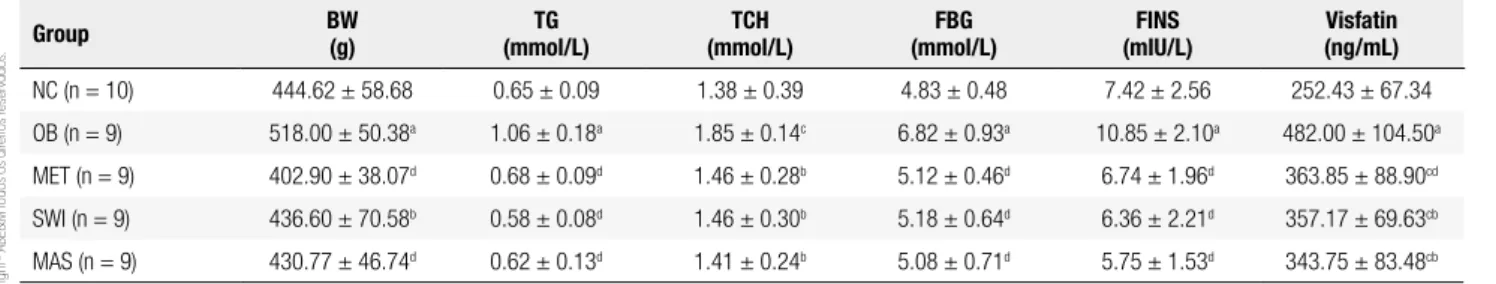
After 6 weeks of treatment, BW, FBG, TG, TCH, FINS, and serum visfatin levels in the group of MET, SWI, and MAS were decreased compared to the OB group (P < 0.01 or P < 0.05, Table 2). Although there was a more sig-niicant change of the biochemical markers in MAS group, the data did not show statistical differences between the MAS group and the MET/SWI group. Thus, an inves-tigation with a larger sample size may be further needed.
Table 1. Comparison of metabolic parameters and visfatin levels among the ive groups before intervention (mean ± SD)
Group BW
(g)
TG (mmol/L)
TCH (mmol/L)
FBG (mmol/L)
FINS (mIU/L)
Visfatin (ng/mL)
NC (n = 10) 431.3 ± 47.22 0.51 ± 0.11 1.32 ± 0.13 4.87 ± 0.84 9.04 ± 3.71 278.03 ± 128.97
OB (n = 10) 523.5 ± 36.87a 1.07 ± 0.25a 1.89 ± 0.24a 7.10 ± 1.21a 17.04 ± 4.15b 476.32 ± 86.25a
MET (n = 10) 520.80 ± 38.91a 0.95 ± 0.27a 1.82 ± 0.23a 6.73 ± 1.55a 21.33 ± 7.99a 548.71 ± 67.97a
SWI (n = 10) 530.40 ± 71.32a 0.93 ± 0.17a 1.77 ± 0.28a 6.95 ± 0.96a 15.82 ± 4.31b 596.24 ± 90.92a
MAS (n = 10) 522.33 ± 59.27a 0.89 ± 0.18a 1.69 ± 0.35a 6.52 ± 1.19a 19.13 ± 7.19a 504.37 ± 83.35a
a P < 0.01, b P < 0.05 vs. Normal Diet group.
Table 2. Comparison of metabolic parameters and visfatin levels among the ive groups after intervention (mean ± SD)
Group BW
(g)
TG (mmol/L)
TCH (mmol/L)
FBG (mmol/L)
FINS (mIU/L)
Visfatin (ng/mL)
NC (n = 10) 444.62 ± 58.68 0.65 ± 0.09 1.38 ± 0.39 4.83 ± 0.48 7.42 ± 2.56 252.43 ± 67.34
OB (n = 9) 518.00 ± 50.38a 1.06 ± 0.18a 1.85 ± 0.14c 6.82 ± 0.93a 10.85 ± 2.10a 482.00 ± 104.50a
MET (n = 9) 402.90 ± 38.07d 0.68 ± 0.09d 1.46 ± 0.28b 5.12 ± 0.46d 6.74 ± 1.96d 363.85 ± 88.90cd
SWI (n = 9) 436.60 ± 70.58b 0.58 ± 0.08d 1.46 ± 0.30b 5.18 ± 0.64d 6.36 ± 2.21d 357.17 ± 69.63cb
MAS (n = 9) 430.77 ± 46.74d 0.62 ± 0.13d 1.41 ± 0.24b 5.08 ± 0.71d 5.75 ± 1.53d 343.75 ± 83.48cb
Cop
yright
© ABE&M t
odos os dir
eit
os r
eser
vados
.
Effects of different treatments on visfatin levels in SAT, PAT and SM
We examined the expression levels of visfatin protein by western blot in different tissues of the ive groups. As shown in igure 1, the expression of visfatin protein in PAT of the obesity group was higher than that of the NC group, which was also observed in SAT and SM (Figure 1B and 1C). In SAT and PAT, visfatin expression levels in the MAS group, MET group, and SWI group were decreased after intervention compared to OB group, specially in the MAS group. However, in the NC, visfatin levels in the MAS group, MET group, and SWI group were decreased with no statistical signiicant difference.
the study include: (a) both metformin treatment and swimming exercise can down-regulate visfatin levels in serum and adipose tissue; (b) neither treatment has effect on visfatin levels in skeletal muscle; (c) both therapies can signiicantly improve insulin sensitivity and glucolipid metabolism.
Adipose tissue participates in the glucose metabolism by secreting various adipokines, which are frequently associated with the action of insulin. Visfatin, a recently discovered adipokine, has been shown to play a role similar to insulin in insulin-sensitive tissues, such as liver, muscle, and fat (8). Visfatin may play a dual role: an endocrine role, by modulating insulin sensitivity in peripheral organs; and an autocrine/paracrine function on visceral adipose tissue, facilitating differentiation and fat deposition (14). Though Pagano (15) have reported decreased plasma visfatin and its messenger RNA in subcutaneous adipose tissue of obese subjects, several other studies have reported elevated blood visfatin levels in patients with T2DM (9,16) or in obese children (17). Consistent with most indings, we noted signiicantly higher visfatin levels in obese rats compared with the controls in our study. Additionally, higher visfatin levels in perirenal adipose tissue were found compared with that in subcutaneous adipose tissue and skeletal muscle, which indicated that visceral adipose tissue was the major source of visfatin. However, the effect of hypoglycemic treatment on visfatin levels has not been thoroughly studied until now. Thus, the effect of metformin and swimming exercise treatments on visfatin levels was observed in our study.
There is limited data about the effect of metformin on adipokines, especially on visfatin. Hsieh and cols. (18) reported that two different metformin formulations had no effect on insulin sensitivity and serum visfatin in patients with T2DM, but patients in this study were under anti-diabetic treatment before enrolment. To avoid this potential drawback, Erdem and cols. (19) designed a study and observed that 12 weeks’ treatment of metformin signiicantly improved glycemic control and insulin sensitivity in 44 newly diagnosed and untreated T2DM patients, but still did not change visfatin levels. Accordingly, a new report demonstrated that insulin-sensitizing modalities induced non-signiicant change of plasma visfatin (20). The insulin-mimetic action of visfatin may be mediated by binding to the insulin receptor via a distinct binding site. In addition, visfatin gene expression has been recently found to be enhanced in macrophages of
5 4.5 4 3.5 3 2.5 2 1.5 1 0.5 0
0B NC MET SWI MAS
PAT SAT MT
□*
□* □*
□*
OB
OB
OB SAT
Visfatin
PAT Visfatin
MT Visfatin
β-actin
β-actin
β-actin
NC
NC
NC MET
MET
MET SW1
SW1
SW1 MAS
MAS
MAS
A
B
C
D
Visfatin/
β
-actin
Figure 1. Effects of different treatments on visfatin levels in subcutaneous adipose tissue (SAT), perirenal adipose tissue (PAT) and skeletal muscle (SM).
(A) Visfatin expression in PAT. (B) Visfatin expression in SAT. (C) Visfatin
expression in SM. (D) Statistical analysis of visfatin levels in the three
tissues after the different treatments. The bar represents the mean ± SD
of triplicate experiments with triplicate samples. ●P < 0.01,▲P < 0.05,
compared with Normal control (NC) group; △P < 0.01,*P < 0.05, compared
with obesity (OB) group; □P < 0.05, compared with Metformin and
Swimming (MAS) group;▽P < 0.05, compared withobesity group in PAT.
DISCUSSION
Cop
yright
© ABE&M t
odos os dir
eit
os r
eser
vados
.
human unstable carotid and coronary atherosclerotic lesions, where the inluence of insulin sensitizers seems to be limited (8,21). The mechanisms cited above indicate that insulin sensitizers may have little effect on visfatin. On the contrary, we found in our study that visfatin levels in serum and adipose tissue decreased after the treatment of metformin. A recent study conducted in patients with polycystic ovary syndrome, a condition with insulin resistance, also showed that visfatin levels decreased compared with the control group after metformin treatment for 3 months (22). It was reported that visfatin secretion was regulated by insulin and glucose by means of the phosphatidylinositol 3 kinase and protein kinase B pathways (23). Other study have shown that metformin has direct actions on adipocytes and skeletal tissue and inhibits plasminogen activator inhibitor-1 in adipose tissue and stimulates AMP-activated protein kinase in mice (19), which may explain what we have observed in the present study. But we did not see any changes in visfatin expression levels in skeletal muscle, neither did Varma and cols. (24). We speculated that low expression levels in skeletal muscle and different detection methods might be responsible for the controversial results. The present study showed that rats on a high-fat diet not only displayed marked increases in body weight, but also more pronounced insulin resistance and dyslipidemia, and 6 weeks of swimming exercise improved these signs of MS. Because exercise training has a profound effect on the prevention and treatment of obesity and diabetes, it was hypothesized that the effects might be mediated by adipocytokines. However, the effect of exercise training on adipokines was not consistent, and exercise-induced changes in visfatin have rarely been described.
Previous studies (25,26) showed a reduction in plasma visfatin after aerobic exercise training in patients with type 2 diabetes and type 1 diabetes compared with matched controls. In addition, exercise training that resulted in weight loss induced signiicant reductions in plasma visfatin in young, overweight Korean women (27). Visfatin was also measured in response to acute exercise bouts where visfatin mRNA expression in SAT was found to be elevated threefold in the hours immediately after exercise (28). These indings demonstrated that both acute and chronic exercise affected the regulation of visfatin. In the present study, we observed that visfatin levels decreased in serum and adipose tissue compared with sedentary controls after 6 weeks of swimming exercise. The exact mechanisms
have not yet been fully elucidated. We also observed that glycemic control and insulin resistance were improved after exercise. It was suggested that visfatin might be sensitive to nutrient availability, and there might be a negative feedback of insulin on glucose-induced visfatin release. Visfatin is expressed by both adipose tissue and skeletal muscle, and evidence of a cross-talk between muscle and adipose tissue is provided by the inding of gene activation in abdominal subcutaneous fat during leg muscle work (29).
Nevertheless, we did not observe any change of visfatin expression levels in skeletal muscle, as hypothesized. Visfatin change levels in the adjunctive therapy of metformin and swimming exercise group were also investigated. Interestingly, we found that decreased visfatin protein expression in adipose tissue was not accompanied by decreased visfatin in circulation compared with metformin and swimming exercise group. It is unclear whether visfatin is secreted into the systemic circulation, as the primary visfatin amino acid sequence does not contain a signal peptide (30). Visfatin may be secreted via an alternative pathway (31). The mechanisms above may explain the different results observed in serum and adipose tissue.
CONCLUSIONS
In conclusion, this investigation demonstrates that both metformin and swimming exercise lead to down-regulation of visfatin expression in adipose tissue and serum, as well as improvement in glycemic and lipid proile, except in skeletal muscle, in high-fat obesity rat model. However, the current study is limited by the sample size, and the duration of swimming exercise may have been too short to result in changes of visfatin in the skeletal muscle. Further studies are needed to clarify the role of visfatin in the pathogenesis of obesity and type 2 diabetes.
Acknowledgments: this study was supported by inancial assistance of the Provincial Education Fund Project.
Disclosure: no potential conlict of interest relevant to this article was reported.
REFERENCES
1. Alberti KG, Zimmet P, Shaw J. The metabolic syndrome--a new worldwide deinition. Lancet. 2005;366:1059-62.
Cop
yright
© ABE&M t
odos os dir
eit
os r
eser
vados
.
3. Fantuzzi G, Faggioni R. Leptin in the regulation of immunity, inlammation, and hematopoiesis. J Leukoc Biol. 2000;68:437-46. 4. Irving AJ, Wallace L, Durakoglugil D, Harvey J. Leptin enhances
NR2B-mediated N-methyl-D-aspartate responses via a mitogen-activated protein kinase-dependent process in cerebellar granule cells. Neuroscience. 2006;138:1137-48.
5. Haider DG, Schaller G, Kapiotis S, Maier C, Luger A, Wolzt M. The release of the adipocytokine visfatin is regulated by glucose and insulin. Diabetologia. 2006;49:1909-14.
6. Beltowski J. Apelin and visfatin: unique “beneicial” adipokines upregulated in obesity? Med Sci Monit. 2006;12:RA112-9. 7. Dogru T, Sonmez A, Tasci I, et al. Plasma visfatin levels in patients
with newly diagnosed and untreated type 2 diabetes mellitus and impaired glucose tolerance. Diabetes Res Clin Pract. 2007;76:24-9. 8. Fukuhara A, Matsuda M, Nishizawa M, Segawa K, Tanaka M,
Kishimoto K, et al. Visfatin: a protein secreted by visceral fat that mimics the effects of insulin. Science. 2005;307:426-30.
9. Chen MP, Chung FM, Chang DM, Tsai JC, Huang HF, Shin SJ, et al. Elevated plasma level of visfatin/pre-B cell colony-enhancing factor in patients with type 2 diabetes mellitus. J Clin Endocrinol Metab. 2006;91:295-9.
10. Wiernsperger NF, Bailey CJ. The antihyperglycaemic effect of metformin: therapeutic and cellular mechanisms. Drugs. 1999; 58 Suppl 1: 31-9; discussion 75-82.
11. King GA, Fitzhugh EC, Bassett DR Jr, McLaughlin JE, Strath SJ, Swartz AM, et al. Relationship of leisure-time physical activity and occupational activity to the prevalence of obesity. Int J Obes Relat Metab Disord. 2001;25:606-12.
12. Gregg EW, Cauley JA, Stone K, Thompson TJ, Bauer DC, Cummings SR, et al. Relationship of changes in physical activity and mortality among older women. JAMA. 2003;289:2379-86. 13. Souza HC, Penteado DM, Martin-Pinge MC, Barbosa Neto O,
Teixeira Vde P, Blanco JH, et al. Nitric oxide synthesis blockade increases hypertrophy and cardiac ibrosis in rats submitted to aerobic training. Arq Bras Cardiol. 2007;89:88-93, 99-104. 14. Sethi JK, Vidal-Puig A. Visfatin: the missing link between
intra-abdominal obesity and diabetes? Trends Mol Med. 2005;11:344-7. 15. Pagano C, Pilon C, Olivieri M, Mason P, Fabris R, Serra R, et al.
Reduced plasma visfatin/pre-B cell colony-enhancing factor in obesity is not related to insulin resistance in humans. J Clin Endocrinol Metab. 2006; 91:3165-70.
16. Retnakaran R, Youn BS, Liu Y, Hanley AJ, Lee NS, Park JW, et al. Correlation of circulating full-length visfatin (PBEF/NAMPT) with metabolic parameters in subjects with and without diabetes: a cross-sectional study. Clin Endocrinol (Oxf). 2008;69:885-93. 17. Davutoglu M, Ozkaya M, Guler E, Garipardic M, Gursoy H,
Karabiber H, et al. Plasma visfatin concentrations in childhood obesity: relationships with insulin resistance and anthropometric indices. Swiss Med Wkly. 2009;139:22-7.
18. Hsieh CH, He CT, Lee CH, Wu LY, Hung YJ. Both slow-release and regular-form metformin improve glycemic control without altering plasma visfatin level in patients with type 2 diabetes mellitus. Metabolism. 2007;56:1087-92.
19. Erdem G, Dogru T, Tasci I, Bozoglu E, Muhsiroglu O, Tapan S, et al. The effects of pioglitazone and metformin on plasma visfatin levels in patients with treatment naive type 2 diabetes mellitus. Diabetes Res Clin Pract. 2008;82:214-8.
20. Kadoglou NP, Tsanikidis H, Kapelouzou A, Vrabas I, Vitta I, Karayannacos PE, et al. Effects of rosiglitazone and metformin treatment on apelin, visfatin, and ghrelin levels in patients with type 2 diabetes mellitus. Metabolism. 2010;59:373-9.
21. Dahl TB, Yndestad A, Skjelland M, Øie E, Dahl A, Michelsen A, et al. Increased expression of visfatin in macrophages of human unstable carotid and coronary atherosclerosis: possible role in inlammation and plaque destabilization. Circulation. 2007;115:972-80.
22. Ozkaya M, Cakal E, Ustun Y, Engin-Ustun Y. Effect of metformin on serum visfatin levels in patients with polycystic ovary syndrome. Fertil Steril. 2010;93:880-4.
23. Haider DG, Holzer G, Schaller G, Weghuber D, Widhalm K, Wagner O, et al. The adipokine visfatin is markedly elevated in obese children. J Pediatr Gastroenterol Nutr. 2006;43:548-9.
24. Varma V, Yao-Borengasser A, Rasouli N, Bodles AM, Phanavanh B, Lee MJ, et al. Human visfatin expression: relationship to insulin sensitivity, intramyocellular lipids, and inlammation. J Clin Endocrinol Metab. 2007;92:666-72.
25. Haider DG, Mittermayer F, Schaller G, Artwohl M, Baumgartner-Parzer SM, Prager G, et al. Free fatty acids normalize a rosiglitazone-induced visfatin release. Am J Physiol Endocrinol Metab. 2006;291:E885-90.
26. Brema I, Hatunic M, Finucane F, Burns N, Nolan JJ, Haider D, et al. Plasma visfatin is reduced after aerobic exercise in early onset type 2 diabetes mellitus. Diabetes Obes Metab. 2008;10:600-2. 27. Choi KM, Kim JH, Cho GJ, Baik SH, Park HS, Kim SM. Effect of
exercise training on plasma visfatin and eotaxin levels. Eur J Endocrinol. 2007;157:437-42.
28. Frydelund-Larsen L, Akerstrom T, Nielsen S, Keller P, Keller C, Pedersen BK. Visfatin mRNA expression in human subcutaneous adipose tissue is regulated by exercise. Am J Physiol Endocrinol Metab. 2007;292:E24-31.
29. Keller C, Keller P, Marshal S, Pedersen BK. IL-6 gene expression in human adipose tissue in response to exercise--effect of carbohydrate ingestion. J Physiol. 2003;550:927-31.
30. Samal B, Sun Y, Stearns G, Xie C, Suggs S, McNiece I. Cloning and characterization of the cDNA encoding a novel human pre-B-cell colony-enhancing factor. Mol Cell Biol. 1994;14:1431-7. 31. Andrei C, Dazzi C, Lotti L, Torrisi MR, Chimini G, Rubartelli A.