Arq Neuropsiquiatr 2005;63(3-A):666-669
N e u romuscular Disease Unit, Department of Neuro l o g y, Hospital Universitário Antonio Pedro da Universidade Federal Fluminense, Niterói RJ, Brazil (UFF): 1Full Professor;2Master Student; 3Radiologist.
Received 4 November 2004, received in final form 2 February 2005. Accepted 1 April 2005.
Dr. Marcos R.G. de Freitas - Rua Gastão Ruch 16 / 1402 - 24220-100 Niterói RJ - Brasil. E-mail: [email protected]
CHRONIC INFLAMMATORY DEMYELINATING
POLYRADICULONEUROPATHY
Two cases with cervical spinal cord compression
Marcos R.G. de Freitas
1, Osvaldo J.M. Nascimento
1, Cristiane N. Soares
2,
Adriana Rocha Brito
2, Romeu Cortes Domingues
3ABSTRACT - Chronic inflammatory demyelinating polyradiculoneuropathy (CIDP) is a peripheral nerve dis-order probably due to an immunological disturb. It evolves either in a steadily progressive or in a relaps-ing and fluctuatrelaps-ing course. Weakness is mainly in the lower limbs proximally and distally. The electro m y o-graphy is demyelinating. The cerebral spinal fluid protein is most of times elevated. Sometimes enlarged nerves are found. There are few cases described with spinal cord compression due to hypertrophic spinal n e rve roots. Two patients (females, 66 and 67 years old) with diagnosis of a long standing CIDP are described. In the first one, the evolution was characterized by remission and relapsing course. The second patient had a chronic and pro g ressive course. These patients presented after a long evolution a cervical spinal cord c o m p ression syndrome due to hypert rophic cervical roots. Neurologists must be aware of the possibility of development of spinal cord compression by enlarged spinal roots in patients with a long standing CIDP.
KEY WORDS: chronic inflammatory demyelinating polyradiculoneuropathy (CIDP), spinal root hypert ro-phy, spinal cord compression.
P o l i rr a d i c u l o n e u ropatia desmielinizante inflamatória crônica: dois casos com síndrome de com-pressão medular
RESUMO - A polirr a d i c u l o n e u ropatia desmielinizante inflamatória crônica (PDIC) é uma afecção dos ner-vos periféricos de natureza autoimune, com evolução por surtos de exacerbação e remissão ou de evolver p ro g ressivo. O acometimento motor é predominante, com fraqueza proximal e distal nos membros infe-riores. A eletroneuromiografia é do tipo desmielinizante com bloqueio de condução nervosa em dois ou mais nervos. Há aumento de proteínas do líquor. Com a evolução da doença pode haver espessamento dos n e rvos distal e/ou proximalmente. Excepcionalmente ocorre compressão da medula espinhal em qualquer segmento por raízes próximas hipert rofiadas. Foram estudadas duas mulheres de 66 e 67 anos re s p e c t i v a-mente com quadro de PDIC de longa evolução. A primeira tinha evolução por surtos e na segunda o evolver era pro g ressivo. Nos dois casos o espessamento proximal dos nervos provocou síndrome de compre s s ã o medular alta. Esta complicação deve ser pensada em casos de PDIC de longa duração.
PA L AV R A S - C H AVE: polineuropatia inflamatória desmielinizante crônica (PIDC),hipert rofia de raízes ner-vosas, compressão medular.
C h ronic inflammatory demyelinating polyradi-culoneuropathy (CIDP) usually presents as a more or less symmetric sensorimotor polyradiculoneuro-pathy with chronic relapsing or remitting or pro-g ressive course. There is no clear estimate of its f re-quency but it may re p resent as many as 10%-30% of previously undiagnosed cases of polyneuropa-t h y1. Usually there is predominance of weakness
with diffuse hyporeflexia or areflexia. The
cere-b rospinal fluid (CSF) demonstrates alcere-buminouscy- albuminouscy-tologic dissociation and nerve conduction studies reveals multifocal conduction slowing, conduction block and temporal dispersion. The nerve biopsy shows primary segmental demyelination and axon-al degeneration with or without inflammatory infiltration and onion bulbs2 , 3. The therapeutic may
includ-Arq Neuropsiquiatr 2005;63(3-A) 667
ing cort i c o s t e roids, plasma exchange and intra-venous immune globulin4. Occasionally the re p e
t-itive demyelination and remyelination with onion bulb formation results in gross enlargement of spi-nal nerves end roots. CIDP is one of the main cau-ses of the hypert rophic neuro p a t h y5. Thickened
peripheral nerves were seen in 11% in one larg e s e r i e s3. Although exceedingly rare, there have been
re c o rded cases of CIDP presenting with spinal cord compression due hypertrophic spinal roots5-13.
We re p o rt two patients with CIDP of long evo-lution with cervical spinal compression due to hypertrophic roots.
CASES
Case 1 –A 66-year-old black woman had re c u rre n t p a resthesias and weakness in hands and feet was first seen in 1986. At that time she had distal tetrapare s i s with abnormal gait, reduced tendon reflexes, proprio-ceptive ataxia and superficial hypoesthesia in her legs. The peripheral nerves were not thickened. Tonus, coor-dination and cranial nerves were normal. An electro d i-agnostic evaluation showed a sensorimotor demyeli-nating polyneuropathy features: absence of sensitive responses, prolonged distal motor latencies and con-duction block in bilateral median and ulnar nerves, seve-re slowing of motor conduction velocity and abolished F waves. Needle electromyography (EMG) showed active d e n e rvation in distal limbs. CSF examination re v e a l e d albuminous-cytologic dissociation and a sural biopsy showed demyelination and remyelination feature s , axonal regeneration and presence of some onion bulbs. T h e re was no duplication in the PMP22 gene. The pati-ent was treated successfully with prednisone; however, t h e re were subsequent relapsing courses. Over a peri-od of 11 years she had been maintained in alternating treatment with steroids and plasma exchange. In 2002 her symptoms worsened. She became tetraparetic and
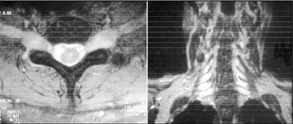
couldnot walk. The tonus was increased in lower limbs and a sensitive cervical level to painful-touch sensation could be found. The tendon reflexes were abolished and there were withdraw reflexes with bilateral Babinski sign. A cervical magnetic resonance image (MRI) re v e a l e d h y p e rt rophy of cervical spinal roots, with spinal com-p ression, enhanced with gadolinium (Fig 1). Stero i d s , imunoglobulin and plasma exchange were given with no improvement.
Case 2 –A 67-year-old woman presented in 1981 a c e rvical pain irradiating to the left arm. She was sub-mitted to a myelography and a cervical spine surg e ry showed hypert rophic cervical roots. In 1983 she had a low back pain radiating to posterior surface of right thigh and weakness of this limb. A laminectomy was performed with some relief of the pain. There is no ref-e rref-encref-e of thref-e nref-eurological ref-examination in this pref-eriod. Five years after she complained of pain and asymmet-ric weakness of all limbs, and stopped walking. No sim-ilar cases in the family are re p o rted. There were distal amyotrophy in all 4 limbs with slight deformity of the left hand, distal and proximal tetraparesis more severe in the lower limbs. Deep tendon reflexes were absent in lower limbs. There were superficial hypoesthesia below the knees and loss of vibratory sense in lower limbs and in the inner aspects of the left fore a rm and hand. The ulnar and posterior auricular nerves were
uni-f o rmly palpable and thick. The CSF showed 1 cell/mm3
and 48 mg/dl of protein. The tonus was increased in the lower limbs. There was bilateral Babinski sign. The left biceps reflex was present and the other tendon reflex-es were absent. A nerve conduction study revealed a generalized slowing of motor conduction and a con-duction block in ulnar and median nerves, the sensory n e rve action potentials were abolished in the sural, ulnar and median nerves and the F waves were prolonged in most nerves. The EMG was not performed. Sural nerve biopsy demonstrated a great loss of fibers, some
668 Arq Neuropsiquiatr 2005;63(3-A)
m a t o ry infiltrates, fibrosis, clusters of regenerated axons as well as thinly myelinated axons and many onion bulbs (Fig 2). Ultrathin sections showed similar features. The patient was put on 60 mg prednisone per day, and 30 days after she could walk without support with gre a t i m p rovement of the strength and sensation. She had been maintained with prednisone and intravenous metil-p rednisolone for 10 years. In 1999 there was worsening of her neurological examination, showing paraplegia with increased muscle tonus, bilateral Babinski sign and diminished superficial and hypoesthesia below C6 lev-el. She died in 1999 of septicemia. A necropsy examina-tion of the spinal cord revealed fusiform swellings of all ventral and dorsal roots. Histological examination s h o w e d that the root enlargements were due to hypert ro p h i c demyelinating neuropathies with onion-bulb form a-tions and cellular infiltration.
DISCUSSION
Our two cases fulfilled clinical, neuro p h y s i o l o g i-cal and pathologii-cal criteria for CIDP diagnosis1 - 3.
T h e re was no family history of Charc o t M a r i e -Tooth (CMT) disease, and the genetic markers for CMT1A in the first patient was absent. Sural nerv e biopsies perf o rmed in both patients showed ima-ges of demyelination and remyelination with onion-bulb formations. In the second patient there was also inflammatory infiltration. There was a h i s t o ryof almost 17 years of relapsing and re m i t-ting courses in both patients. Treatment over sev-eral years with cort i c o s t e roids resulted in an une-quivocal improvement. The patients presented ma-ny years after the onset, cervical spinal cord com-p ression with tetracom-paralysis, hycom-pert o n i c i t y, bilat-eral Babinski sign and a sensitive level. Our patient number 2 presented with radicular signs since the
beginning of the disease, first in cervical level and two years after in lumbar level. The most striking f e a t u re was the diffuse, marked enlargement of peripheral roots, demonstrated in one case by MRI and the other by necropsy studies. They had an unusual clinical picture of cervical spinal cord com-pression determined by CIDP.
Spine MRI is a valuable addition to the diagnos-tic armamentarium in CIDP. Enlarged spinal roots may be identified in patients being investigated for demyelinating neuropathy as in our first case. N e rve root enhancement with gadolinium, seen in our patient, is sometimes found in inflammato-ry demyelinating neuropathies5,9,11.
When CIDP evolves slowly and its pro g re s s i o n is more insidious it may be difficult to distinguish it from a hypert rophic CMT. There are few cases of here d i t a ry hypert rophic neuropathy pro d u c i n g spinal cord compression syndromes14,15.The
infor-mation described by Symmons and Blackwwod in the first case re p o rt e d1 5does not allow confident
d e t e rmination of whether the neuropathy was a c q u i red or genetically determined. In our first ca-se the molecular genetic studies discloca-sed no dupli-cation at 17p11.2. Although we had not done DNA studies in our second patient, the relapsing and fluctuating course and the inflammatory infiltrates in the nerve biopsy confirm the diagnosis of CIDP. The nerve enhancement with gadolinium seen i n patient one, although nonspecific, probably distin-guish clinically active CIDP from genetically deter-mined hypert rophic neuro p a t h y, where the ro o t s do not enhance5 , 9 , 1 1 , 1 6. Both our patients had
impro-ved in the course of the disease with cort i c o s-teroids.
Arq Neuropsiquiatr 2005;63(3-A) 669
The diff e rential diagnosis of hypert rophy ro o t s should be done also with neurofibromatosis that could be associated with spinal compression syn-d ro m e s1 3. The long course of disease (16 years in
case 1 and 17 years in case 2) was an evident deter-minant for the evolution of hypert ro p h y, causing spinal compression. Recently it has been re p o rt-e d1 7the beneficial effect of interf e ron1a in
pati-ents with CIDP that are refractory to the conven-tional treatment, although controlled and rando-mized studies are needed to confirm the eff e c t i v e-ness of this treatment. Although decompre s s i v e laminectomy was not done in our patients, we a g ree that it is an acceptable option of tre a t m e n t in cases with spinal cord compression4,5.
Although CIDP is a common disorder of the pe-ripheral nerve system, the radicular and the spinal c o m p ression due to this syndrome is seldom re l a t-e d5 - 1 3. In case of patients with CIDP with enlarg e d
n e rves and minimal symptoms and signs of spinal c o rd involvement it is necessary to perf o rm MRI studies of the spine and try to modify the tre a t-ment to prevent the functional deterioration. Our cases and other observations already mentioned suggest that new forms of treatment are needed in cases of spinal cord compression due do enlar-ged spinal roots in CIDP.
REFERENCES
1. Kissel JT, Mendell JR. Chronic inflammatory demyelinating polyradicu-l o n e u ro p a t h y. In Kissepolyradicu-l JT, Mendepolyradicu-lpolyradicu-l JR, Cornbpolyradicu-lath DR (Eds). Diagnosis and management of peripheral nerves. New York: Oxford Univ Pre s s , 2001:173-191.
2. Dyck PJ, Lais AC, Ohta M, et al. Chronic inflammatory polyradicu-loneuropathy. Mayo Clin Proc 1975;50:621-637.
3. Barohn RJ, Kissel JT, Warmolts JR, Mendell JR. Chronic inflammatory demyelinating polyradiculoneuropathy: clinical characteristics, course and recommendations for diagnostic criteria. A rch Neurol 1989;46: 878-884.
4. M i d roni G, Dyck PJ. Chronic inflammatory demyelinting polyradicu-l o n e u ropathy: unusuapolyradicu-l cpolyradicu-linicapolyradicu-l features and terapeutic re s p o n s e s . Neurology 1996;46:1206-1212.
5. Schady W, Goulding PJ, Lecky BRF, King RHM, Smith CML. Massive nerve root enlargement in chronic inflammatory demyelinating polyneuropathy. J Neurol Neurosurg Psychiatry 1996; 61:636-640. 6. Aïdi M El Aloui Faris, Amarti A, Belaïdi H, et al.
Polyradiculoneuro-pathie inflammatoire démyélinisante chronique hypertrophique avec atteinte des nerfs crâniens: à propos de deux observations. Rev Neuro l (Paris) 2002;158:819-823.
7. De Silva RN, Willison HJ, Doyle D, Weir AI, Hadley, Thomas AM. Nerve root hypertrophy on chronic inflammatory demyelinating polyneu-ropathy. Muscle Nerve 1994;17:168-170.
8. Duart e J, Cruz Marti nez A, Rodriguez F, Mendoza A, Sempere A P, Claveria LE. Hypertrophy of multiple cranial nerves and spinal roots in chronic inflammatory demyelinating neuro p a t h y. J Neurol Neuro s u rg Psychiatry 1999;67:685-687.
9. Duggins AJ, McLeod JG, Pollard JD et al. Spinal root and plexus hyper-t rophy in chronic inflammahyper-tory demyelinahyper-ting polyneuro p a hyper-t h y. Brain 1999;122:1383-1390.
10. G i n s b e rg L, Platts AD, Thomas PK. Chronic inflammatory demyelinat-ing polyneuropathy mimickdemyelinat-ing a lumbar spinal stenosis syndrome. J Neurol Neurosurg Psychiatry 1995;59:189-191.
11. Mizuno K, Nagamatsu M, Hattori N, et al. Chronic inflammatory demy-elinating polyradiculoneuropathy with diffuse and massive peripher-al nerve hypertrophy: distinctive clinicperipher-al and magnetic resonance imag-ing features. Muscle Nerve 1998;21:805-808.
12. Oguz B, Oguz KK, Cila A, Tan E. Diffuse spinal and intercostal nerve involvement in chronic inflammatory demyelinating polyradiculoneu-ropathy: MRI findings. Eur Radiol 2003:13(S4):L230-234.
13. Pytel P, Rezania K, Soliven B, Frank J, Wollmann R. Chronic inflamma-tory demyelinating polyradiculoneuropathy (CIDP) with hypertro p h i c spinal radiculopathy mimicking neuro f i b romatosis. Acta Neuro p a t h o l 2003;105:185-188.
14. Rosen SA, Wang H. Cornblath DR, Uematsu S, Hurko O. Compre s s i o n s y n d romes due to hypertrophic nerve roots in hereditary motor sen-sory neuropathy type I. Neurology 1989;39:1173-1177.
15. Symonds CP, Blackwood W. Spinal Cord compression in hypertro p h i c neuritis. Brain 1962;85:251-259.
16. M o rgan GW, Barohn RJ, Bazan C III, King RB, Klucznik RP. Nerve ro o t enhancement with MRI in inflammatory demyelinating polyradicu-loneuropathy. Neurology 1993;43:618-620.