1N e u rophysiology Department, MD, International Center of Neural Science and Rehabilitation, North Lake SARAH Hospital, Brasilia D F, Brazil;2MD, PhD, University of Brasilia School of Medicine Pos-Graduation Program, Brasilia DF, Brazil. Senior Fellow Researc h e r. Received 12 September 2005, received in final form 23 January 2006. Accepted 5 April 2006.
D r. Paulo Sérgio Azeredo Henriques Filho - SMLN Trecho 06 - Chácara 232 / casa 02 - 71540-060 Brasília DF - Brasil. E-mail: pazere d o@ linkexpress.com.br
ABNORMALITIES IN AUDITORY EVOKED POTENTIALS
OF 75 PATIENTS WITH ARNOLD-CHIARI
MALFORMATIONS TYPES I AND II
Paulo Sergio A. Henriques Filho
1, Riccardo Pratesi
2ABSTRACT -Objective:To evaluate the frequency and degree of severity of abnormalities in the auditory pathways in patients with Chiari malformations type I and II. Method:This is a series-of-case descriptive study in which the possible presence of auditory pathways abnormalities in 75 patients (48 children and 27 adults) with Chiari malformation types I and II were analyzed by means of auditory evoked potentials evaluation. The analysis was based on the determination of intervals among potentials peak values, absolute latency and amplitude ratio among potentials V and I. Results:Among the 75 patients studied, 27 (36%) disclosed Arnold-Chiari malformations type I and 48 (64%) showed Arnold-Chiari malformations type II. F i f t y - t h ree (71%) of these patients showed some degree of auditory evoked potential abnormalities. Te s t s w e re normal in the remaining 22 (29%) patients. Conclusion:A u d i t o ry evoked potentials testing can be c o n s i d e red a valuable instrument for diagnosis and evaluation of brain stem functional abnormalities in patients with Arnold-Chiari malformations type I and II. The determination of the presence and degree of severity of these abnormalities can be contributory to the prevention of further handicaps in these patients either through physical therapy or by means of precocious corrective surgical intervention.
KEY WORDS: Arnold-Chiari malformations type I and type II, auditory evoked potentials.
A n o rmalidades nos potenciais evocados auditivos de 75 pacientes com os tipos I e II das mal-formações de Arnold-Chiari
RESUMO -Objetivo:Avaliar a freqüência e grau de comprometimento das vias auditivas em tronco cere-bral por meio de potencial evocado auditivo, em pacientes afetados por malformações de Arn o l d - C h i a r i de tipos I e II. Método:Foi efetuado um estudo descritivo de tipo série de casos, sendo selecionados 75 pacientes (48 crianças e 27 adultos) nos quais foi realizada avaliação dos potenciais evocados das vias audi-tivas, com base à determinação dos valores dos intervalos entre picos de potenciais, da latência absoluta e da razão entre as amplitudes dos potenciais V e I. Resultados:E n t re os 75 pacientes avaliados, 27 (36%) a p resentavam malformações de Arnold-Chiari de tipo I e 48 (64%) apresentavam malformações de tipo II. Em 53 (71%) do total de pacientes os potenciais evocados auditivos mostraram algum grau de anormali-dade. Os testes foram normais nos restantes 22 (29%). Conclusão:O potencial evocado auditivo pode ser considerado valioso instrumento para o diagnóstico e avaliação da gravidade das anormalidades funcionais de tronco cerebral em pacientes port a d o res de malformações de Arnold-Chiari de tipo I e II. Esta avaliação pode contribuir de maneira significativa não somente no diagnóstico como também na prevenção de ulte-r i o ulte-res lesões, pela adoção de medidas pulte-reventivas, tanto poulte-r meio de fisioteulte-rapia como poulte-r inteulte-rv e n ç ã o cirúrgica precoce.
PALAVRAS-CHAVE: malformação de Arnold-Chiari de tipo I e de tipo II, potenciais evocados auditivos.
Neural tube defects affect 3 children out of 1000 b o rnalive1. Among these anomalies are the
malfor-mations described by Chiari in 1891 and almost con-c u rrently by Arnold, two pioneers in the study of sucon-ch defects in human beings2. There are four types of
Chiari malformations: type I is identified by hern i a-tion of cerebellar tonsils and medulla oblongata, t h rough the foramen magnum. Type II is
Chiari malformations and is generally associated with a somber prognosis in terms of severe disability and early death. Anatomically is characterized by the asso-ciation of high cervical or occipital encephalocele and the presence of many of the abnormalities usually found in the second type. Finally the type IV, that is the less complex of Chiari malformations, is solely e x p ressed by a variable degree of cerebellar hypoplsia. Although patients with Arnold-Chiari I malform a-tion are generally asymptomatic at birth, they may eventually develop pro g ressive brain stem dysfunc-tion during their lives. This dysfuncdysfunc-tion can be pro-duced by traumatic, vascular or inflammatory episo-des or by any other kind of damaging events. Patients with type II Arnold-Chiari malformation are gener-ally symptomatic since birth but the above mentioned deleterious episodes can aggravate their pre - e x i s t-ing condition3 , 4. Due to their position in the brain
stem the auditory pathways are frequently injure d . In Arnold-Chiari malformations these injuries may include all segments of the auditory system such as the distal and proximal regions of the cochlear nerv e , and the auditory pathways running through the superior region of the bulb, the superior and inferi-or region of the pons, and the inferiinferi-or region of the mesencephalon5.
T h e re is an evident diff e rence in the period of life during which patients with Arnold-Chiari malform a-tions I or II will look for medical assistance. Due to the benignity of their clinical picture patients with type I will eventually appear at rehabilitation cen-ters during their adult life. That is not the case of pa-tients with type II malformations that, in general, are b rought to medical attention during the first years of their life due to the precocity and severity of their symptoms mainly characterized by an early function-al limitation of movements and the function-almost constant p resence of myelomeningocele with the consequent u r i n a ry bladder disord e r. This is the most pro b a b l e explanation for the diff e rence in the number of pa-tients with type I and II Arnold-Chiari malform a t i o n s found in our study. The analysis of brain stem audito-ry-evoked potentials is frequently utilized to detect abnormalities in the auditory pathways and several authors have applied this method in the evaluation of patients with Arnold-Chiari malformations type II. On the other hand, auditory-evoked potentials stu-dies focusing on the abnormalities of the auditory pathways of patients with Arnold-Chiari malform a-tion type I are rare.
Studies focusing on the abnormalities in the audi-t o ry paaudi-thways of paaudi-tienaudi-ts wiaudi-th Arnold-Chiari malfor-mations are scanty in the literature, this fact being
p a rticularly true in the case of Arnold-Chiari malfor-mation type I. Trying to contribute to a more detailed knowledge on brain stem functioning of these pa-tients, the aim of the present study was to evaluate the frequency and degree of severity of the abnor-malities in the auditory pathways in patients with A rnold-Chiari malformations type I and II. A more detailed knowledge of the characteristics of their a b n o rmalities and of their future evolution could be c o n t r i b u t o ry to the management of these patients, t rying to prevent further handicaps, both thro u g h physical therapy or by means of a precocious corre c-tive surgical intervention.
METHOD
This is a series-of-case descriptive study, carried out after the approval of the Ethic Committee of the SARAH Rehabili-tation Hospitals Network as well as the approval of the re l-atives and/or the patients involved in the study. The sam-ple size, of this study was composed by 75 subjects with Ar-nold-Chiari malformation. The data were collected and analyzed between march 2002 and march 2003. Individuals without Arnold-Chiari malformation were excluded fro m the selection after interviews and clinical or radiological examinations, as well as patients with encephalic injuries, n e u ropathies or those using medications such as, for exam-ple aminoglycosides, in order to avoid any undue interf e r-ence in the analysis of the data. All patients underw e n t a u d i t o ryevoked potentials test during the morning and in waking state.
The data were collected with a Medtronic instru m e n t and Key-point software (Medtronic Functional Diagnostics A/S, Denmark) and utilizing a Key-point software (Ve r s i o n 3.0, of the Medtronic Functional Diagnostics A/S). Auditory potentials were re c o rded through gold re c o rding electro d e s which were attached to the skin with collodion, and filled with conductive jelly. The electrodes were placed on Cz scalp position and on the left and right mastoid pro c e s s . The register of the auditory potentials w as obtained uti-lizing the three following channels: the M1-Cz (M for mas-toid process), the M2-Cz and the M1-M2. We re used two limiting filters, one inferior to 2 Hz and one superior to 3 KHz. The frequency of the stimulus was equal to 11 Hz and its intensity was of 100dB. The stimulus modality was the r a refaction click, with mask of the oposite side. To evoke the potentials headphones were placed over both the pati-e n t ’s pati-ears. Thpati-e guidpati-elinpati-es of thpati-e Intpati-ernational Fpati-edpati-eration of Clinical Neuro p h y s i o l o g y6w e re strictly applyied in the evaluation of the auditory evoked potentials. The absolute l a t e n c y, the latency among peaks and the amplitude ratio among potentials V and I were analyzed6.
The software SPSS for Windows software, (standard ver-sion, # 10.0.1, SPSS Inc. 1999)7was used in the statistical analysis, being evaluated the frequency of discrete and con-tinuous variables. The median value of the central tenden-cy was calculated in the analysis of continuous variables.
RESULTS
aged 2 to 16, mean age 7, and 27 adults, aged 19 to 70, mean age 38) were analyzed, 27 disclosing type I and 48 showing type II Chiari malformations. Among the patients with the type II 47 (98%) were childre n and adolescent, and among the type I 25 (92%) were adult. Fifty-five (71%) of these patients showed some degree of auditory evoked potential abnormalities. Tests were normal in the remaining 20 (27%) patients. Among these 55 patients that showed abnormal audi-t o ry evoked poaudi-tenaudi-tials 21 (38.2%) peraudi-tained audi-to pa-tients with Chiari type I malformation and 34 (61.8%) to patients with type II malformation.
In Table 1, can be seen detailed data concerning the potentials values in relation with the two diff e r-ent types of Chiari malformation.
In Table 2 can be seen the number of abnorm a l tests and the corresponding affected segment of the auditory pathway.
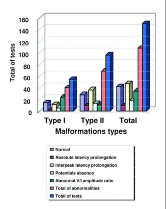
In Graphic 1 the diff e rent abnormalities found in the auditory pathways can be visualized as: abnor-mal amplitude ratio between potentials V and I; ab-solute latency prolongation; interpeak latency I-III, I-V, III-V; and absence of potential I, III or V.
DISCUSSION
The absence of patients with Chiari malform a-tions type III and IV in our study can be explained by the diff e rence in structural abnormalities and in clin-ical manifestations that are characteristic of each type of malformation. The severity and complexity of the m a l f o rmations found in type III is generally incompa-tible with an extended life span. On the other hand, the usual benignity of the abnormalities and the lack of serious clinical manifestations found in patients Table 1. Absolute latency central tendency, interpeak latency and amplitude ratio between the potentials V and I.
Absolute latency central tendency Interpeak potential latency values Potentials V/I
Types of Chiari I III V I-III III-V I-V V / I
malformations
Type I 1.50±0.12 3.70±0.32 5.6±0.35 2.20±0.28 2.00±0.27 4.15±0.30 2.0±4.32 Type II 1.50±0.14 4.0±0.31 6.0±0.35 2.50±0.32 2.05±0.26 4.50±0.32 1.37±0.75
I, III, V: potentials generated by components of the auditory pathways; I-III, III-V and I-V: interpeak latency (ms); potentials V/I: amplitude ratio between the potentials I and V (µV).
Table 2. Number of abnormal tests, distributed according to the type of Chiari malformation and auditory pathway aff e c t -ed segment.
Affected segment Chiari type I Chiari type II Total
Segment 1 30 19 49
Segment 2 10 35 45
Segment 3 3 14 17
Without
abnormalities 11 28 39
Total 54 96 150
Tests: comprehend the sum of right and left auditory evoked poten-tials; Segment 1: part of the cochlear nerve near the cochlea (deficit defined by the absolute latency prolongation or by the absence of the potential I); Segment 2: include the segment localized between the cochlear nerve near the cochlea and the transition part of the bulb and the pons (deficit defined by the absolute latency prolongation of the potential III, and or by the interpeak latency I-III prolongation, and or by the absence of the potential III; Segment 3: part localized between the lower and upper portion of the pons (deficit defined by the absolute latency prolongation of the potential V , and or by the interpeak laten-cy I-V and III-V prolongation, and or by the absence of the potential V).
with type IV malformation drastically decrease their p resence in hospital settings. These characteristic clin-ical patterns are probably the cause of the absence of patients with malformations type III and IV in our s t u d y. In this study was observed a pre d o m i n a n c e (98%) of children and adolescent among the patient with the type II, and predominance (92%) of adults among patients with the type I. This discrepancy is explained by the greater complexity of the malfor-mation type II, that due to its more severe clinical re p e rcussion will need an earlier medical attention8.
In the study of auditory evoked potentials abnor-malities we choose to proceed with a segmental ana-lysis of the auditory pathways since discrete gro u p s of neurons, as generators of specific potentials, as is the case of potentials III and V, are still
undetermin-e d5 , 6 , 9 , 1 0. Analysis of the absolute latency values in
patients with Chiari malformation type II disclosed a tendency to increased median value of potentials III and V when compared to normal values (Table 1). The most plausible explanation for this discre p a n c y is that it is probably due to the association of type II brainstem abnormalities and the sensory-neural mat-uration delay typically found in this kind of malfor-mation5,11,12.
The amplitude ratio between potentials V and I showed a higher median value in patients with mal-f o rmation type I (Table 1). This alteration is sugges-tive of higher frequency of peripheral injuries of the a u d i t o ry pathways, namely, sensory neural deafness and or loss of higher frequency components of these pathways. These disorders can appear alone or can be seen in association with other abnormalities. An higher frequency of these peripheral disorders of au-d i t o ry pathways in patients with type I malform a t i o n can be explained by the timing in which the patients have been evaluated and by the natural history of the malformation that, during the adult life, tend to manifest functional re p e rcussions consequent to even-tual vascular or traumatic events, leading to stre t c h-ing of the cochlear nerve distally to the brain stem.
The involvement of the components responsible for higher frequencies transmission could be explain-ed by the possible alteration of the cere b rospinal flu-id circulation consequent to brain stem compre s s i o n by herniated cerebellar tonsils at the foramen mag-num level, with repercussion on endolymphatic flu-id dynamic and resultant hydrops and cochlear nerv e dysfunction.
In 68 of the 96 tests performed on patients with type II malformation (Table 2), the abnormality ob-s e rved waob-s a conduction deficit in the caudal ob-
seg-ment of the cochlear nerve. In the present study this segment has been the most frequently affected por-tion of the auditory pathways, a result that diff e r from the study of Nishimura et al.5that reported as
the most affected region the one localized between the segments of the pons. This diff e rence is easily explained by diff e rences in age-group of the two studies since the sample of the former authors includ-ed patients with type II Arnold-Chiari malform a t i o n s , during the first year of their lives. It is known that this age-group is more prone to alterations of this p o rtion of pons. These abnormalities, when occur-ring duoccur-ring the first year of life, result in an incre a s e d p robability of brain stem dysfunction. In addition, a p rolonged time of maturation of the auditory path-ways may occur in children with Arnold-Chiari mal-f o rmation. The auditory pathways omal-f these childre n only reach their normal latency values around 8 to 9 years of age whereas subjects without this malform a-tion reach normal latency values around their fourt year of life5 , 1 1. These facts could explain the smaller
frequency of these alterations in older patients.
Another possible explanation for the diff e re n c e in findings between the two studies can be due to the methodology applied on the present study since all tests showing isolated prolongations of the inter-peak latency III-V were disre g a rded, being consid-e rconsid-ed abnormal only whconsid-en prconsid-esconsid-ent thconsid-e association of an abnormal relation of amplitude V/I and or the lengthening of the latency between peak I-V. In Nishimura et al.5study prolongation of the isolated
interpeak latency III-V was adopted as the parame-ter of abnormality.
Among the 150 tests perf o rmed were detected 17 alterations due to conduction deficit between the caudal segment and the rostral segment of the pons ( Table 2). This abnormality was identified by the pro-longation of the latency between peaks III-V in asso-ciation with a prolongation of the latency between peaks I-V and or an abnormal amplitude ratio V/I, smaller then 0.5 microvolts or yet, by the absence of the potential V. In two cases was observed a conduc-tion deficit of the above menconduc-tioned segment, which was identified by the abnormal amplitude ratio. This fact is not described in previous articles about Arn o l d -Chiari malformations and auditory pathways conduc-tion deficits5 , 1 1 , 1 3. It is relevant to point out that the
alte-rations in the medulla oblongata, whose more fre-quently re p o rted alterations are secondary to trau-ma during life, which can be easily visualized in MRI1 4.
The majority of cases among the 17 cases of con-duction deficits identified by the above described cri-teria, are re p resented by patients with Arn o l d - C h i a r i m a l f o rmation type II. This fact re i n f o rce the state-ments that alterations at this level, or more precise-l y, at the precise-leveprecise-l of the pons segment of the brain stem, a re alterations consequent todysgenesis,being more complex in patients with malformations type II when c o m p a red to patients with type I malform a t i o n s2 , 3.
Analyzing the added results of all variables (Table 2 and Graphic 1) that pointed to a conduction deficits of the auditory pathways, according to the levels or segments of those pathways, a higher frequency of alterations at the peripheral level in patients with m a l f o rmation type I can be noted. In patients with malformation type II, the higher frequency of alter-ations was registered above the cochlear nerve and below or at the level of the caudal segment of the pons. These alterations reinforce the data previous-ly discussed about the complexity of these malfor-mations. In the type I the cochlear nerve is the most f requently stru c t u re affected by traumatic injury dur-ing the life, whereas in type II the medulla oblonga-ta and pons are the preferred oblonga-targets.
Comparing the short latency evoked potentials alterations of the auditory pathways between mal-f o rmations type I and II, a small dimal-fmal-f e rence in perc e n-tage was re g i s t e red in the results, that is, 69% of type I patients and 63% of type II patients presented such a b n o rmalities. This slight diff e rence between the fre-quency of alterations in type I and II malformations could be explained by the evaluation of the several segments of the pathway, in general evaluated at d i ff e rent moments of the natural history of their mal-f o rmations, when the re s e a rch is held at the hospi-tal level. The patients with type I generally seek re h a-bilitation centers assistance when there is some func-tional abnormality on the stru c t u reof their nerv o u s system, something that generally occurs during their adult life, when they discover the syndrome. Now, patients with type II malformation, due to their pre-cocious functional limitation of their movements, and due to the presence of urinary bladder disord e r s related to the almost constant presence of myelome-ningocele are generally seen earliers at the rehabil-itations centers, almost always during their infancy or childhood. This can explain the reason for the ma-jority of patients in this study being children.
In conclusion, the analysis of short latency audito-ry evoked potentials perf o rmed in the present study,
contributed to the demonstration of several degre e s of brain stem abnormalities among patients with type I and II Chiari malformations. The frequency of these abnormalities, in both types of Chiari malfor-mation was not very large. The increased frequency of abnormal findings, among the patients with type II malformation was probably related to the com-plexity of this type of malformation.
The analysis of the amplitude ratio between the potentials V and I, in addition with the other crite-ria that define abnorm a l i t y, allows the identification of a greater number of alterations in the auditory pathways functions of patients with Arn o l d - C h i a r i m a l f o rmations types I and is a valuable tool to incre a-se the quality and precision of the diagnosis.
Acknowledgment– The authors thank the authori-zation of this re s e a rch at the SARAH Rehabilitation Hospitals Network. To doctors: Aloysio Campos da Paz, MD PHD P resident of this Network, José Carlos Dias Ferreira, MD PHD Chief of the Neurophysiology Service, Eliana Va l v e rd e M a g ro Borigatto, MD PHD Chief of the Pediatric Serv i c e and Régis Tavares, MD Chief of the Neurosurgical Service.
REFERENCES
1. Trimble BK, Baird PA. Congenital anomalies of the central nervous sys-tem: incidence in British Columbia, 1952-72. Teratology 1978;17:43-50. 2. Koehler PJ. Historical vignette. Chiari’s description of cerebelar ectopy (1891), with a summary of Cleland’s and Arnold’s contributions and some early observations on neural-tube defects. J Neurosurg 1991;75: 823-826.
3. Gilbert JN, Jones KL, Rorke LB, Chernoff GF, James HE. Central nerv-ous system anomalies associated with meningomyelocele, hydro-cephalus, and the Arnold-Chiari malformation. Reappraisal of theo-ries re g a rding the pathogenesis of posterior neural tube closure defects. Neurosurgery 1986;18:559-564.
4. Dyste GN, Menezes AH. Presentation and management of pediatric Chiari malformation without myelodysplasia. Neurosurgery 1988;23: 589-597.
5. Nishimura T, Mori K, Uchida Y, Ohira T, Tamura K. Brain stem audi-tory-evoked potentials in meningomyelocele: natural history of Chiari II malformations. Child’s Nerv Syst 1991;7:316-326.
6. Pratt H, A m i n o ff M, Nuwer MR, Starr A. Short-latency auditory evoked potentials. In Deuschl G, Eisen A (eds). Recommendations for the prac-tice of clinical neurophysiology: guidelines of the International Fede-ration of Clinical Neuro p h y s i o l o g y. 2n ded. Electroencephalogr Clin Neurophysiol 1999;52(Suppl):69-77.
7. SPSS for Windows. Standard version. Release 10.0.1. SPSS Inc.1999. 8. Carmel PW, Markesbery WR. Early description of the A r n o l d - C h i a r i
malformation: the contribution of John Cleland. Neuro s u rgery 1972; 37:543-547.
9. Nuwer MR, A m i n o ff M, Goodin D, et al. IFCN recommended stan-d a rstan-ds for brain-stem austan-ditory evokestan-d potentials: report of an IFCN committee. Electroencephalogr Clin Neurophysiol 1994;91:12-17. 10. Chiappa KH, Hill RA. Brain stem auditory evoked potentials:
inter-p retation. In Chiainter-pinter-pa KH (ed). Evoked inter-potentials in clinical medicine. 3rded. Philadelphia: Lippincott-Raven, 1997:199-268.
11. Fujii M, Tomita T, McLone DG, Grant JA, Stack CV, Mori K. Develop-mental normo-maturation of brainstem auditory evoked potentials in children with asymptomatic meningo-myelocele during the first year of life. Child’s Nerv Syst 1997;13:147-153.
12. Sperling NM, Franco RA J r, Milhorat TH. Otologic manifestations of Chiari I malformation. Otol Neurotol 2001;22:678-681.
13. Barnet AB, Weiss IP, Shaer C. Evoked potentials in infant brainstem s y n d rome associated with Arnold-Chiari malformation. Dev Med Child Neurol 1993;35:42-48.