*Correspondence: Khalid Hussain. University College of Pharmacy. University of the Punjab. Lahore-54000, Pakistan. E-mail: [email protected], [email protected]
A
vol. 52, n. 1, jan./mar., 2016 <http://dx.doi.org/10.1590/S1984-82502016000100004
Effect of cellulose acetate phthalate and polyethylene glycol on
physical properties and release of theophylline from microcapsules
Amjad Hussain
1, Ahmad Mehmood Mumtaz
2, Muhammad Sohail Arshad
3, Nasir Abbas
1,
Abida Latif
1, Rahat Shamim
1, Nadeem Irfan Bukhari
1, Khalid Hussain
1,*1University College of Pharmacy, University of the Punjab, Lahore, Pakistan, 2Ministry of Health Islamabad, Pakistan, 3Department of Pharmacy, Bahauddin Zakariya University, Multan, Pakistan
The present study describes the development of theophylline microcapsules by a non-solvent addition method and the efect of plasticizer addition on microencapsulation. The release was studied in distilled water and the data were analysed by various mathematical models for determining the mechanism of release. Prepared microcapsules were found to be spherical, free lowing and having more than 80% entrapped drug. The polymer - cellulose acetate phthalate and plasticizer - polyethylene glycol was considered to be afecting the properties of microcapsules including drug release (time for 50% drug release, T50). The formulation with the highest proportion of polymer and without plasticizer (F3) showed the slowest release with T50 = 4.3 h, while the formulation with lower proportion of polymer
and 20% (w/w) plasticizer (F13 &14) showed the fastest release of drug with T50 values of 1.2 h and
1.3 h, respectively. The drug release from most of the formulations was found to be following Higuchi model. It is concluded from the results of the present study that cellulose acetate phthalate signiicantly afects the sustained release of the drug in water, whereas the addition of polyethylene glycol slightly enhances the drug release.
Uniterms: Theophylline/microcapsules. Microencapsulation. Cellulose acetate phthalate. Polyethylene
glycol. Microcapsules/mechanism of release. Drugs/release.
O presente estudo descreve o desenvolvimento de microcápsulas de teoilina pelo método sem adição de solvente e o efeito da adição de plastiicante na microencapsulação. A liberação foi estudada em água destilada e os dados foram analisados por vários modelos matemáticos para determinação do mecanismo de liberação. As microcápsulas preparadas mostraram-se esféricas, livres de corrente e com mais de 80% de fármaco encapsulado. O polímero - ftalato de acetato de celulose e o plastiicante - polietileno glicol – afetaram as propriedades das microcápsulas, incluindo a liberação do fármaco (tempo para liberação de 50% do fármaco, T50). A formulação com a maior proporção de polímero e sem plastiicante (F3) se
mostrou como a de liberação mais lenta, com T50 = 4,3 h, enquanto as formulações com menor proporção
de polímero e 20% de plastiicante (m/m) (F13 &14) apresentaram a liberação mais rápida do fármaco, com T50 de 1,2 h e 1,3 h, respectivamente. A liberação do fármaco para a maioria das formulações seguiu
o modelo de Higuchi. Concluiu-se, dos resultados do presente estudo, que o ftalato do acetato de celulose afeta signiicativamente a liberação controlada do fármaco em água, enquanto que a adição de polietileno glicol aumenta ligeiramente a liberação do fármaco.
Unitermos: Teoilina/microcápsulas. Microencapsulação. Ftalato de acetato de celulose. Polietilenoglicol.
INTRODUCTION
Theophylline is an effective bronchodilator used for the treatment of chronic asthma and obstructive lung disease (Hendeles, Weinberger, 1983). The drug is under a narrow therapeutic index and has rapid and uniform absorption throughout the gastrointestinal tract (Ly, 1997). The conventionally used multiple dosing regimens of this drug results in adverse reactions that affect the gastrointestinal tract and the central nervous system. This is caused by the fluctuations of drug concentration in blood during the dosing intervals (Milavetz et al., 1984).
Sustained-release theophylline formulations offer the advantage of less frequent dosing as well as minimum fluctuations in serum concentration during the dosing intervals as compared to conventional dosage forms. Thus, theophylline could be a candidate for a sustained release formulation.
The microencapsulation technique is an efective method for the preparation of oral sustained release dosage forms because the encapsulated drug is easier to administer and shows better compliance (Jalsenjak et al., 1976). Microcapsules are manufactured by various techniques including removal, hot-melt, solvent-evaporation, spray-coating, pan-coating, spray drying, phase-separation and coacervation (Madan, 1978). The non-solvent addition method has been frequently used by many researchers because it ofers several advantages over the others particularly, coacervation and spray drying (Pongpaibul and Whitworth, 1986). Moreover, it is less time consuming and can be done at room temperature in the absence of water, thus suitable for water-sensitive active pharmaceutical ingredients.
For microencapsulation of drugs, cellulose acetate phthalate (CAP) has served as a polymer (Beyger and Nairn, 1986). It is an enteric polymer and is in need of a plasticizer in microencapsulation to improve its ilm forming characteristics. For such purposes, polyethylene glycol (PEG) is a frequently used (Rao, Diwan, 1997).
The present study was launched to prepare theophylline microcapsules using non-solvent addition method, and to evaluate the efect of diferent blends of the polymer and the plasticizer on the release of theophylline from microcapsule formulations.
MATERIAL AND METHODS
Material
Anhydrous theophylline (Beijing Infoark Taee, China), polyethylene glycol 6000 (PEG-6000) and
cellulose acetate phthalate (China) were obtained from Pacific Pharmaceuticals (Pvt.) Ltd. Lahore, Pakistan. Magnesium stearate (E. Merck, Germany) was obtained from Flow Pharmaceuticals (Pvt.) Ltd. Analytical grade acetone, n-hexane and liquid parain (E. Merck, Germany) were purchased from the local market.
Preparation of microcapsules
Fifteen microcapsule theophylline formulations were prepared according to the composition shown in Table I using a method described by (Ahmad et al., 2012) with some modiications. Briely, the polymer solution was prepared by soaking 2.0 g of CAP in 15 mL of acetone for 1 h in a beaker, covered with aluminum foil to avoid solvent evaporation, and then the contents were stirred for 10 to 15 min. A ixed amount (1.0 g) of theophylline was dispersed separately in varying amount of this polymer solution (Table I). Then each of the mixtures was mixed with 200 mL of liquid parain by stirring in a round bottom lask. The resulting dispersions were cooled to 10 °C. To each of the dispersions, 0.5 g of magnesium stearate was added while continuously stirring (locally made paddle) at 700 to 800 rpm to prevent aggregation. Then 30 mL of n-hexane (as non-solvent) was added to the above dispersions. After 5 min, microcapsules were separated by iltration and washed twice with 50 mL of n-hexane and dried overnight at room temperature in desiccators. In the formulations F4 to F15, PEG was added in CAP solution in a ratio of 10, 20, 30 and 40% (Table I). The PEG was added while stirring the polymer solution.
Determination of percentage yield
The yield of theophylline microcapsules was determined by the equation given as follows (Equation 1):
Equation 1
Particle size
Measurement of flowability
The angle of repose was measured using funnel with ixed-base cone. Carr’s index was established using the tapped and bulk density. The tapped density was determined by loading 2 g of microcapsules in a 10 mL measuring cylinder. The cylinder was tapped by dropping it three times freely from a height of 5 cm onto the desktop. The measured volume was recorded and bulk density was calculated using equation, d = m/v, where d, m and v are tapped density, weight and volume of microcapsules, respectively.
Determination of drug content
Standard stock solution (100 µg/mL) was prepared by dissolving 10 mg of theophylline in 10 mL of sodium hydroxide solution (0.1 N) in a 100 mL volumetric lask and then making up the volume with distilled water. The working standard solutions having a concentration 20, 40, 60, and 80 μg/mL were prepared by diluting the stock solution with distilled water.
Microcapsules of each formulation equivalent to 10 mg were dissolved in 10 mL of sodium hydroxide solution (0.1 N) in a 100 mL volumetric lask and volume was made up with distilled water. The working sample solution of each of the samples was prepared by taking
40 mL of the stock solution in a 100 mL volumetric lask and making up the volume with distilled water.
All the standard and sample solutions were analyzed in triplicate using spectrophotometer at 271.2 nm. The contents of theophylline of microcapsule formulations were determined from the calibration curve using the linear regression equation.
In-vitro dissolution test
Dissolution tests were performed in 900 mL deionized water using USP basket method-II with rotation speed of 100 rpm at 37 ± 0.5°C. Microcapsules equivalent to 100 mg theophylline was placed into baskets. The aliquots of 10 mL were withdrawn at regular intervals (15, 30, 60, 90, 120, 150 min) and replaced with an equal volume of fresh medium. All the samples were iltered, diluted suiciently and the UV absorbance was determined at λmax of 271.2 nm. All the samples were analyzed in triplicate (n =3) and the contents of theophylline were determined from the calibration curve.
Time for 50% of drug released (T50)
T50 - time taken by any formulation to release 50%
of its active contents in the dissolution media - was based on the release proile of each formulation taking the time against 50% release.
Release kinetics analysis
Three kinetic models such as zero order (Equation.3), irst order (Equation 4) and Higuchi model (Equation 5) were implemented to analyze in-vitro release data to ind the equation which best described the data based on highest R2.
Qt =K1t Equation 2
lnQt = ln Qo – K2t Equation 3
Q = K3 (t)0.5 Equation 4
where Q is release percentage at time t, and K1, K2 and K3
are the rate constants of zero order, irst order and Higuchi model, respectively.
For the mechanism of drug release, the Ritger-Peppas model (Equation 6) was also employed (Higuchi, 1963).
Mt/M∞ = kt
n Equation 5
where Mt/M∞ is the fraction of drug released at time t, k is a TABLE I - Composition of formulations with drug, polymer and
plasticizer
Formulation Polymer
CAP. (g)
Plasticizer (g)
CAP to PEG Ratio
F1 1 0 1 : 0
F2 2 0 2 : 0
F3 3 0 3 : 0
F4 1 0.1 1 : 0.1
F5 2 0.1 2 : 0.1
F6 3 0.1 3 : 0.1
F7 1 0.2 1 : 0.2
F8 2 0.2 2 : 0.2
F9 3 0.2 3 : 0.2
F10 1 0.3 1 : 0.3
F11 2 0.3 2 : 0.3
F12 3 0.3 3 : 0.3
F13 1 0.4 1 : 0.4
F14 2 0.4 2 : 0.4
F15 3 0.4 3 : 0.4
proportionality constant, which accounts for the structural and geometrical properties of the polymer matrix, and n is the difusional exponent indicative of the mechanism of drug release (Washington, 1996).
RESULTS AND DISCUSSION
Microcapsules of theophylline were prepared using diferent blends of CAP as polymer and PEG as plasticizer. The yield of all formulations was above 75% (Table II). The developed microcapsules were characterized in term of particle size, density, lowability, drug content, release and time for 50% drug release (T50%).
Physical properties of formulations
T h e p h y s i c a l p r o p e r t i e s o f t h e o p h y l l i n e microcapsules have been provided in Table II. The particle size of formulations decreased upon increasing the polymer ratio in F1 to F3. The higher contents of the polymer increased in viscosity of polymeric solution producing smaller sized microcapsules. These indings are consistent with that reported earlier (Sanghvi, Nairn, 1991; Song et al., 2005). However, in formulations containing plasticizer (F4 to F15), the particle size increase (Table II) with the concentration of the plasticizer. This increase in particle size was caused by plasticizer induced moisture uptake by the polymer wall (Wan et al., 1993). The angle of
repose ≤ 25º of all the microcapsules formulations indicate good lowability which is also depicted in the value of Carr index i.e. <15 in all microcapsule formulations. All the formulations have exhibited good drug entrapment eiciency, greater than 80% in all formulations, except F7 which might not be the real efect.
Drug release
Five standard solutions of theophylline were analysed in triplicate and the absorbance of these solutions was plotted against the respective concentration to construct the calibration curve. The linear regression equation determined from the calibration curve was found to be Y= 0.0047X + 0.0874 (R2 =0.9923). This equation
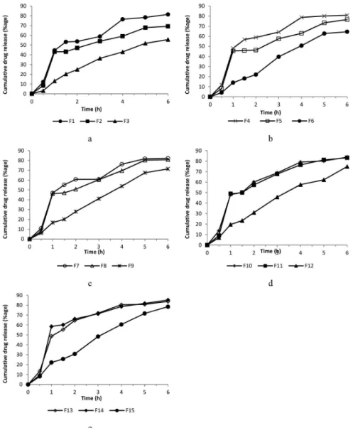
was used to calculate the drug contents realsed from the microcapsule formulations at different time intervals. As shows in Figure1, the release of theophylline from microcapsules decreased gradually by increasing drug to polymer ratio of 1:1 to 1:3 in formulation F1 to F3 (Figure 1). This efect is ascribed to the better covering of drug particles by the polymer (Goosen et al., 1985). The F3 showed only 12% release while F13 and F14 exhibited approximately 50% release in the first one hour. The formulations F13 and F14 released the drug most rapidly from all the microcapsule formulations (Figure 1). The inclusion of plasticizer produced channels in the polymer ilm of microcapsules (Palomo et al., 1996). In this study
TABLE II - Physical properties of theophylline microcapsule formulations
Formulation Drug/Polymer
ratio
Plasticizer (%)
Mean particle size (µm)
Density (Tapped)
Drug content/ Yield (%)
Carr Index
Angle of repose
F1 1:1 0 318 0.476 79.2 4.55 20
F2 1:2 0 257 0.429 85.9 3.16 21
F3 1:3 0 266 0.512 97.9 2.50 22
F4 1:1 10 499 0.421 79.3 1.16 22
F5 1:2 10 512 0.481 87 3.49 23
F6 1:3 10 242 0.530 93 3.85 22
F7 1:1 20 320 0.487 73.2 3.53 23
F8 1:2 20 302 0.476 79.5 3.45 24
F9 1:3 20 297 0.434 92.4 4.17 23
F10 1:1 30 344 0.500 85.5 6.98 22
F11 1:2 30 289 0.487 78.5 4.65 25
F12 1:3 30 314 0.481 88 2.35 21
F13 1:1 40 394 0.465 80.9 6.52 22
F14 1:2 40 259 0.459 84.8 3.33 23
channeling effect of plasticizer caused the increased release from microcapsules. The time of 50% drug release was prolonged almost linearly with an increase in drug to polymer ratio from F1 to F3 (Table III). The higher amount of polymer provided thick coating thus caused slow release of drug (Jalil and Nixon, 1990). However, T50 for formulations containing plasticizer was found
to be decreasing as compared to formulations without plasticizer. This was due to the swelling and hydration of the polymer ilm and development of pores on the wall of microcapsules secondary to leach of the plasticizer (Merkle, Speiser, 1973).
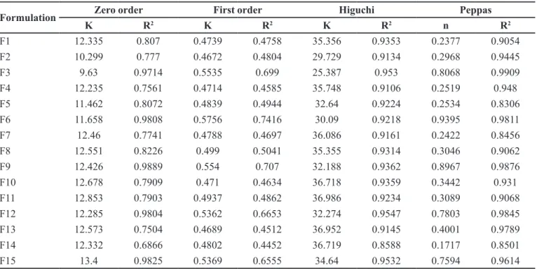
Kinetics analysis of release study
The release data were submitted to zero order, irst
order, Higuchi’s and Peppas model (Reddy et al., 2003, Wu et al., 2003). The release model parameters for all the formulations are shown in Table IV. The release of formulation F1 and F2 followed the Higuchi square root model with R2 values 0.9353 and 0.9134, respectively. The
values of n from Peppas model for these formulations were 0.2377 and 0.2968, respectively (n < 5), which indicated that the drug release for F1 and F2 followed the Fickian difusion (Li et al., 2006, Hossain et al., 2007). The release from F3 followed zero order (R2 =0.9714) and the n value
of Peppas model was 0.8068 (n > 5) which indicated a non-Fickian or anomalous release. These results were in agreement with the indings of previous studies (Ritger, Peppas, 1987; Majid Khan, Zhu, 1999).
The increase of 10 to 40% concentration of plasticizer in formulation F4 to F15 did not essentially
change the mechanism of drug release as all the formulations with drug to polymer ratio 1:1 and 1:2 (with or without plasticizer) followed the Higuchi model and the mechanism according to Peppas model was Fickian diffusion. The formulation with drug to polymer ratio
1:3 (with or without plasticizer) followed the zero order release with non-Fickian or anomalous release mechanism.
It appeared that all the formulations followed the kinetics, which could not be explained by a single model. Kinetic proile during the irst hour was usually the first order, which probably represented the non-encapsulated fraction of drug. Then another profile (Higuchi or zero order) after 1 h indicated the release of the drug by difusion, erosion or by the combination of both mechanisms as shown by ‘n’ values). Possibly there is more than one mechanism involved, and it seems that the efect of polymer concentration is more pronounced as compared to the percentage of plasticizer in each formulation. This argument is supported by the experimental data indicating more or less similar release profiles of the formulations (F3, F6, F9, F12 and F15) having an increasing concentration of the plasticizer and keeping drug to polymer ratio constant (1:3).
CONCLUSION
In this study, the microcapsules of theophylline were prepared using a non-solvent addition method. The microcapsules have shown better entrapment eiciency of drug and other processing properties. It is also concluded that the rate of drug release is inversely related to the polymer concentration. The addition of plasticizer slightly enhances the drug release.
TABLE III - Time for 50% drug release and cumulative %age of drug released in 6 h
Formulation T50 (h) (±SD) Total drug released
in 6 h (%)
F1 2.4 ± 0.1 81
F2 3.2 ± 0.15 69
F3 4.8 ± 0.2 56
F4 2.2 ± 0.1 81
F5 2.8 ± 0.1 77
F6 4.2 ± 0.2 64
F7 2.4 ± 0.15 82
F8 2.6 ± 0.15 81
F9 3.6 ± 0.2 71
F10 2.2 ± 0.1 84
F11 2.4 ± 0.1 83
F12 3.6 ± 0.15 75
F13 1.2 ± 0.1 84
F14 1.3 ± 0.1 85
F15 3.4 ± 0.2 78
TABLE IV - Kinetic values obtained from diferent plots of theophylline microcapsule formulations
Formulation Zero order First order Higuchi Peppas
K R2 K R2 K R2 n R2
F1 12.335 0.807 0.4739 0.4758 35.356 0.9353 0.2377 0.9054
F2 10.299 0.777 0.4672 0.4804 29.729 0.9134 0.2968 0.9445
F3 9.63 0.9714 0.5535 0.699 25.387 0.953 0.8068 0.9909
F4 12.235 0.7561 0.4714 0.4585 35.748 0.9106 0.2519 0.948
F5 11.462 0.8072 0.4839 0.4944 32.64 0.9224 0.2534 0.8306
F6 11.658 0.9808 0.5756 0.7416 30.09 0.9218 0.9395 0.9811
F7 12.46 0.7741 0.4788 0.4697 36.086 0.9161 0.2422 0.8456
F8 12.551 0.8226 0.499 0.5041 35.355 0.9314 0.3046 0.9062
F9 12.426 0.9889 0.554 0.707 32.188 0.9362 0.8967 0.9876
F10 12.678 0.7909 0.471 0.4634 36.718 0.9359 0.3442 0.931
F11 12.853 0.7903 0.4937 0.4862 36.986 0.9234 0.3089 0.9068
F12 12.285 0.9804 0.5362 0.6653 32.274 0.9547 0.7803 0.9845
F13 12.573 0.7504 0.4689 0.4512 36.952 0.9145 0.4001 0.9789
F14 12.332 0.6866 0.4802 0.4452 36.719 0.8588 0.1717 0.8501
ACKNOWLEDGMENTS
The authors are thankful to M/S Flow Pharmaceuticals (Pvt.) Ltd., Sheikhupura Road, Lahore, Pakistan and M/S Paciic Pharmaceuticals (Pvt.) Ltd. Lahore, Pakistan for gifting materials and extending laboratory facilities for this study.
REFERENCES
AHMAD, M.; AKHTAR, N.; MURTAZA, G.; HUSSAIN, S.W. Formulation development and in vitro evaluation of theophylline microcapsules. Pak. J. Pharm. Sci., v.25, n.1, p.15-19, 2012.
BEYGER, J.W.; NAIRN, J.G. Some factors affecting the microencapsulation of pharmaceuticals with cellulose acetate phthalate. J. Pharm. Sci., v.75, n.6, p.573-578, 1986.
GOOSEN, M.F.A.; O’SHEA, G.M.; GHARAPETIAN, H.M.; CHOU, S.; SUN, A.M. Optimization of microencapsulation parameters: semipermeable microcapsules as a bioartiicial pancreas. Biotechnol. Bioeng., v.27, n.2, p.146-150, 1985.
HENDELES, L.; WEINBERGER, M. Theophylline. A” state of the art” review. Pharmacotherapy, v.3, n.1, p.2-44, 1983.
HIGUCHI, T. Mechanism of sustained‐action medication: theoretical analysis of rate of release of solid drugs dispersed in solid matrices. J. Pharm. Sci., v.52, n.12, p.1145-1149, 1963.
JALIL, R.; NIXON, J. Biodegradable poly (lactic acid) and poly (lactide-co-glycolide) microcapsules: problems associated with preparative techniques and release properties. J. Microencapsul., v.7, n.3, p.297-325, 1990.
JALSENJAK, I.; NICOLAIDOU, C.F.; NIXON, J. The in vitro
dissolution of phenobarbitone sodium from ethyl cellulose microcapsules. J. Pharm. Pharmacol., v.28, n.12,
p.912-914, 1976.
LY, J.P.-H. Development of an oral microspherical formulation for bimodal in vitro release of theophylline. Toronto, 1997.
87 p. [Thesis of Master of Science Degree - Graduate Department of Pharmaceutical Sciences - University of Toronto].
MADAN, P. Microencapsulation I. Phase separation or coacervation. Drug Dev. Ind. Pharm., v.4, n.1, p.95-116,
1978.
MAJID, K.G.; ZHU, J.B. Studies on drug release kinetics from ibuprofen–carbomer hydrophilic matrix tablets: inluence of co-excipients on release rate of the drug. J. Control. Release,
v.57, n.2, p.197-203, 1999.
MERKLE, H.; SPEISER, P. Preparation and in vitro evaluation of cellulose acetate phthalate coacervate microcapsules. J. Pharm. Sci., v.62, n.9, p.1444-1448, 1973.
MILAVETZ, G.; WEINBERGER, M.; VAUGHAN, L. Dose dependency for absorption and elimination rates of theophylline implications for studies of bioavailability.
Pharmacotherapy, v.4, n.4, p.216-220, 1984.
PALOMO, M.; BALLESTEROS, M.; FRUTOS, P. Solvent and plasticizer inluences on ethylcellulose-microcapsules. J. Microencapsul., v.13, n.3, p.307-318, 1996.
PONGPAIBUL, Y.; WHITWORTH, C. Preparation and in vitro
dissolution characteristics of propranolol microcapsules.
Int. J. Pharm., v.33, n.1/3, p.243-248, 1986.
RAO, P.R.; DIWAN, P.V. Permeability studies of cellulose acetate free films for transdermal use: influence of plasticizers. Pharm. Acta Helv., v.72, n.1, p.47-51, 1997.
REDDY, K.R.; MUTALIK, S.; REDDY, S. Once-daily sustained-release matrix tablets of nicorandil: formulation and in vitro evaluation. AAPS PharmSciTech, v.4, n.4, art.61, p.1-9, 2003.
RITGER, P.L.; PEPPAS, N.A. A simple equation for description of solute release II. Fickian and anomalous release from swellable devices. J. Control. Release, v.5, n.2, p.37-42,
1987.
SANGHVI, S.P.; NAIRN, J.G. Phase diagram studies for microencapsulation of pharmaceuticals using cellulose acetate trimellitate. J. Pharm. Sci., v.80, n.4, p.394-398,
1991.
SONG, M.; LI, N.; SUN, S.; TIEDT, L.R.; LIEBENBERG, W.; DE VILLIERS, M.M. Efect of viscosity and concentration of wall former, emulsiier and pore-inducer on the properties of amoxicillin microcapsules prepared by emulsion solvent evaporation. Farmaco, v.60, n.3, p.261-267, 2005.
WASHINGTON, C. Drug release from microparticulate systems. In: BENITA, S., ed. Microencapsulation: methods
and industrial applications. New York: Marcel Dekker,
1996. p.155-181. (Drugs and the Pharmaceutical Sciences, v.73).
WU, P.C.; HUANG, Y.B.; CHANG, J.I.; TSAI, M.J.; TSAI, Y.H. Preparation and evaluation of sustained release microspheres of potassium chloride prepared with ethylcellulose. Int. J. Pharm., v.260, n.1, p.115-121, 2003.
Received for publication on 27th March 2015