This study assessed the pH, radiopacity, antimicrobial effect, cytotoxicity and biocompatibility of endodontic filling materials for primary teeth. Zinc oxide eugenol (ZOE), Vitapex and Calen paste thickened with zinc oxide (ZO) were evaluated in comparison to an experimental MTA-based material. Radiopacity was tested using a graduated aluminum stepwedge with a digital sensor (n=5). The materials pH was recorded at 1, 4, 12 h; 1, 3, 7, 15 and 30 days (n=5). Direct contact test was used to assess the antimicrobial efficacy against Enterococcus faecalis after 1, 4, 12, 24 h (n=5). Cytotoxicity assay used MTT test for cell viability after incubation for 1, 3 and 7 days (n=5). For biocompatibility test, Wistar rats had received implants containing each material (n=5). The biopsied tissues were histologically analyzed after 15, 30 and 60 days. The results of radiopacity, pH, antimicrobial capacity and cytotoxicity were analyzed using ANOVA and Tukey tests. The histological data were submitted to Kruskal-Wallis test. The experimental material presented the lowest radiopacity (3.28 mm Al) and had a pH>12.0 throughout the test period. The experimental material showed the highest antibacterial effect, killing over 99.97% bacteria in 4 h. Vitapex presented the highest cell viability. Initially, biocompatibility test showed moderate to severe inflammation in all groups. After 60 days, Calen+ZO group showed moderate inflammation, while the others showed predominantly mild inflammatory reaction. The present results demonstrated that the experimental MTA-based material exhibited satisfactory behavior regarding the studied properties. Additional in vivo studies are necessary for a better evaluation of the material.
Physicochemical and Biological
Evaluation of Endodontic Filling
M a t e r i a l s f o r P r i m a r y T e e t h
Katerine Jahnecke Pilownic1, Ana Paula Neutzling Gomes1, Zhe Jun Wang2, Luiza Helena Silva Almeida3, Ana Regina Romano1,Ya Shen2, Anelize de Oliveira Campello Felix4, Markus Haapasalo2, Fernanda Geraldo Pappen1
1Graduate Program in Dentistry, UFPel - Universidade Federal de Pelotas, Pelotas, RS, Brazil 2Dental School, University of British Columbia, Vancouver, BC, Canada 3Dental School, AVANTIS University, Balneário Camboriú, SC, Brazil 4Central Vivarium, UFPel - Universidade Federal de Pelotas, Pelotas, RS, Brazil Correspondence: Fernanda Geraldo Pappen, R. Gonçalves Chaves, 457, 507, 96015-560 Pelotas, RS, Brasil. Tel +55-53-3222-4162. e-mail: [email protected]
Key Words: endodontics; deciduous tooth; root canal filling materials.
Introduction
An ideal endodontic filling material for primary teeth must have several properties that make it suitable for use. It should have a resorption rate similar to that of the primary root, be harmless to the periapical tissues and permanent tooth germ, readily resorb if pressed beyond the apex, and flow easily into the complex anatomy of primary root canals. It should also have strong antiseptic properties to avoid contamination during manipulation and to inhibit the growth of microorganisms (1).
Zinc oxide eugenol (ZOE) have been widely used as endodontic filling material for primary teeth (2). The potentially adverse characteristics of ZOE include irritation to tissue and triggering of inflammatory and foreign-body reactions if extruded into the periapical tissue (1). ZOE has limited antimicrobial action, and it tends to resorb at a slower rate than the roots of the primary teeth (2,3). When forced beyond the apex, it may affect the eruption of permanent teeth (4). Therefore, iodoform-containing materials, or calcium hydroxide pastes have substituted the use of ZOE.
Some studies have also suggested the use of calcium hydroxide associated to zinc oxide, as well as calcium hydroxide and iodoform, in order to improve the chances
of success in root canal therapy of primary teeth (1,5). Iodoform pastes have better resorbability than ZOE has, and are less likely to cause a foreign body reaction. They also have potent germicidal properties (1). Among the beneficial properties of calcium hydroxide are its biocompatibility, antibacterial activity, induction of mineralized tissue formation and easy resorption (1).
Recently, a resorbable ready-to-use experimental mineral trioxide aggregate (MTA)-based endodontic filling material for primary teeth was developed (Angelus, Londrina, PR, Brazil), containing approximately 30% MTA. According to the manufacturer, the experimental material is composed by ester glycol salicylate, titanium dioxide, calcium tungstate, silicon dioxide, toluene sulfonamide and calcium silicate. One of the differences between experimental MTA-based material and MTA-Fillapex, a MTA-based sealer for permanent teeth, is the higher proportion of salicylates and resin components in MTA-Fillapex. The use of a ready-to-use endodontic material is desirable in pediatric dentistry, since it can reduce the time of dental appointment. Additionally, materials which need to be manipulated may have their proportions changed interposing the clinical performance of material.
Endodontic filling materials for primary teeth
single material which has all the desired physical, chemical, antimicrobial and biological requirements for endodontic filling material for primary teeth. It is important to highlight that the use of a filling material with all requirements would improve the outcome of post-endodontic treatment in primary teeth (5).
This study was undertaken to evaluate the radiopacity, pH, antimicrobial capacity, cytotoxicity and biocompatibility of an experimental MTA-based material in comparison to other endodontic filling materials commonly used for primary teeth.
Material and Methods
Four endodontic materials were tested: Group I, experimental MTA-based material, composed by ester glycol salicylate, titanium dioxide, calcium tungstate, silicone dioxide, toluene sulfonamide and 30% calcium silicate (Angelus, Londrina, PR, Brazil); Group II, zinc oxide eugenol cement (ZOE) (S.S.White Artigos Dentários Ltda., Rio de Janeiro, RJ, Brazil); Group III, a premixed calcium hydroxide and iodoform paste (Vitapex) (Neo Dental International Inc., Federal Way, WA, USA); Group IV, 1.0 g of a commercial calcium hydroxide and polyethylene glycol-based paste (Calen) (S.S.White, Rio de Janeiro, RJ, Brazil) thickened with 1.0 g zinc oxide (Biodinâmica Química e Farmacêutica Ltda., Ibiporã, PR, Brazil) (5,6).
pH Measurement
Suspensions of endodontic filling materials in sterile water were prepared at a concentration of 100 mg/mL as described by Zhang et al (7). The pH was determined in 4 mL of each suspension, stored in closed 10-mL BD Falcon test tubes with deionized water (Nalgene®, Brand Products, Rochester, NY, USA) eliminating the effects of environmental factors until the measurement time, along the experimental time. Before measurements, the suspensions were mixed by vortexing for 30 s. The pH was recorded at1, 4, 12 h; 1, 3, 7, 15 and 30 days periods adapted from Carvalho et al. (8). The pH value was measured with a sympHony pHmeter (VWR International, Radnor, PA, USA) previously calibrated with pH 4.0, 7.0 and 10.0 standard solutions. Distilled water was used as a control. The mean values and standard deviation were calculated and recorded. Five samples per group were prepared, measurements were performed in triplicate.
Radiopacity Measurement
Radiopacity assay were performed according to ISO 6876/2001 specifications. Acrylic plates (22×45×1 mm), containing wells measuring 1 mm in depth and 10 mm in diameter were used to standardize the samples. The specimens were placed on a digital radiographic sensor
(Vista ScanPlus, Dür Dental AG, Bietigheim-Bissingen, Germany) next to a graduated aluminum stepwedge with thickness ranging from 0.5 to 5mm (in 0.5 mm increments) and exposed using a Timex 70C x-ray unit (Gnatus Ltda, Ribeirão Preto, SP, Brazil) operating at 70 kV, 8 mA. The exposure time was 0.2 s, and the focus-film distance was 30 cm. The obtained images wer,e imported into Adobe Photoshop 12.0.4 software (Adobe Systems Incorporated, San Jose, CA, USA) and saved in TIFF format.
The radiographic density values obtained were converted into mm Al. The equal-density tool of the software was used to identify areas of equal density. The means and standard deviations of the grey levels (pixel density) of the aluminum stepwedge and the specimens were obtained in a standardized central area of 1.5 mm2. Five samples per group were evaluated, and the results were analyzed by calculating the means of five measurements per sample.
Antibacterial Assay
The methodology used to assess the antibacterial assay
was the same described by Zhang et al. (7). Enterococcus
faecalis (ATCC 29212) was used for the direct contact test (DCT).The bacteria were cultured and incubated overnight in air at 37 ˚C on Tryptic Soy Agar (TSA) (Becton, Spark, MD, USA) plates for the experiments. After checking for purity, E. faecalis was suspended in sterile water and adjusted to a density of 3.2 ×10⁷ colony-forming units (CFU)/mL by using
a spectrophotometer (Bioespectro, Curitiba, PR, Brazil) at 405 nm. The bacterial suspension was used immediately after the concentration adjustment.
Suspensions of each material were prepared in sterile water at concentrations of 100 mg/mL. Bacterial and material suspensions were mixed in equal volumes of 500 µL each. After incubation at room temperature for 1, 4, 12 and 24 h, the mixtures were serially ten-fold diluted and three droplets of 20 µL from each mixture were cultured on TSA plates. The bacterial survival was assessed after incubation for 24-48 h at 37 ˚C, when colonies on the plates were counted and the CFU/mL was calculated. Pure sterile water served as a positive control. All experiments were performed in quintuplicate.
Cytotoxicity Assay
K.J. Pilownic et al
µg/mL streptomycin, 0.25 µg/mL fungizone and 10% fetal bovine serum (Gibco).
The materials specimens were prepared under aseptic conditions. The surface area to volume ratio used for extract preparation was approximately 250 mm²/mL. One milliliter of extract was drawn from each well after incubation at 37º °C and 100% relative humidity for 24 h. Each extract was divided into 6 aliquots to obtain 5 parallel experimental groups and 1 background group. Extracts were serially diluted 1:1 with DMEM to achieve five concentrations of each extract. DMEM incubated for 24 h without materials was used as a control.
Human gingival fibroblast suspension (110 µL/well) was seeded to 96-well flat bottomed tissue culture plates (Nunc, Roskilde, Denmark) at a density of 5x10³ cells/100 µL and incubated for 24 h to achieve attachment of the cells before adding the extracts. After incubation for 1, 3 and 7 days with the extract (9), cell viability was determined by methyl-thiazol-tetrazolium (MTT) assay by using a CellTiter 96Assay kit (Promega, Madison, WI, USA) according to the manufacturer’s instructions. Cell viability was calculated by using the following formula: 100(a-b)/c, where “a” and “b”, were optical density (OD) values from test wells and background wells, respectively, and c was the mean OD value from control wells. The background absorbance was subtracted from values. The experiments were performed in quintuplicate.
Biocompatibility Test
The Institutional Research Animal Ethics Committee approved this study (Protocol #8977; 02/04/14). All procedures in animals were carried out in accordance with institutional guidelines for animal care and use.
Tested materials were inserted into sterile polyethylene tubes measuring 10 mm with 1.0 mm internal diameter (Abbott). Each tube had one of the ends closed with gutta-percha and this side was used as negative control. The filled tubes were implanted on animal’s dorsal subcutaneous tissue.
Fifteen Wistar rats (Rattus norvegicus; age 6 months; weight ~250 g) were used in this study. The animals’ tails were marked for individual identification. The rats were housed in plastic cages (two per cage) placed in ventilated racks (Alesco, Monte Mor, SP, Brazil) at 22 °C with a 12 h light⁄dark cycle (lights on at 7:00 am, off at 7:00 pm). During the experiments, the rats were provided with a standard diet of rat chow (Nuvilab, Colombo, PR, Brazil) and filtered water ad libitum (10).
The animals were anesthetized using ketamine (80 mg/ kg) and xylazine (4 mg/kg) (SDC ECommerce, Marilia, SP, Brazil). Four 2-cm incisions were made on the shaved backs in a head-to-tail orientation using a #15 Bard Parker blade
(Lamedid Solidor, Osasco, SP, Brazil). Each animal received four implants, two in the scapular region and two in the caudal region, into the spaces created by blunt dissection, according to a previously established placement order and site rotation. The tissue closure was performed using a 4-0 silk suture (Johnson & Johnson, São José dos Campos, SP, Brazil). Sterile instruments and aseptic techniques were used throughout the experiments. To aid recovery, paracetamol (0.06 mg/g-1day-1) was added to their drinking water for 72 h (10).
The animals were euthanized at 15, 30 and 60 days after the procedures (n=5 per group at each time point). They were anesthetized intraperitoneally with chloral hydrate (350 mg/kg) (Sigma Aldrich, St. Louis, MI, USA) and physiological saline (Sigma Aldrich). Then, 10% paraformaldehyde in 0.1 M phosphate buffer (pH 7.4) (Sigma Aldrich) was perfused transcardially. The tubes and the surrounding tissues were removed and fixed in 10% buffered formalin (pH 7.0) (Sigma Aldrich). The specimens were paraffin embedded, sectioned serially to 3-mm slices, and stained with hematoxylin and eosin.
A previously calibrated experienced pathologist who was blinded to sample group assignment performed histological evaluation of the H&E-stained sections. Slices were analyzed with a light microscope (RM2235; Leica, São Paulo, SP, Brazil), using 40, 100, 200, and 400x magnification. The pathologist performed descriptive analysis and assigned scores for inflammatory response and fibrous capsule organization adjacent to endodontic materials.
Tissue reactions in contact with the materials were scored as absent (no or few inflammatory cells); mild (less than 25 inflammatory cells); moderate (25 to 125 inflammatory cells); severe (125 or more inflammatory cells). Fibrous capsules were classified as thin if the thickness was <150 mm, or thick if >150 mm. The presence of giant cells and calcification was also recorded. In a random order, only one control in each animal was evaluated.
In order to test the intra-examiner variability, Kappa coefficient was obtained using a sample of 15 sections. The intra-examiner weighted Kappa was calculated separately for the presence of inflammatory cells (k=0.81, p<0.001), fibers thickness (k=0.878, p<0.001) and calcification (k=1.00, p<0.001).
Statistical Analysis
Endodontic filling materials for primary teeth
level of p<0.05. The results of biocompatibility were analyzed by the Kruskal-Wallis and Mann-Whitney test at a 5% significance level.
Results
pH Measurement
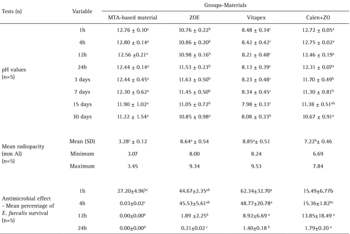
Table 1 shows the average pH values according to the experimental periods for each material tested. The pH of the experimental MTA-based endodontic material was similar to that of Calen+ZO within the experimental period. After 30 days, experimental MTA had the highest pH while Vitapex had the lowest pH.
Radiopacity Measurement
The results of the radiopacity test are presented in Table 1. Vitapex and ZOE presented higher radiopacity in comparison with the other tested materials (p<0.05). Experimental MTA-based endodontic material had the lowest radiopacity value of 3.28 mm Al.
Antibacterial Assay
The results of the DCT are shown in Table 1. The experimental MTA-based filling material killed more
bacteria than the other materials. It eliminated over 99.9% of the E. faecalis cells after 4 h of exposure. After 24 h of exposure, all other materials eliminated more than 98% of the bacteria.
Cytotoxicity Assay
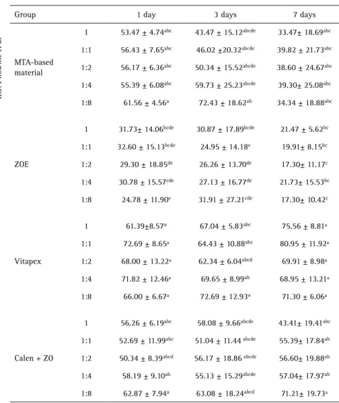
The MTT test showed that fibroblast cell viability was significantly associated with the different endodontic filling materials tested (p<0.001), but it was not correlated to incubation time or extract concentrations (p>0.05).
Cells exposed to extracts from Vitapex showed the highest viabilities at all extract concentrations, whereas cells exposed to ZOE extracts displayed the lowest viabilities (p<0.001). The cells exposed to the MTA-based material and those exposed to Calen+ZO showed no statistical difference in viability (p=0.115) (Table 2).
Biocompatibility Test
Representative samples of each group are demonstrated in Figure 1. On day 15, thick fibrous capsule formation and moderate inflammatory reaction were observed with all Vitapex and experimental MTA-based material specimens. In ZOE and Calen+ZO groups, the intensity of inflammatory
Table 1. Mean and standard deviation of pH, antimicrobial effect and radiopacity for the tested materials
Tests (n) Variable
Groups-Materials
MTA-based material ZOE Vitapex Calen+ZO
pH values (n=5)
1h 12.76 ± 0.10a 10.76 ± 0.22b 8.48 ± 0.34c 12.72 ± 0.05a
4h 12.80 ± 0.14a 10.86 ± 0.20b 8.42 ± 0.42c 12.75 ± 0.02a
12h 12.56 ±0.21a 10.98 ± 0.16b 8.21 ± 0.48c 12.46 ± 0.19a
24h 12.44 ± 0.14a 11.53 ± 0.23b 8.13 ± 0.39c 12.31 ± 0.07a
3 days 12.44 ± 0.45a 11.63 ± 0.50b 8.23 ± 0.48c 11.70 ± 0.49b
7 days 12.30 ± 0.62a 11.45 ± 0.50b 8.34 ± 0.45c 11.30 ± 0.81b
15 days 11.90 ± 1.02a 11.05 ± 0.72b 7.98 ± 0.33c 11.38 ± 0.51ab
30 days 11.22 ± 1.54a 10.85 ± 0.98a 8.08 ± 0.33b 10.67 ± 0.91a
Mean radiopacity (mm Al) (n=5)
Mean (SD) 3.28c ± 0.12 8.64a ± 0.54 8.85a± 0.51 7.22b± 0.46
Minimum 3.07 8.00 8.24 6.69
Maximum 3.45 9.34 9.53 7.84
Antimicrobial effect - Mean percentage of E. faecalis survival (n=5)
1h 27.20±4.96bc 44.67±2.35ab 62.34±32.70a 15.49±6.77b
4h 0.03±0.02c 45.53±5.61ab 48.77±20.78a 15.36±1.82bc
12h 0.00±0.00b 1.89 ±2.25b 8.92±6.69 a 13.85±18.49 a
24h 0.00±0.00d 0.21±0.02 c 1.40±0.18 b 1.79±0.20 a
K.J. Pilownic et al
reaction was moderate to severe (p=0.001) (Table 3). The Vitapex group showed a large capsule extension, with evident presence of macrophage foam cells. A reduction in the thickness of the fibrous capsule was evident from day 15 onward. On day 30, mild inflammatory cell infiltration was observed in MTA-based material group, while the other materials induced moderate inflammation mostly (p=0.001).
After 60 days, the inflammatory reaction was mild in most specimens of ZOE, experimental MTA-based material and Vitapex, while most specimens from Calen+ZO group showed moderate inflammatory infiltrate (p=0.001).
Discussion
The biomechanical endodontic treatment of primary teeth remains far from ideal, especially due to their
anatomical features and difficulties in obtaining a good radiographic view of the apex for determining the working length. The use of endodontic filling materials with antibacterial activity is beneficial to reduce the number of microorganisms remaining in the endodontic space. Furthermore, due to the physiological root resorption process, materials used in endodontic treatment of primary teeth should be harmless to the periapical tissues and permanent tooth germs (1).
While zinc oxide-eugenol cement (ZOE) has been widely used (1-3), due to ZOE’s irritating potential and low resorption capacity, materials containing iodoform and
or calcium hydroxide (Ca(OH)2) have been recommended
specially due to their antibacterial activity, biocompatibility and easy resorption (11). The association of zinc oxide and Ca(OH)2 (6,7) or Ca(OH)2 and iodoform (11) had been also
advocated. However, materials which need to be manipulated may have their proportions changed interposing their clinical performance (5).
Calcium silicate materials, as Mineral Trioxide Aggregate (MTA), have been widely used for dental applications such as root-end filling, root-repair cements, pulp capping and in the composition of root canal sealers (12), mainly due to its biocompatibility and bioactivity (13,14). Conventional MTA has some disadvantages, such as reduced working time, long setting time and difficulty of handling it to fill the root canals (14). Consequently, modifications in its original formulation have been suggested in order to improve its physicochemical and mechanical properties m a i n t a i n i n g i t s e x c e l l e n t biological characteristics.
MTA-based sealers, such as MTA-Fillapex, were introduced in attempt to combine biological and sealing properties. However, it has demonstrated insufficient biocompatibility effects on bone and subcutaneous connective tissue (15,16). In addition, the antibacterial activity of MTA-based sealers remains a limitation, since the material presents an
Table 2. Cell viability of extracts with various concentrations derived from the tested materials after cell culture for 1, 3 and 7 days. The results show mean percentage and standard deviation of three parallel experiments performed in triplicate
Group 1 day 3 days 7 days
MTA-based material
1 53.47 ± 4.74abc 43.47 ± 15.12abcde 33.47± 18.69abc
1:1 56.43 ± 7.65abc 46.02 ±20.32abcde 39.82 ± 21.73abc
1:2 56.17 ± 6.36abc 50.34 ± 15.52abcde 38.60 ± 24.67abc
1:4 55.39 ± 6.08abc 59.73 ± 25.23abcde 39.30± 25.08abc
1:8 61.56 ± 4.56a 72.43 ± 18.62ab 34.34 ± 18.88abc
ZOE
1 31.73± 14.06bcde 30.87 ± 17.89bcde 21.47 ± 5.62bc
1:1 32.60 ± 15.13bcde 24.95 ± 14.18e 19.91± 8.15bc
1:2 29.30 ± 18.85de 26.26 ± 13.70de 17.30± 11.17c
1:4 30.78 ± 15.57cde 27.13 ± 16.77de 21.73± 15.53bc
1:8 24.78 ± 11.90e 31.91 ± 27.21cde 17.30± 10.42c
Vitapex
1 61.39±8.57a 67.04 ± 5.83abc 75.56 ± 8.81ª
1:1 72.69 ± 8.65ª 64.43 ± 10.88abc 80.95 ± 11.92ª
1:2 68.00 ± 13.22ª 62.34 ± 6.04abcd 69.91 ± 8.98ª
1:4 71.82 ± 12.46ª 69.65 ± 8.99ab 68.95 ± 13.21ª
1:8 66.00 ± 6.67ª 72.69 ± 12.93a 71.30 ± 6.06ª
Calen + ZO
1 56.26 ± 6.19abc 58.08 ± 9.66abcde 43.41± 19.41abc
1:1 52.69 ± 11.99abc 51.04 ± 11.44 abcde 55.39± 17.84ab
1:2 50.34 ± 8.39abcd 56.17 ± 18.86 abcde 56.60± 19.88ab
1:4 58.19 ± 9.10ab 55.13 ± 15.29abcde 57.04± 17.97ab
1:8 62.87 ± 7.94a 63.08 ± 18.24abcd 71.21± 19.73ª
Endodontic filling materials for primary teeth Figure 1. Subcutaneous tissue reactions within the different experimental groups. Hematoxylin and eosin staining, 10. A: ZOE - 15days - Thin fibrous capsule formation with moderate inflammatory cell infiltration; B: Experimental MTA - 15days - Thick fibrous capsule formation and moderate inflammatory cell infiltration; C: Vitapex - 15days - Very thick fibrous capsule formation and intense infiltrate inflammatory associated with material particulae; D: Calen+ ZO - 15days - Thick fibrous capsule formation and intense infiltrate inflammatory; E: Control - 15 days - Thin fibrous capsule formation and mild inflammatory cell infiltration; F: ZOE - 30 days - Thin fibrous capsule without inflammatory cells after 30 days; G: Experimental MTA - 30 days - Thin fibrous capsule formation and mild infiltrate inflammatory;H: Vitapex - 30 days -Thick fibrous capsule formation and intense infiltrate inflammatory; I: Calen+ ZO - 30 days - Thin fibrous capsule with mild infiltrate inflammatory; J: Control - 30 days - Thin fibrous capsule with mild inflammatory cell infiltration; K: ZOE - 60 days - Thin fibrous capsule with mild to moderate inflammatory cell infiltration; L: Experimental MTA - 60 days - Thin fibrous capsule surrounding the tube without inflammatory cells; M: Vitapex - 60 days - Thin fibrous capsule surrounding the tube with few chronic inflammatory cells; N: Calen+ ZO - 60days - Thin fibrous capsule surrounding the tube with mild inflammatory cell infiltration; O: Control - 60days - Thin fibrous capsule surrounding the tube without inflammatory cells after 60 days.
Table 3. Percentage of tissue reaction according to different groups and different evaluation times
Variables
MTA-based
material (%) ZOE (%) Vitapex (%) Calen+ZO (%) Control group (%)
15 30 60 15 30 60 15 30 60 15 30 60 15 30 60
Inflammatory reaction
Absent - - - 100
Mild - 80 80 - 20 80 - - 100 - - 20 100 100
Moderate 100 20 20 80 80 20 100 100 - 80 100 80 - -
Severe - - - 20 - - - 20 - -- - -
-Fibrous capsule
Thin - 80 100 - 20 80 - - 100 - - 100 100 100 100
Thick 100 20 - 100 80 20 100 100 - 100 100 - - -
-Giant Cells 60 20 - 40 - 20 40 60 - 60 20 - - -
-K.J. Pilownic et al
antibacterial activity before setting that is not sustained after setting (17). The present study is the first one in the literature to report the use of MTA in the composition of endodontic filling materials for primary teeth. The experimental MTA-containing material tested in this study is a resorbable ready-to-use material, which contains 30% calcium silicate and had been developed as an alternative to be used in endodontic filling for primary teeth, whereas there is no single material on the market, which fulfills all the desirable physical, chemical and biological properties.
Endodontic filling materials should have sufficient radiopacity to allow for a clear distinction between the materials and surrounding anatomic structures (18). The radiopacifying agent of the experimental MTA-based material is calcium tungstate and even though it has the lowest radiopacity, it is in accordance with that required as per ISO 6876/2001, which recommends the minimal of 3 mm Al for root canal filling materials.
Materials that have strong antibacterial properties also tends to have high cytotoxicity (9). The experimental MTA-based material in the present study showed a strong antibacterial effect, but also good low cytotoxicity. Calcium in its hydroxide form is the main chemical compound released from conventional MTA (12). The antimicrobial action of experimental MTA-based material is affected by the liberation of hydroxyl ions, given the high pH. Despite having the highest value of pH, Calen thickened with zinc oxide showed low antimicrobial action, according to the DCT. Measurement of the pH values and the effectiveness in killing E. faecalis indicates that there are factors other than pH that influence the antibacterial activity of the materials (7).
In this study, the traditional endodontic filling material, ZOE, presented good antibacterial effect against E. faecalis. These results contradict those found by using agar diffusion test (ADT) (2). However, frequently the results of studies which use ADT are not reliable, since the inhibition zones may be more related to the materials’ solubility and diffusibility in agar than to their actual efficacy against the microorganisms (19). On the contrary, the DCT measures the effect of direct and close contact between the organisms and the material. However, this methodology also presents recognized limitations of results, since it had used the DCT in a single-specie model, instead of a polymicrobial biofilm model.
MTT colorimetric assay is widely used for evaluating the cytotoxicity of materials, and is routinely used to test dental and endodontic filling materials in cell culture systems (9,10). As a limitation, however, MTT assays may underestimate cellular damage and detect cell death only at the stage of apoptosis when cellular metabolism is substantially reduced (20). Our results showed similar
cytotoxicity of experimental MTA-based material and Calen+ZO. The major benefits of conventional MTA include biocompatibility, antimicrobial action, and the fact that it promotes regeneration of tissues when it is placed in contact with the dental pulp or peri-radicular tissues (12,18). However, so far, it has not been used as part of the composition of an endodontic filling material for primary teeth.
Fibroblast viability was higher among cells exposed to Vitapex. This finding is in agreement with those of Chen et al. (10). The high cytocompatibility of Vitapex may be due to calcium ions, which are coated with silicon oil to reduce leaching. Contrary to our findings, Wright et al. (21) reported that iodoform-containing materials could cause considerable tissue necrosis, presenting higher cytotoxicity than ZOE does. Eugenol, which is a phenolic compound, in ZOE is regarded as an irritant (2,21). In this study, ZOE showed higher cytotoxicity than all the other tested materials. Despite this, the high clinical success rates of ZOE may be because this material does not easily flow into the narrow and tortuous root canals of primary teeth and is not frequently extruded into the periapical tissues (1,4).
Every tested material was well tolerated by the subcutaneous tissues. This statement is justified by the capsule extension was thicker on day 15 and it declined by over the time, in all experimental groups. It was possible to observe collagen fiber formation at the area in contact with the all tested materials. Sanders et al. (22) described that there is a meaningful relationship between inflammation and fibrous capsule thickness. The decreasing capsule thickness is a sign of biocompatibility and the deferred harmful effects of a material are considered more important than its initial effects in biocompatibility tests (22).
After 60 days, the inflammatory reaction was absent in control group; mild in most specimens of ZOE, experimental MTA-based material and Vitapex, while most specimens from Calen+ZO group still showed moderate inflammatory infiltrate. When used alone, Calen paste present recognized biocompatibility (23), thus the persistent inflammation observed in Calen+ZO group may be attributed to the higher concentration of zinc in the material, since zinc has the capacity to influence in the inflammatory process for reducing phagocytic capacity of macrophages and to interfere in the membrane of the lysosomes (24).
Endodontic filling materials for primary teeth
main requirements for the indication of an endodontic filling material for primary teeth. Calcific precipitation was observed around implantation sites in all experimental groups, but more frequently in Calen+ZO specimens. The production of calcific structures in subcutaneous investigations is a sign of osteoinductivity of the material (26). The osteoinductivity and conductivity the materials may be attributed to the release of calcium and phosphorous as well as the formation of hydroxyapatite crystals over the material (23). Although the conventional MTA is well known by its excellent biocompatibility, antimicrobial action, dentinogenic and osteogenic potentials, mineralizing and good sealing properties (25) the composition of experimental MTA-based material differs from the traditional MTA. Our results demonstrated an optimal tissue reaction to the experimental MTA-based material, what had occurred probably due to the presence of MTA in its composition and the absence of toxic components.
This study comprises in vitro and in vivo tests performed in animals, and due to its limitations, the results cannot be extrapolated directly to the clinical practice. Additionally, due to the fact that Calen + ZO and Vitapex are soluble pastes, which does not set; while ZOE and MTA-based material are cements, the sample standardization to assess properties such as solubility, dimensional change, flow and setting time could not be performed.
The present results demonstrated that the experimental MTA-based material exhibited satisfactory behavior
regarding the studied properties. Additional in vivo
studies, however, are necessary for a better evaluation of the material.
Resumo
Este estudo avaliou o pH, radiopacidade, efeito antimicrobiano, citotoxicidade e biocompatibilidae de materiais obturadores de dentes decíduos. Foram avaliados o cimento de óxido de zinco e eugenol (OZE), Vitapex e pasta Calen espessada com óxido de zinco (OZ), comparativamente a um material experimental à base de MTA. A radiopacidade foi testada usando uma escala milimetrada de alumínio, com um sensor digital. O pH dos materiais também foi avaliado nos períodos de 1, 4, 12 h; 1, 3, 7, 15, e 30 dias (n=5). O teste de contato direto foi utilizado para avaliação da atividade antimicrobiana dos materiais conta uma cepa de Enterococcus faecalis após incubação por 1, 4, 12, e 24 h (n=5). Para avaliação da citotoxicidade foi utilizado o teste de MTT para avaliação da viabilidade celular. No teste de biocompatibilidade, ratos Wistar receberam implantes subcutâneos contendo cada material obturador (n=5). Após biópsia, os tecidos foram submetidos à avaliação histológica em períodos de 15, 30 e 60 dias. Os resultados de radiopacidade, pH, ação antimicrobiana e citotoxicidade foram analisados usando os testes ANOVA e Tukey. Os dados histológicos foram submetidos ao teste de Kruskal-Wallis. O material experimental apresentou a menor radiopacidade (3.28 mm Al) e apresentou um pH superior a 12,0 durante todo o período experimental. O material experimental apresentou o maior efeito antimicrobiano, eliminando mais de 99,97% das bactérias em um período de 4 h. O Vitapex permitiu uma maior viabilidade celular em comparação aos demais materiais avaliados. Inicialmente, os testes de biocompatibilidade demonstraram inflamação de moderada a severa em todos os grupos. Após 60 dias, apenas o grupo Calen+ OZ apresentou inflamação moderada, enquanto os outros
materiais demonstraram predominantemente a indução de inflamação leve. Os resultados demonstraram que o material experimental à base de MTA exibiu comportamento satisfatório com relação às propriedades avaliadas. Estudos adicionais, in vivo, são necessários para uma melhor avaliação do material.
References
1. Mortazavi M, Mesbahi M. Comparison of zinc oxide and eugenol, and Vitapex for root canal treatment of necrotic primary teeth. Int J Paediatr Dent 2004;14:417-424.
2. Reddy S, Ramakrishna Y. Evaluation of antimicrobial efficacy of various root canal filling materials used in primary teeth: a microbiological study. J Clin Pediatr Dent 2007;31:193-198.
3. Al-Ostwani AO, Al-Monaqel BM, Al-Tinawi MK. A clinical and radiographic study of four different root canal fillings in primary molars. J Indian Soc Pedod Prev Dent 2016;34:55-59.
4. Coll JA, Sadrian R. Predicting pulpectomy success and its relationship to exfoliation and succedaneous dentition. Pediatr Dent 1996;18:57-63. 5. Segato RA, Pucinelli CM, Ferreira DC , Daldegan A de R, Silva RS,
Nelson-Filho P, Silva LA. Physicochemical properties of root canal filling materials for primary teeth. Braz Dent J 2016;27:196-201.
6. Pilownic KJ, Carvalho CN, Romano AR, Morgental RD, Shen Y, Haapasalo M, et al.. Antibiofilm activity of five different endodontic filling materials used in primary teeth using confocal laser scanning microscopy. Pediatr Dent 2017;39:145-149.
7. Zhang H, Pappen FG, Haapasalo M. Dentin enhances the antibacterial effect of mineral trioxide aggregate and bioaggregate. J Endod 2009;35:221-224.
8. Carvalho CN, Wang Z, Shen Y, Gavini G, Martinelli JR, Manso A, et al.. Comparative analyses of ion release, pH and multispecies biofilm formation between conventional and bioactive gutta-percha. Int Endod J 2016;49:1048-1056.
9. Zhou HM, Shen Y, Wang ZJ, Li L, Zheng YF, Häkkinen L, et al.. In vitro cytotoxicity evaluation of a novel root repair material. J Endod 2013;39:478-483.
10. Chen CW, Huang TH, Kao CT. Comparison of the biocompatibility between 2 endodontic filling materials for primary teeth. Chin Dent J 2005;24:28-34.
11. Ding SJ, Kao CT, Shie MY, Hung C Jr, Huang TH. The physical and cytological properties of white MTA mixed with Na2HPO4 as an accelerant. J Endod 2008;34:748-751.
12. Parirokh M, Torabinejad M. Mineral trioxide aggregate: a comprehensive literature review - Part III: clinical applications, drawbacks, and mechanism of action. J Endod 2010;36:400-413.
13. Reyes-Carmona JF, Felippe MS, Felippe WT. Biomineralization ability and interaction of mineral trioxide aggregate and white Portland cement with dentin in a phosphate-containing fluid. J Endod 2009;35:731-736.
14. Scarparo RK, Haddad D, Acasigua GA, Fossati AC, Fachin EV, Grecca FS. Mineral trioxide aggregate-based sealer: analysis of tissue reactions to a new endodontic material. J Endod 2010;36:1174-1178.
15. Tavares CO, Bottcher DE, Assmann E, Kopper PM, de Figueiredo JA, Grecca FS, et al. Tissue reactions to a new mineral trioxide aggregate-containing endodontic sealer. J Endod 2013;39:653-657.
16. Assmann E, Bottcher DE, Hoppe CB, Grecca FS, Kopper PM. Evaluation of bone tissue response to a sealer containing mineral trioxide aggregate. J Endod 2015;41:62-6.
17. Jafari F, Samadi Kafil H, Jafari S, Aghazadeh M, Momeni T. Antibacterial activity of MTA Fillapex and AH 26 root canal sealers at different time intervals. Iran Endod J 2016;11:192-197.
18. Anthonappa RP, King NM, Martens LC. Is there sufficient evidence to support the long-term efficacy of mineral trioxide aggregate (MTA) for endodontic therapy in primary teeth? Int Endod J 2013;46:198-204. 19. Editorial Board of the Journal of Endodontics. Wanted: a base of
evidence. J Endod 2007;33 :1401-1402.
K.J. Pilownic et al
21. Wright KJ, Barbosa SV, Araki K, Spangberg LS. In vitro antimicrobial and cytotoxic effects of Kri 1 paste and Zinc oxide-eugenol used in primary tooth pulpectomies. Pediatr Dent 1994;16:102-106.
22. Sanders JE, Rochefort JR. Fibrous encapsulation of single polymer depends on their vertical dimension in subcutaneous tissue. J Biomed Mat Res 2003;67:1181-1187.
23. Leonardo MR, Hernandez ME, Silva LA, Tanomaru-Filho M. Effect of a calcium hydroxide-based root canal dressing on periapical repair in dogs: a histological study. Oral Surg Oral Med Oral Pathol Oral Radiol Endod 2006;102:680-685.
24. Pinto DN, Sousa DL, Rocha RB, Moreira JJ. Eighteen-month clinical and radiographic evaluation of two root canal-filling materials in
primary teeth with pulp necrosis secondary to trauma. Dent Traumatol 2011;27:221-224.
25. Khashaba RM, Moussa MM, Chutkan NB, Borke JL. The response of subcutaneous connective tissue to newly developed calcium phosphate-based root canal sealers. Int Endod J 2011;44:342-352. 26. Moretton TR, Brown CE Jr, Legan JJ, Kafrawy AH. Tissue reactions
after subcutaneous and intraosseous implantation of mineral trioxide aggregate and ethoxy benzoic acid cement. J Biomed Mater Res 2000;52:528-533.