RBCCV 44205-1440 DOI: 10.5935/1678-9741.20130007
Correção da ruptura de cordas tendíneas na insuiciência mitral degenerativa pelo emprego de cordas
padronizadas de pericárdio bovino
Surgical repair of chordae tendineae rupture
after degenerative valvular regurgitation using
standardized bovine pericardium
Francisco Gregori Júnior
1, Moacir Fernandes de Godoy
2, Celso Otaviano Cordeiro
3, Alexandre
Noboru Murakami
3, Rogerio Teruya
3, Sergio Shigueru Hayashi
4, Wallace Kohata de Aquino
5,
Luiz Eduardo Gallina
61. Associate professor, Head of the Cardiac Surgery Discipline of the Londrina State University, Londrina, PR, Brazil – Idealizer of the study; Data collection; discussion, writing.
2. Adjunct professor and Full Professor at Cardiology and Cardiovascular Surgery Department at São José do Rio Preto Medical School (Famerp), Teaching Adjunct professor at Famerp, São José do Rio Preto, SP, Brazil – Statistical Analysis/Discussion.
3. Surgeon at Hospital Evangélico de Londrina and Hospital João de Freitas de Arapongas, Londrina, PR, Brazil –Data collection; discussion.
4. Cardiologist at Hospital Evangélico de Londrina, Londrina, PR, Brazil – Data collection; discussion.
5. Cardiologist at Hospital Evangélico de Londrina, Londrina, PR, Brazil – Discussion.
6. Cardiologist at Hospital João de Freitas de Arapongas, Arapongas, PR, Brazil – Discussion.
This study was carried out at Surgical Clinics at Londrina State University (UEL), Hospital Evangélico de Londrina and Hospital Regional João de Freitas de Arapongas, Paraná, Brazil. Cardiology and Cardiovascular Surgery Department at São José do Rio Preto Medical School (Famerp), São José do Rio Preto, SP, Brazil.
Correspondence address Moacir F. Godoy
Rua Garabed Karabashian, 570 – São José do Rio Preto, SP, Brazil – Zip code: 15070-600
E-mail: mf60204@gmail.com
Article received on January 27th, 2013 Article accepted on January 29th, 2013 Abstract
Objective: To evaluate clinically and by Doppler Echocardiography 22 patients submitted to mitral valve repair after valvular regurgitation using standardized bovine pericardium chordae.
Methods: The patients had degenerative mitral regurgitation. Fourteen (63.6%) patients were male and the age ranged from 19 to 76 years (mean 56.8 ± 13.8 years). The strings of bovine pericardium treated with glutaraldehyde were reinforced in its transverse ends forming a trapezoid.
Results: One patient (4.5%) died in the immediate postoperative period with in low cardiac output syndrome and three (13.6%) in the late postoperative period. One patient (4.5%) was reoperated. The actuarial curves for survival free of death from cardiovascular causes and free
from reoperation for patients who left the hospital (21), showed rates of 82.0 ± 9.8% and 83.9 ± 10.4% at 70 months postoperatively, respectively. Seventeen patients (77.3%) are alive with native valves. Of the 17 patients alive with native valves 16 (94.1%) were in functional class I. The Doppler Echocardiography postoperatively (mean 41 months, 4-70 months), showed no mitral regurgitation in 11 (64.7%) patients and mild regurgitation in ive (29.4%).
Conclusion: The technique of standard cords of bovine pericardium implantation to replace chordae tendineae of the mitral valve in patients with degenerative mitral regurgitation showed satisfactory results.
Resumo
Objetivo: Avaliar, clinicamente e pelo ecodopplercardiograma, o funcionamento da valva mitral em 22 pacientes submetidos à correção do reluxo valvar com substituição das cordas tendíneas nativas por cordas padronizadas de pericárdio bovino.
Métodos: Os pacientes apresentavam insuiciência mitral degenerativa. Quatorze (63,6%) pacientes eram do gênero masculino e a idade variou de 19 a 76 anos (média 56,8±13,8 anos). As cordas de pericárdio bovino foram tratadas com
glutaraldeído, com reforço de suas extremidades transversais formando um trapézio.
Resultados: Um (4,5%) paciente faleceu no pós-operatório imediato em síndrome de baixo débito cardíaco e três (13,6%) no pós-operatório tardio. Uma (4,5%) paciente foi reoperada. As curvas atuariais de sobrevivência livre de óbitos por causa cardiovascular e livres de reoperações para os pacientes que deixaram o hospital (21) demonstraram taxas de 82,0±9,8% e 83,9±10,4%, aos 70 meses de pós-operatório, respectivamente. Dezessete (77,3%) pacientes estão vivos com a própria valva. Dos 17 pacientes vivos com a própria valva 16 (94,1%) estão em classe funcional I. O ecodoppler pós-operatório (média de 41 meses; 4 a 70 meses) demonstrou ausência de regurgitação mitral em 11 (64,7%) pacientes e regurgitação discreta em cinco (29,4%).
Conclusão: A técnica de implante de cordas padronizadas de pericárdio bovino para substituição de cordas tendíneas da valva mitral em pacientes com insuiciência mitral degenerativa demonstrou resultados bastante satisfatórios.
Descritores: Insuiciência da valva mitral. Músculos papilares. Cordas tendinosas. Pericárdio.
Abbreviations, acronyms & symbols
INTRODUCTION
If not the most frequent, surely one of the most im-portant causes of mitral regurgitation has been ruptured chordae, present in all etiologies, especially in degenera-tive. The chordae rupture, leading to prolapse of the cor-responding lealet, were surgically treated at the beginning of surgery with cardiopulmonary bypass by McGoon [1], by plication of the prolapsed portion of the lealet.
Initial results were good, but in the long-term led to the decreased mobility of the lealet, especially the anterior. Merendino et al. [2] proposed the resection of the posterior lealet of the mitral valve, thus eliminating the prolapse and suturing edge to edge this lelet, preceded by plication of the mitral ring. This technique has been used to the pres-ent day, with excellpres-ent results; limited, however, to rupture of the posterior lealet [3]. Poor results with resection of the anterior lealet of the mitral valve led Carpentier et al. [4] to introduce the technique of transferring strings of the posterior lealet to the anterior lealet of the mitral valve in cases of ruptured chordae.
The creation of new chordae from tissue lap of the anterior lealet was proposed by Gregori et al. [5], for correction of prolapse in cases of rupture of the anterior lealet chordae. However, this technique has been reserved for those cases where there is exuberant tissue, which is common in the degenerative disease. Since January 1991,
we employed a technique for correction of prolapse of the anterior lealet of the mitral valve secondary to ruptured chordae, with good results. It consists in providing chordae from the partial transfer of the tricuspid valve to the mitral valve [6]. The valve prolapse has been treated by other techniques that try to avoid any kind of restriction on the mobility of the anterior lealet of the mitral valve, such as the making of bovine pericardium [7] or polytetraluoroethylene (PTFE) [8] artiicial chordae.
Since 2006, we have used a prosthesis for replacement of ruptured tendineae chordae [9,10]. It deals with bovine pericardium premolded chordae preserved in glutaraldehyde. The technique is similar to the partial transfer of the tricuspid valve to the mitral valve for the supply of new chordae [6]. The aim of this study is to present the postoperative outcome of a consecutive series of patients with degenerative mitral regurgitation secondary to ruptured chordae, who had undergone reconstructive surgery with implantation of bovine pericardium premolded chordae treated in glutaraldehyde and anti-calcifying agents.
METHODS
We prospectively studied 22 patients undergoing mitral valve failure repair surgery of degenerative cause, from May 1996 to May 2012. These patients had mitral valve prolapse by chordal rupture leading to regurgitation. The patients
LA MVA FE MMTG NYHA
PD2VE PTFE
VDFVE
Left atrium diameter Mitral valve area Ejection fraction
Mean Mitral Trasvalvar Gradient New York Heart Association Left ventricular end-diastolic pressure Polytetraluoroethylene
underwent consecutively surgery by the same surgeon, at Hospital Evangélico de Londrina (Londrina, PR, Brazil) and Hospital João de Freitas de Arapongas (Arapongas, PR, Brazil), by the technique of replacing the ruptured chordae tendineae by implantation of bovine pericardium premolded chordae treated in glutaraldehyde and anti-calcifying agents (Braile Biomédica, São José do Rio Preto, SP, Brazil).
The clinical preoperative data of patients are shown in Table 1. Fourteen (63.6%) patients were male and eight (36.4%) were female. The age of patients ranged from 19 to 76 years, mean 56.8 ± 13.8 years.
All patients had mitral regurgitation of degenerative etiology, and fibroelastic degeneration in 16 (72.7%) patients and Barlow syndrome in six (27.3%). Fifteen (68.2%) patients were in functional class III and seven (31.8%) in class IV according the New York Heart Association (NYHA).
Auscultation of the mitral regurgitation has shown murmur in all patients, being severe in 13 (59.1%) and very
severe in nine (40.9%). Atrial ibrillation was present in four (18.2%) patients. The clinical diagnosis was conirmed by
echocardiography and hemodynamic Doppler then when surgery was indicated. Five (22.7%) patients had tricuspid regurgitation, one (4.5%), atrial septal defect and one (4.5%), chronic coronary failure.
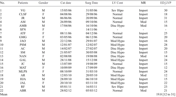
The preoperative hemodynamic data are shown in Table 2. On hemodynamic examination, performed in 21 patients,
mitral regurgitation was moderate in ive (23.8%) and severe
in 16 (76.2%) patients. As the left ventricle contractility observed by ventriculography we found: normal in eight (38.1%) patients, mild hypokinesia of the left ventricle in
ive (23.8%), moderate in six (28.6%) and severe hypokinesia
in two (9.5%). The average left ventricular end-diastolic pressure (DP2LV) was 19.8 mmHg (range 12-31 mmHg).
Table 1. Preoperative clinical data.
No.
1 2 3 4 5 6 7 8 9 10 11 12 13 14 15 16 17 18 19 20 21 22 Mean ± SD
Patients VG CLSF JR AM AMB SFN ATF GMG JAO PSM AC SM NAN GAL JC MAT MLPS AR HJA JAL RF ABR Gender M F M M M F F F M M M M F M M F F M M F M M Age(years) 61 51 59 58 53 23 68 69 61 19 72 76 45 55 66 53 62 53 55 70 62 58 56.8 ± 13.8
Diag IM IM IM IM IM IM IM IM IM IM IM IM IM IM IM IM IM IM IM IM IM IM Assoc Diag TI -IC -TI CoF TI -TI TI -Etiol FD FD FD FD FD FD FD FD FD FD FD BARLOW FD FD BARLOW FD FD BARLOW FD BARLOW BARLOW BARLOW FC IV III III III III III III III IV IV III IV III III IV III III IV IV III III III SMM ++++ +++ +++ +++ +++ +++ +++ +++ ++++ ++++ +++ ++++ +++ +++ ++++ +++ +++ ++++ +++ ++++ ++++ ++++
M = Male, F = Female, FC = Functional Class (NYHA) = Diag = diagnosis, Assoc Diag = Associate diagnosis, Etiol = Etiology, SMM =
Systolic mitral murmur, MF = Mitral failure, TI =Tricuspid Insuficiency, IC = Interatrial communication, CoF = Coronary Failure, FD
In all patients, two changes were always present, ruptured chordae and dilatation of the mitral valve annulus. In 13 (59.1%) patients, chord rupture was on the anterior
lealet of the mitral valve, in eight (36.4%), in the posterior lealet and in one (4.5%) in both lealets.
Prosthesis
The bovine pericardium premolded chordae were per-formed in monoblock (Braile Biomédica, São José do Rio Preto, SP, Brazil), trapezoid-shaped, with enhanced bovine pericardium or Dacron in their upper and lower beams. The chordae are premolded in number from ive to seven, with lengths ranging from 20 to 35 mm. Its width is 2 mm and are distant from each other by 3 mm. The standardiza-tion of the chordae was conirmed by the use of steel me -ters corresponding to the size of the prostheses. The peri-cardium was treated with 0.5% glutaraldehyde, subjected to anti-calciication treatment with glutamic acid and pre -served in 4% formaldehyde solution. Strength and
durabil-ity tests showed levels of rupture of about 15 kg/cm2 [11].
The prostheses implanted were in numbers 35, 30, 25 and 20 in one (4.5%), 12 (54.5%), seven (31.8%) and two (9.1%) patients, respectively. The size of the graft was determined based on the distance from the top of the papillary muscle
to the free edge of the lealet in its original position, not
prolapsed. Thirteen (59.1%) patients received prostheses
to the anterior lealet, eight (36.4%) to the posterior lealet
and one (4.5%) for both. The implant prosthesis begins with setting the lower part of the trapezoid at the top of the papillary muscle associated with the chordae using a U-section 5-0 polypropylene, anchored in a Dacron pad. In sequence, the larger beam of the trapezoid will be sutured on the free edge of
the compromised mitral lealet with interrupted sutures using 5-0 polypropylene. The prosthesis with ive to seven chordae
in their original form can be reduced up to two chordae if necessary. It can also have its upper stem (larger) divided, and thus can be both tops sutured to the anterior and posterior
lealets while correcting occasional prolapse of two lealets. Table 2. Preoperative hemodynamic data.
No. 1 2 3 4 5 6 7 8 9 10 11 12 13 14 15 16 17 18 19 20 21 22 Mean Patients VG CLSF JR AM AMB SFN ATF GMG JAO PSM AC SM NAN GAL JC MAT MLPS AR HJA JAL RF ABR Gender M F M M M F F F M M M M F M M F F M M F M M Cat date 15/05/06 04/08/06 06/06/06 26/09/06 17/04/06 -08/11/06 03/05/06 22/12/06 12/01/07 14/02/07 21/05/07 02/08/08 28/11/08 13/07/09 10/09/09 08/10/09 12/03/10 28/09/10 20/10/10 05/01/11 28/02/12 Surg Date 31/05/06 29/08/06 20/09/06 09/10/06 16/10/06 14/11/06 04/12/06 06/12/06 29/01/07 12/02/07 27/02/07 30/05/08 19/08/08 15/12/08 19/08/09 17/09/09 31/03/10 20/05/10 06/10/10 03/11/10 16/03/11 05/03/12 LV Cont Sev Hipo Normal Normal Normal Disc Hypo -Normal Normal Mod Hypo Mod Hypo Disc Hypo Disc Hypo Disc Hypo Mod Hypo Normal Disc Hypo Normal Mod Hypo Mod Hypo Mod Hypo Sev Hipo Normal MR Import Import Import Mod Mod -Import Import Import Import Import Import Mod Import Import Import Import Mod Import Import Import Mod
ED2LVP
30 18 31 15 16 -25 20 16 28 20 15 14 24 15 12 24 12 15 22 23 21 19.8 [12 to 31]
Associated repair techniques (Table 3)
Annuloplasty - The dilatation of the mitral annulus, present in all cases, was corrected with the use of Gregori-Braile® prosthetic ring [12]. In association, in two (91%)
cases, was performed plicature of the mitral annulus near the posteromedial commissure using technique by Wooler et al. [13]. This is a hemi-ellipse prosthesis industrialized and commercially available (Braile Biomédica, São José do Rio Preto, SP, Brazil). Its shape resembles the Carpentier ring
without the anterior segment, and presents a modiication
in its right half. This is a correction of the ring curve. Thus, the prosthesis is hemi-ellipse-shaped, and the right curvature
is rectiied. The material used is 316 stainless steel, medical grade coated with a layer of silicone rubber, and inally by
Dacron. It presents in various sizes, according to various dimensions of the mitral annulus. Choosing the ideal
ring was based solely on the distance between the ibrous
trigones, which generally correspond to the projections of the commissures in mitral annulus of the patient, regardless of the anteroposterior diameter.
The ixation of the prosthesis in the mitral annulus was
performed usingh U-section 2-0 polyester thread in the mitral annulus 1-2 mm apart and then on the outside of the prosthetic ring. The two thresholds which correspond to the ends of the prosthesis are applied to the mitral annulus, at the height of the
commissures of the projection, so that after the ixation of the prosthesis, the posterior lealets moves to the anterior, and what
is important, the posteromedial portion moves more sharply in the anterior-lateral directon, thus correcting existing dilation. In two (9.1%) patients, annuloplasty was performed according Wooler et al. [13] to correct the excessive dilation of the posterior annulus of the mitral valve near the posteromedial commissure.
Table 3. Surgical data.
No. 1 2 3 4 5 6 7 8 9 10 11 12 13 14 15 16 17 18 19 20 21 22 Mean Patients VG CLSF JR AM AMB SFN ATF GMG JAO PSM AC SM NAN GAL JC MAT MLPS AR HJA JAL RF ABR Surg Date 05/31/06 08/29/06 09/20/06 10/09/06 10/26/06 11/14/06 12/04/06 12/06/06 01/29/07 02/12/07 02/27/07 05/30/08 08/19/08 12/15/08 08/19/09 09/17/09 03/31/10 05/20/10 10/06/10 11/03/10 0316/11 03/05/12 Annulus Ring Ring Ring Ring Ring Ring Ring Ring Ring Ring Ring Ring Ring Ring Ring Ring Ring Ring Ring Ring+ WOOLER Ring Ring + WOOLER
Techniques
DeVega
SHORT GREGORI CHORDAE RES. CALCIF SHORT. CHORDAE FRATER/CARP
SHORT. CHORDAE CARP/ IAC CLOS MERENDINO + RES. ANT LEAF
SHORT. CHORDAE FRATER MERENDINO + MMDA SHORT. CHORDAE CARP+DeVega MERENDINO + SHORT GREGORI CHORDAE
MERENDINO ENL POST LEAF
-SHORT. CHORDAE FRATER -MERENDINO -DeVega
MERENDINO SHORT. CHORDAE CARP + DeVega MERENDINO Chordae No. A 30 A 20 A 30 P 30 P 30 P 20 P 30 P 30 A 30 P 35 A 25 A+P 30 A 25 A 30 P 25 A 25 A 25 P 30 A 25 A 30 A 25 A 30 CBT 94 98 98 92 117 90 87 75 100 81 182 120 71 99 97 67 82 100 85 86 121 151 100 TMA 41 63 66 52 62 27 48 26 58 44 123 61 37 52 57 22 42 41 34 51 59 79 52
Surg Date = Date of surgery, Annulus = Type of annuloplasty, CBT = Cardiopulmonary bypass time, TMA = Time of myocardial anoxia,
Short = Shortenings, RES = Resection, Calcif = Calciication, Carp = Carpentier, AntLeaf = Anterior lealet, Post leaf = Posterior lealet, MMDA = Mammary/anterior descending coronary artery, Enl post leaf = Enlargement of posterior lealet, A = Anterior, P = Posterior,
Chordal shortening - The chordal shortening was performed in eight (36.4%) patients. In two (9.1%) of these patients, chordal shortening was performed according to Carpentier et al. [14] with longitudinal incision of the papillary muscle and burial intrapapillar stretched chord. In two (9.1%) patients was used the method described by shortening according Gregori Jr. et al. [15]. We performed a small incision in the anterior
lealet near the elongated cords beam being pulled through the incision and then ixed to the surface of the anterior lealet. In four (18.2%) patients, we used the technique by Frater et al. [7], or that is, ixing the elongated chordae on the inner face of the anterior lealet. Minor shortenings
are achieved with this technique. Eventually, we use the three methods described for the shortening of elongated chordae in the same patient.
Leaflet resection - In eight (36.4%) patients, we
performed resections of lealets, being quadrangular in the posterior lealet [2] in seven patients and triangular in anterior lealet in one [1]. We avoid this last procedure in the anterior lealet because it could lead to mitral stenosis by decreasing lealet mobility. After quadrangular resection of the posterior lealet and posterior lealet prolapse correction, the two edges of the lealet were approximated and sutured
using 5-0 polypropylene sutures.
Lealet enlargement - In one (4.5%) patient, we expanded the posterior lealet of the mitral valve using a bovine
pericardial patch to thereby facilitate better coaptation of
the posterior lealet to the anterior.
Other Associated Surgical Techniques
Four (18.2%) patients with functional tricuspid
insuficiency underwent annuloplasty according to the
technique described by DeVega [16], which consists in encircling the tricuspid annulus to flee the area corresponding to the passage of the conduction beam near the atrioventricular node. One (4.5%) patient had their interatrial communication closed and another (4.5%) underwent myocardial revascularization with internal thoracic artery to the anterior interventricular branch of the left coronary artery.
Postoperative Clinical Evaluation
Hospital and late mortality, the rate of reoperation due to failure of reconstructive surgery, and the postoperative morbidity were observed. The surviving patients (17) were clinically assessed at a median time after surgery of 47 months (range 4-70 months). As preoperatively,
patients were classiied according to the clinical status according to the NYHA classiication of symptoms.
On physical examination, during this same period, the auscultation of the mitral valve was assessed for the presence of murmurs.
EchoDopplercardiographic assessment
Seventeen patients underwent Doppler echocardiography to assess left ventricular function, left atrial diameter, mitral valve area, mean gradient, end-diastolic left ventricular volume and the degree of valvular regurgitation. The classification of Doppler echocardiographic mitral regurgitation was based on the extent and magnitude of the regurgitant jet in the left ventricular systole. We assessed the following indices: ejection fraction (EF) of the left ventricle, left atrium diameter (LA), mitral valve area (MVA), mitral mean gradient (GTVM) and left ventricular end-diastolic volume (LVEDV). The mean postoperative assessment of the 17 surviving patients was 47 months (range 4-70 months).
Statistical Analysis
Actuarial survival curve free of death and actuarial survival free of reoperation were obtained for total (22) of patients and for the 21 patients who were discharged from hospital and outpatients [17].
RESULTS
The postoperative clinical data are presented in Table 4. There was one death in the immediate postoperative period (4.5%) in low cardiac output syndrome in 70-year-old patients, diagnosed with mitral and associated tricuspid
insuficiency and carrier of Barlow syndrome. The mitral
annuloplasty ring consisted of using Gregori-Braile ring and tricuspid annuloplasty was performed using DeVega technique [6].
There were no thromboembolic events or hemolysis
postoperatively. Four patients who had atrial ibrillation
preoperatively continued with arrhythmia in the postoperative period. Two (9.1%) patients required reoperation at 35 and 46 months for mitral valve replacement, the latter died in
low cardiac output syndrome in the irst hours after arriving
at the Intensive Care Unit. In both patients, the chordae shortening technique was associated and in none of them the bovine pericardium chordae were compromised implanted
in the posterior lealet. There was evolution of ibroelastic degeneration, especially involving the anterior lealet of the mitral valve that received no artiicial chordae.
Two patients died, one month and 12 months postoperatively, suddenly, being previously in functional class I, one without mitral regurgitation and one with slight
relux at Doppler echocardiography. In the irst patient, ventricular ibrillation on mobile ICU’s electrocardiogram
with native valves. Sixteen (94.1%) of 17 surviving patients are in functional class I and one (5.9%) in functional class II (NYHA). Fifteen (88.2%) patients had no murmurs in the mitral valve, and two (11.8%), there was presence of
slight systolic murmur, one (5.9%) of them associated with a diastolic murmur at the apex. Actuarial survival free of death of 22 patients from cardiovascular causes demonstrated survival probability at 70 months of 78.3 ± 10.1% (Figure 1).
Table 4. Postoperative clinical data.
No. 1 2 3 4 5 6 7 8 9 10 11 12 13 14 15 16 17 18 19 20 21 22 Patients VG CLSF JR AM AMB SFN ATF GMG JAO PSM AC SM NAN GAL JC MAT MLPS AR HJA JAL RF ABR Gender M F M M M F F F M M M M F M M F F M M F M M Age (years) 61 51 59 58 53 23 68 69 61 19 72 76 45 55 66 53 62 53 55 70 62 58 Surg Date 05/31/06 08/29/06 09/20/06 10/09/06 10/16/06 11/14/06 12/04/06 12/06/06 01/29/07 02/12/07 02/27/07 05/30/08 08/19/08 12/15/08 08/19/09 09/17/09 03/31/10 05/20/10 10/06/10 11/03/10 03/16/11 03/05/12 Evol Date 12 M 06/16/12 05/15/12 07/18/12 06/19/12 10/22/09 04/20/12 08/05/10 06/11/12 06/11/12 07/20/12 07/20/12 06/12/12 03/13/12 07/05/12 01/03/12 06/04/12 06/15/12 07/11/12 -1 M 07/04/12 Evol
LAT SUDDEN DEATH GOOD GOOD GOOD GOOD MVR REOP GOOD LAT DEAT TVM REOP
GOOD GOOD GOOD GOOD GOOD GOOD GOOD GOOD GOOD GOOD GOOD IMMED DEATH LOS
LAT DEAT VF GOOD FC -I I I I -I -I II I I I I I I I I I -I
Evol = Evolution, Evol Date = Date of Evolution, FC = Functional Class, = SMM = Systolic mitral murmur, DMM = Diastolic mitral murmur, M = Male, F = Female, Lat Death = Late Deat, Immed Deat = Immediate Death, Reop = Reoperation, MVR = Mitral Valve Replacement, VF = Ventricular Fibrillation, LOS = Low output syndrome.
SMM -0 0 0 0 0 -0 0 + + 0 0 0 0 0 0 0 0 -0 DMM -0 0 0 0 0 -0 0 + 0 0 0 0 0 0 0 0 0 -0
Fig. 1 - Actuarial survival curve of the patients (22). It is observed that 78.3 ± 10.1% of the patients are still alive after 70 months of follow-up
The actuarial survival of patients who left the hospital and remain under outpatient treatment (21) demonstrated survival probability at 70 months of 82.0 ± 9.8% (Figure 2).
The Doppler echocardiographic assessment was performed in the 17 surviving patients, between the months of January and July 2012, with a mean postoperative follow-up of 47 months (range 4-70 months). Left ventricular function measured by EF showed a mean of 0.59 (range 0.27 to 0.76). The average diameter of the left atrium was 4.5 cm (ranging from 3.0 to 6.9 cm). The mean MVS assessed was 2.9 cm2 (range 1.6 to 4.5 cm2), and satisfactory in all except
one (1.6 cm2) patient. The mean GTVM was 3.3 mmHg
(range 1-9 mmHg), with negligible values in all patients except one. The average LVFDV was 141 ml (range 83-247 ml), which is normal in 12 (70.5%).
The analysis of valve competence showed no mitral regurgitation in 11 (64.7%) of the 17 surviving patients
assessed, slight regurgitation in ive (29.4%) and mild/
moderate in one (5.9%). Therefore, satisfactory competence of the mitral valve was observed in 94.1% of cases. Mild tricuspid regurgitation was observed in four (23.5%) patients and mild regurgitation of the aortic valve in four (23.5%). The actuarial reoperation-free after implantation of standardized bovine pericardium chordae treated with glutaraldehyde and non-calcifying agent for replacement of ruptured chordae tendineae of the mitral valve was at 70 months postoperatively, 83.9 ± 10.4% (Figure 3).
The actuarial reoperation-free for the 21 patients who were discharged and followed as outpatients at 70 months postoperatively showed rate of 83.9 ± 10.4% (Figure 4).
DISCUSSION
Several techniques have been described over the years aiming at mitral valve reconstruction. Therefore, a thorough knowledge of anatomical pathology is necessary. The mitral
valve is composed by an incomplete ibrous ring that goes from the right ibrous trigone to the left ibrous trigone - its
anterior portion is occupied by the ring of the aortic valve and it was previously thought that it did not dilate until Hueb et al. [18] showed pathological changes of this portion of the mitral valve in hearts with ischemic and degenerative disease. Anyway, changing that part or not, the annular dilatation should be corrected - and this is the last step of the reconstruction, restoring the original shape of the ring which is of a vat, culminating with a perfect approximation
of the anterior and posterior lealets and commissures.
There are six valves composing the mitral valve, one anterior and three posterior (posterolateral, posteromedial and posterior itself), besides the two commissures
containing lealets and chordae attached to the papillary
muscles and/or the free wall the left ventricle. During left ventricular systole, the mitral valve contracts, similarly to a sphincter - takes the form of a kidney while relieving the
outlow of the left ventricle. In the dilated left ventricle
(either in volume overload or cardiomyopathy), the ring expands subsequently acquiring the ovoid form, but more intensely at right next to the posteromedial commissure.
Hence the reason we have used the ring developed by us [12] that with the shape of a hemi-ellipse approximates
the posterior lealets to the anterior lealet, and because it
has correction on its right side, approximates this part of the mitral annulus, more often dilated. Even being open, in
its anterior portion, the upper ends of the prosthesis, ixed Fig. 3 - Actuarial survival curve of the surgical method, or that
is, the implanted prosthesis in the mitral valve (22 patients). It is observed that 83.9 ± 10.4% of the prostheses are free from dysfunction at 70 months postoperatively
along the trigone, prevent annular dilatation on that site,
occupied by the ibrous ring of the aortic valve. Stretching
and rupture of chordae are the most common complications
in degenerative mitral valve. Changes in the lealets also
occurs, with sagging and proliferation of connective tissue and may be exaggerated, as in Barlow syndrome (six patients in this study), much harder to be treated than in
ibroelastic degeneration, when there is plenty of tissue.
Faced with cases of chordae rupture, surgeons commonly have different behavior if it is located in the
anterior lealet of the mitral valve. The chordae rupture of the anterior lealet is a major challenge, since in these cases the valve failure is usually signiicant. The anterior lealet has basically function of excursion and the posterior lealets
have function of containing and support to the anterior
lealet. Valve prolapse by chordae rupture are easier to be treated when present in the posterior lealet.
The repair by Merendino et al. [2], have been used successfully for a long time, often solving the problem often in such cases. Contrary to what the literature shows, in our cases, correction of chordae rupture of the anterior
lealet was more frequent than in the posterior lealet.
The surgical technique to be used depends on the level of commitment and the location of the damaged chordae. There are several procedures that can be adopted in chordal
rupture. Transfering chordae from the posterior lealet to the anterior lealet of the mitral valve proposed by Carpentier et
al. [4] has been used, including in our department.
Constructing a neocord through the removal of a lap of the anterior lealet of the mitral valve when lowered on top of
the papillary muscle, replacing ruptured chordae tendineae is an alternative technique [5], but its use is restricted to
patients with the anterior lealet well developed, which does
not occur in rheumatic, ischemic or even in endocarditis diseases. It is therefore almost exclusively of degenerative disease. Partial transfer of the tricuspid valve (posterior
lealet in most cases) to the mitral valve, providing chordae to the anterior lealet, was proposed by Gregori et al. in 1992
[6], with very satisfactory results.
These techniques require handling of leaflets and chordae with normal anatomy and, in general, are feared by surgeons that initiate in reconstructive surgery of the mitral valve. Synthetic and biological materials have been used for the replacement of chordae. The PTFE threads, proposed by David et al. [8] have been shown to be more frequently used in the world. A recent study using PTFE
showed excellent results at ive and 10 years postoperatively,
with no reoperation in 93.3% and 81.7%, respectively [19]. However, the use of these techniques requires enormous degree of subjectivity, requiring a high degree of individual
skill. Dang et al. [20] described a simpliied method for
using PTFE, slightly easing its implementation.
With biological materials, Frater et al. [7] were the irst
to employ the bovine pericardium for chordal replacement with satisfactory initial results. Their study was interrupted
due to fear of calciication over the years. However, in their
original study, two groups of patients undergoing implant of PTFE and bovine pericardium chordae were compared. It should be emphasized that a pericardial measured 4 mm wide, with not being premolded chordae, nor standardized by instruments (meters). In addition, follow-up time was longer in the group which used bovine pericardium than in the group that used PTFE.
Even so, there was no signiicant difference in long-term evolution of the two groups, nor calciication of bovine pericardium implanted. Calciication is really a problem
and should be matter of concern. Many efforts have been made to improve the durability of the bovine pericardium with the introduction of new chemical reagents as used in our case, the glutamic acid. It has been very frequent the use of bovine pericardium in cardiovascular surgery. Its
use is common in the manufacture of prostheses, in oriice
occlusions in congenital heart defects, reconstructions of the ventricular outflow tract, in ventricular repair after repair of left ventricular aneurysms, among other procedures. In mitral valve surgery, the pericardium has been used as a rope to maintain the tension between the top of the papillary muscles and the mitral valve in valve
replacement, with signiicant improvement in ventricular
function [21].
The use of standardized bovine pericardium prosthesis makes the procedure easier and therefore fast, objective
and reproducible. Laboratory tests of the artiicial chordae
showed a rupture level in 15 kg/cm2. It must be remembered
that in a patient with high blood pressure of 140 mmHg in systole of his left ventricle, the tension to which the chordae are subjected is approximately 0.5 kg/cm2, therefore thirty
times smaller. The chordae implantation technique is similar to that described by us in case of partial transfer
of the tricuspid valve (posterior lealet, usually) to the
mitral valve [6]. In this study, in all patients was performed annuloplasty using rigid open Gregori-Braile ring [12], preceeded in both cases, of Wooler annuloplasty [13], along the posteromedial commissure. Clinical improvement was observed in surviving patients, both with respect to functional class (16 of them in functional class I and one in functional class II) and the excellent auscultation of mitral valve (15 with no murmursand two with discrete systolic murmur in the mitral focus, one of them associated with a diastolic murmur at the apex).
ventricular hypokinesis, in addition to high left ventricular end diastolic pressure. The Doppler echocardiography has been the best method for postoperative assessment of mitral valve and, in the patients of this study, implantation of standard bovine pericardial chordae associated with open rigid annuloplasty and other techniques presented postoperative quite satisfactory. We attribute these results to
a signiicantly greater coaptation line between the lealets,
since extensive resections are avoided.
The postoperative data of this prospective study
conirmed the clinical indings observed. Except in one case,
the mean transvalvular gradients were normal, in over 70% of cases, left ventricular end-diastolic volume was normal and the mitral valve area was satisfactory. Satisfactory results were also observed in the analysis of mitral valve competence in view of the absence of regurgitation or only
mild mitral regurgitation in 16 of 17 patients. The beneit of the artiicial chordae implantation is more evident in cases of ruptured chordae of the anterior lealet, in which
resection should be avoided because it may damage its main function, which is excursion. However, it is still interesting
its application in the posterior lealet. Falk et al. [22]
corroborated this statement in a prospective randomized study comparing the use of PTFE chordae with resection
of the posterior lealet prolapse.
Thus, the good late results are technique-dependent. A prospective analysis of our cases is valid because the series is consecutive, the patients had mitral valve lesions of the same etiology (degenerative) and underwent surgery by the same surgeon. The actuarial survival curves free of death and reoperation showed probability of survival at 70
months postoperatively for more than 80%, conirming our
previous publications [9,10]. Over 90% of the 17 surviving patients showed competent mitral valve postoperatively,
and none showed signs of calciication of the implanted
prosthesis. Furthermore, in no patient with events of death
or reoperation was conirmed dysfunction of the bovine
pericardium prosthesis preserved in glutaraldehyde for the replacement of ruptured chordae tendineae.
The early death in the irst surgery and early death
in a patient who had undergone surgery for mitral valve replacement were due to low cardiac output syndrome, with prostheses functioning normally. In the two patients who died suddenly in the late postoperative period, the
most likely cause would have been ventricular ibrillation
- documented in a mobile ICU. In the rupture of mitral valve chordae, patients have usually in severe cases, acute pulmonary edema. With good postoperative follow-up, with respect to the patient who had undergone surgery for mitral valve replacement, during surgery was observed that the mitral valve showed severe degeneration of the
anterior lealet, being with good aspect the implant of bovine pericardium chordae in the posterior lealet.
CONCLUSION
Despite a follow-up not extensive, the outcomes obtained, both clinical and Doppler echocardiographic,
allow us to afirm that clinical outcome after use of the
technique of chordae replacement by premolded chordae of bovine pericardium preserved in glutaraldehyde are satisfactory in patients with impaired mitral degenerative etiology.
REFERENCES
1. McGoon DC. Repair of mitral insuficiency due to ruptured
chordae tendineae. J Thorac Cardiovasc Surg. 1960;39:357-62.
2. Merendino KA, Thomas GI, Jesseph JE, Herron PW, Winterscheid LC, Vetto RR. The open correction of rheumatic mitral regurgitation and/or stenosis; with special reference to regurgitation treated by posterormedial annuloplasty utilizing a pump-oxygenator. Ann Surg. 1959;150(1):5-22.
3. Pomerantzeff PAM, Brandão CMA, Rossi EG, Cardoso LF, Tarasoutchi F, Grimberg M, et al. Quadrangular resection without ring annuloplasty in mitral valve repair. Eur Cardiovasc Surg. 1997;2:271-3.
4. Carpentier A, Relland J, Deloche A, Fabiani JN, D'Allaines C, Blondeau P, et al. Conservative management of the prolapsed mitral valve. Ann Thorac Surg, 1978;26(4):294-302.
5. Gregori F Jr, Takeda R, Silva S, Façanha L, Meier MA. A
new technique for mitral insuficiency caused by ruptured chordae of the anterior lealet. J Thorac Cardiovasc Surg,
1988;96(5):765-8.
6. Gregori F Jr, Cordeiro C, Croti UA, Hayashi SS, Silva SS, Gregori TE. Partial tricuspid valve transfer for repair of mitral
insuficiency due to ruptured chordae tendineae. Ann Thorac
Surg, 1999;68(5):1686-91.
7. Frater RW, Gabbay S, Shore D, Factor S, Strom J. Reproducible replacement of elongated or ruptured mitral valve chordae. Ann Thorac Surg. 1983;35(1):14-28.
8. David TE, Bos J, Rakowski H. Mitral valve repair by
replacement of chordae tendineae with polytetraluorethylene
sutures. J Thorac Cardiovasc Surg. 1991;101(3):495-501.
9. Leal JCF, Gregori Jr. F, Galina LE, Thevenard RS, Braile
DM. Avaliação ecocardiográica em pacientes submetidos
10. Gregori F Jr, Leal JC, Braile DM. Premolded bovine pericardial chords for replacement of ruptured or elongated chordae tendineae. Heart Surg Forum. 2010;13(1):E17-20.
11. Braile DM, Ardito RV, Pinto GH, Santos JLV, Zaiantchik M, Souza DRS, et al.. Plástica mitral. Rev Bras Cir Cardiovasc. 1990;5(2):86-98.
12. Gregori F, Silva SS, Hayashi SS, Aquino W, Cordeiro C, Silva LR. Mitral valvuloplasty with a new prosthetic ring.
Analysis of the irst 105 cases. Eur J Cardiothorac Surg.
1994;8(4):168-72.
13. Wooler GH, Nixon PG, Grimshaw VA, Watson DA. Experience with the repair of the mitral valve in mitral in competence. Thorax. 1962;17:49-57.
14. Carpentier A, Deloche A, Dauptain J, Soyer R, Blondeau PH, Piwinica A, et al. A new reconstructive operation for correction
of mitral and tricuspid insuficiency. J Thorac Cardiovasc Surg.
1971;61(1):1-13.
15. Gregori Júnior F, Silva S, Façanha L, Cordeiro C, Aquino W, Moure O. Preliminary results with a new technique for repairing elongated chordae tendineae of the anterior mitral valve leaflet. J Thorac Cardiovasc Surg. 1994;107(1):321-3.
16. DeVega NF. La anuloplastia selectiva, regulable y permanente. Rev Esp Cardiol. 1972;25:555-6.
17. Kaplan EL, Meier P. Non-parametric estimation from incomplete observations. J Am Statist Assoc. 1958;53:457-8.
18. Hueb AC, Jatene FB, Moreira LF, Pomerantzeff PM, Kallás E, Oliveira SA. Ventricular remodeling and mitral valve
modiications in dilated cardiomyopathy: new insights from
anatomic study. J Thorac Cardiovasc Surg. 2002;124(6):1216-24.
19. Kobayashi J, Sasako Y, Bando K, Minatoya K, Niwaya K, Kitamura S. Ten-year experience of chordal replacement
with expanded polytetraluoroethylene in mitral valve repair.
Circulation. 2000;102(19 Suppl 3):III30-4.
20. Dang NC, Stewart AS, Kay J, Mercando ML, Kruger KH,
Topkara VK, et al. Simpliied placement of multiple artiicial
mitral valve chords. Heart Surg Forum. 2005;8(3):E129-31.
21. Gomes OM, Pitchon M, Barros MVL. Utilização de cordas tendíneas de pericárdio bovino em cirurgia da valva mitral. Coração. 1990;2:20-2.
22. Falk V, Seeburger J, Czesla M, Borger MA, Willige J, Kuntze T,
et al. How does the use of polytetraluoroethylene neochordae
for posterior mitral valve prolapse (loop technique) compare
with lealet resection? A prospective randomized trial. J Thorac
Cardiovasc Surg. 2008;136(5):1205.
23. Gregori Junior F. Conservative surgical management of
mitral insuficiency: an alternative approach. Rev Bras Cir