RBCCV 44205-1417 DOI: 10.5935/1678-9741.20120092
Gene expression of endothelin receptors in
replaced rheumatic mitral stenotic valves
Expressão gênica de receptores de endotelina em valvas mitrais reumáticas estenóticas substituídas
Sydney Correia Leão
1, Fernanda Maria Silveira Souto
2, Ricardo Vieira da Costa
3, Thaisa de Fatima
Almeida Rocha
4, Yolanda Galindo Pacheco
5, Tania Maria de Andrade Rodrigues
61. Medical student. Laboratory of Molecular Anatomy, Department of Morphology, Biological and Health Science Center, Sergipe Federal University, UFS, São Cristóvão, SE, Brazil (Bolsista PICVOL). Editing and proofreading of the manuscript.
2. Medical student. Laboratory of Molecular Anatomy, Department of Morphology, Biological and Health Science Center, Sergipe Federal University, UFS, São Cristóvão, SE, Brazil. Data collection, translation and revision of the manuscript.
3. Fellow of PROBP Master graduate program, Department of Medicine, Biological and Health Science Center, Sergipe Federal University, UFS, Aracaju, SE, Brazil. Data collection and editing of the manuscript. 4. Medical student. Laboratory of Molecular Anatomy, Department of
Morphology, Biological and Health Science Center, Sergipe Federal University, UFS, São Cristóvão, SE, Brazil. Translation and revision of the manuscript.
5. Titular professor, Anatomy Department, Medicine Faculty, Brasília University, Brasília, DF, Brazil. Data collection, review of the inal manuscript.
6. Adjunct professor, Department of Morphology, Biological and Health
Science Center, Sergipe Federal University, UFS, São Cristóvão, SE, Brazil. Data collection and editing of the manuscript.
Work carried out at Laboratory of Molecular Anatomy, Department of Morphology, Biological and Health Science Center, Sergipe Federal University, UFS, São Cristóvão, SE, Brazil.
Correspondence address Sydney Correia Leão
Marechal Rondon Avenue – Jardim Rosa Elze – Professor José Aloísio de Campos University City – São Cristovão, SE, Brazil.
E-mail: [email protected]
Financial support: National Consoling of Research (CNPq) [Universal Announcement MCT/CNPq grant number 14/2008; Universal, Process grant number 472808/2008-7, TMAR].
Article received on June 24th, 2012 Article accepted on November 12th, 2012 Abstract
Objectives: Rheumatic fever is a highly prevalent disease in Brazil, and it poses a major public health problem. It is the leading cause of acquired heart disease in childhood and adolescence. The aim of this study was to evaluate the gene expression of ET-3 and its receptors, in replaced rheumatic mitral valves.
Methods: We studied the gene expression of endothelin-3 (ET-3) and its receptors, endothelin receptor A and endothelin receptor B (ETr-A and ETr-B), in the rheumatic mitral valves of 17 patients who underwent valve replacement surgery. The samples also underwent a histological analysis.
Results: Our data showed that almost all patients, regardless of individual characteristics such as gender or age, expressed the endothelin receptor genes, but did not express the genes for ET-3. In quantitative analysis, the
ETr-A/GAPDH mean ratio was 33.04 ± 18.09%; while the ETr-B/GAPDH mean ratio was 114.58 ± 42.30%. Regarding histopathological individual features, the frequency of
ibrosis is 100%, 88.23% of mononuclear iniltrate, 52.94% of neovascularization, 58.82% of calciication and absence of ossiication.
Conclusion: The presence of receptors ETr-A and ETr-B in rheumatic mitral valves suggests its interaction with the system of circulating endothelins, particularly ETr-B (known for acting in the removal of excess endothelin) detected in a greater proportion, which could explain the lack of expression of endothelin in rheumatic mitral valve, process to be elucidated.
Abbreviations, Acronyms & Symbols
bFGF Basic ibroblast growth factor ET-3 Endothelin type 3
ETr-A Endothelins receptors type A ETr-B Endothelins receptors type B ETr-C Endothelins receptors type C ETrs Endothelins receptors ETs Endothelins
LAM Laboratory of Molecular Anatomy NO Nitric oxide
PASP Pulmonary artery systolic pressure RF Rheumatic fever
RT-PCR Reverse transcription polymerase chain reaction UFS Universidade Federal de Sergipe
VEGF Vascular endothelial growth factor
INTRODUCTION
Rheumatic fever (RF) is a serious public health problem and a strong indicator of poverty and poor health services in developing countries. It is a rheumatic and
inlammatory disease with an autoimmune origin. It recurs
in response to Streptococcus pyogenes (Group A beta-hemolytic Strep, GAS) infection. This agent is responsible for approximately 15.6 million cases of rheumatic heart disease annually across the globe, with 282,000 new cases and 233,000 deaths each year. As a result, health systems incur high costs, paying for the tests, surgeries and hospitalizations required to treat the complications of this condition. For instance, approximately 3 million patients per year are hospitalized due to congestive heart failure [1-4].We can divide the manifestations of rheumatic fever in acute and chronic. Acute rheumatic fever affects several sites such as the skin (erythema marginatum), the basal ganglia (chorea of Syndeham) and heart (rheumatic carditis). The involvement of cardiac valves (especially mitral valve) is extremely common in chronic rheumatic heart disease.
There are two types of valve dysfunction on rheumatic
disease: stenosis and insuficiency. These two types of
dysfunction are not mutually exclusive. A patient with mitral stenosis may remain asymptomatic for long periods of time despite the gradual decrease in cardiac output and increase in pulmonary vascular resistance, which may eventually lead to a vascular morphofunctional change [5-7]. The symptoms usually depend on the effective valve area involved and the tissue damage level (obtained via a regular echocardiogram), although this correlation is not always reliable. In advanced disease, all of these events
result from inlammatory damage to the tissue that is accompanied by neovascularization and the calciication
of the mitral apparatus, which was formerly an avascular structure [8].
Increased peripheral vascular resistance is a key event in the development of heart failure, and endothelin is one of the most potent vasoconstrictors involved in this disease
[9], and stimulates the secretion also of inlammatory
cytokines [10]. The endothelins (ETs) are potent peptides formed by a chain of 21 amino acids. Some of their properties are well known, such as vascular tone control in both vascular and non-vascular tissues. Currently, there
are three identiied isopeptides encoded by three different
genes: ET-1, ET-2 and ET-3. The pharmacological effects of ETs indicate the existence of three subtypes of receptors
Resumo
Objetivos: A febre reumática é uma doença altamente prevalente no Brasil, e representa um importante problema de saúde pública. É a principal causa de cardiopatia adquirida na infância e adolescência. O objetivo deste estudo foi avaliar a expressão gênica de ET-3 e seus receptores, em valvas mitrais reumáticas substituídas. Métodos: Estudamos a expressão gênica de endotelina-3 (ET-3) e de seus receptores,
receptor da endotelina A e receptor da endotelina B (ETr-A e ETr-B), nas valvas mitrais reumáticas de 17 pacientes que se submeteram à cirurgia de troca valvar. As amostras também foram submetidas à análise histológica.
Resultados: Nossos dados mostraram que praticamente todos os pacientes, independentemente de características individuais, como sexo ou idade, expressaram os genes de receptores de endotelina, porém não expressaram os genes para ET-3. Na análise quantitativa, a média da proporção ETr-A/GAPDH foi de 33,04 ± 18,09%; enquanto que a média da proporção ETr-B/GAPDH foi de 114,58 ± 42,30%. Em relação às características histopatológicas individuais, a
frequência de ibrose foi de 100%, iniltrado mononuclear de 88,23%, neovascularização de 52,94%, calciicação de 58,82% e houve ausência de ossiicação.
Conclusão: A presença de receptores ETr-A e ETr-B em valvas mitrais reumáticas sugere sua interação com o sistema de endotelinas circulantes, particularmente ETr-B (reconhecido por atuar na remoção do excesso de endotelina), detectado em maior proporção, o que poderia explicar a ausência da expressão de endotelina em valva mitral reumática, processo a ser elucidado.
(ETrs): type A (ETr-A), type B (ETr-B) and type C (ETr-C)
[11]. ETs and ETrs exhibit different afinities, and ETr-A
acts as a vasoconstrictor in the smooth muscle layer (tunica media) of the arterial wall [12].
The importance of ETs and its receptors for the pathogenesis of many diseases has been the subject of intense research since its discovery [13-15]. Those diseases that involve excessive vasoconstriction and cell proliferation have been an especial focus. Over the last decade, a growing volume of research has been conducted on the actions of endothelin and its receptors, exploring its unique pharmacological response and its possible correlation with cardiovascular disease [16]. The correlation between the role of endothelin in the cardiovascular system and its
pro-inlammatory activity has made it important to determine
whether ETs participates in the pathophysiology of heart injury. However, severe pulmonary hypertension, either as
a inal outcome of those diseases that involve excessive
vasoconstriction and cell proliferation or as perpetuating
them, may also affect the inluence of ETs [17].
Therefore, in this study we evaluated the expression of ET-3 and its receptors, ETr-A and ETr-B, in the rheumatic mitral valves of patients who underwent valve replacement surgery. Moreover, histological analysis in the valves was performed.
METHODS
The University Hospital Ethics and Human Research Committee of the Federal University of Sergipe approved this project, which was assigned the number 0105.0.107.000-09. In addition, all experimental protocols were conducted according to Declaration of Helsinki and had signed the Informed Consent form. A histological analysis of the mitral valves was performed. The study group comprised seventeen patients (mean age 37.7 ± 13.7 years) with serious rheumatic mitral stenosis (mean valve area of 1.0 ± 0.28 cm2 and mean pulmonary artery
systolic pressure (PASP) of 45.82 ± 6.45 mmHg) (Table 1) who underwent surgical treatment between the months of June 2009 and March 2010. These patients were six male and eleven female adults. The most common clinical manifestations of them were dyspnea (94.11%), chest pain (35.29%), and tachypnea (47.05%) (Table 1).
Seventeen valves were collected from the Cardiothoracic Surgery Service at Cirurgia Hospital in Aracaju, Sergipe, Brazil. After the valves had been collected, samples were sent to the Laboratory of Molecular Anatomy (LAM) at the Morphology Department, Sergipe Federal University (Universidade Federal de Sergipe, UFS), where they were stored and subjected to a molecular and histological analysis.
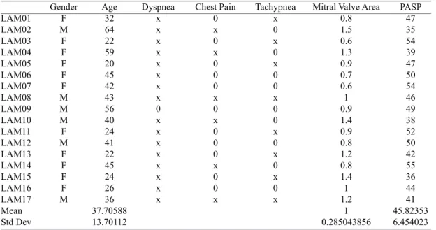
Table 1. Echocardiographic data of patients.
LAM01 LAM02 LAM03 LAM04 LAM05 LAM06 LAM07 LAM08 LAM09 LAM10 LAM11 LAM12 LAM13 LAM14 LAM15 LAM16 LAM17 Mean Std Dev Gender F M F F F F F M M M F M F F F F M Age 32 64 22 59 20 45 42 43 56 40 24 41 22 45 24 26 36 37.70588 13.70112 Dyspnea x x x x x x x x 0 x x x x x x x x Chest Pain 0 x 0 x 0 0 0 x 0 x 0 0 0 x 0 0 x Tachypnea x 0 x 0 x 0 0 x 0 0 x 0 x 0 x 0 x
Mitral Valve Area 0.8 1.5 0.6 1.3 0.9 0.7 0.6 1 0.9 1.4 0.9 0.8 1.2 0.8 1.4 1 1.2 1 0.285043856 PASP 47 35 54 39 47 50 54 46 49 38 52 50 42 55 36 44 41 45.82353 6.454023
Molecular analysis
Immediately after removal, valve tissue obtained was fragmented into three roughly equal segments (named
M1, M2 and M3), the irst near the mitral annulus, second
in the intermediate region and the third at the end in contact with the chordae tendineae. This division intended differentiation in regions that are macroscopically distinct as to their involvement by rheumatic disease. After that, each sample valve was submerged in RNAlater® stabilizer
solution (Applied Biosystems/Ambion, Austin, Texas, USA) for 24 hours at room temperature (25°C). Next, the excess solution was discarded, and the samples were stored at -80°C. The mitral fragments (100 mg) were submitted to a total RNA extraction protocol using the SV
RNA Puriication Kit (Promega®, Madison, Wisconsin, USA). Then, each sample of total RNA was quantiied by
spectrophotometry (UV-1601-UV® Spectrophotometer Shimadzu Corporation). For this purpose, it was
performed dilution of 4 μL de RNA in 196 μL of Milli-Q water totalizing 200 μL of solution. The samples were
placed in an appropriate cuvette and then analyzed on the spectrophotometer. The absorbance values obtained were analyzed using the following formula: [RNA (µg/mL)] = 40 x A260 x dilution / 1000 [18]. A purity assessment was performed using the ratio of the absorbance values obtained at 260 nm and 280 nm (A260/A280); only values between 1.6 and 2.6 were considered viable.
The total cDNA was obtained from each analyzed sample via reverse transcription polymerase chain reaction (RT-PCR) analysis. We used the protocol provided by the manufacturer of the Reverse Transcriptase
IMPROM-II™ Kit (Promega®, Madison, Wisconsin, USA). From
the cDNAs obtained, we ampliied the target fragment
using PCR [19,20]. The primers used in this technique
were ET-3 (Endothelin 3), ETr-A (receptor type A) and ETr-B (Receptor Type B) (Table 2). The constitutive gene GAPDH (Glyceraldehyde-3-phosphate dehydrogenase) was analyzed as a sample control (Table 2). The programs
used for ampliication were optimized using different
annealing temperature combinations in accordance with mean temperature data supplied by Promega®. The
ampliication products were analyzed via electrophoresis
on 2% agarose gel stained with ethidium bromide (1 mg/ ml) and sample buffer (bromophenol blue) 6x. They were
viewed using the photo documentation system (Kodak Gel
Logic 100® Imaging System, Eastman Kodak Corporation,
Rochester, NY, USA). Quantitative analysis of each
sample was performed with software ImageJ (National
Institute of Health, Bethesda, MD, USA). Quantiication
was performed by counting the average number of pixels of each sample (including GAPDH sample). Thereafter, it was performed a ratio between the average number of pixels in each sample by the amount found in GAPDH sample (ETr-A/GAPDH and ETr-B/GAPDH ratio).
Histological analysis
All of the excised, fragmented valves were formalin
ixed in a neutral 10% solution (pH 7.0). They were then submitted to decalciication, embedded in parafin and cut
using a microtome (Hacker Edge SL-200® Microtome,
Winnsboro, USA) at a 4 µm thickness. Hematoxylin and eosin staining was then performed. Each segment
was examined for the presence of ibrosis, calciication, ossiication, angiogenesis and mononuclear cells.
Statistical analysis
For statistical analysis, it was perfomed measures of central tendency and variance. We also examined the correlation between the results obtained for the mean ratio ETr-A/GAPDH and ETr-B/GAPDH. For correlation analysis, we used Pearson’s correlation, with the
signiicance level of 5% (P value< 0.05). RESULTS
Regarding RNA extraction of mitral valves, the average
concentration of nucleic acid was 7.20 ± 5.60 ng/μl. The
absorbance value obtained at 260 nm was 0.18 ± 0.14 and absorbance at 280 nm was 0.09±0.06. Ratio between A260 and A280 (A260/280) was 1.81 ± 0.16 (Table 3). The expression of endothelin and its receptors in the mitral valves using the PCR of the cDNA is showed in the Figure
1. As shown in the Figure 1A, there was ampliication of
the 480 bp fragment corresponding with the expected ET-3 amplicon in only one sample. Figures 1B and 1C show the results for the ETr-A and ETr-B primers, respectively. Interestingly, we observed that for ETr-A and ETr-B,
Table 2. List of primers prepared for molecular analysis.
Name of Oligonucleotide ET-3 Sense
ET-3 Antisense
ETr-A Sense
ETr-A Antisense
ETr-B Sense
ETr-B Antisense
GAPDH HUMAN Sense
GAPDH HUMAN Antisense
Sequency
5’ CCA AAC TCT GGA CGT CAG CAG 3’
5’ ATT TCC TGC ATG AAA CCG GAG 3’
5’ TTC AGA CTT CGC CAG ACA GA 3’
5’ CAA GCA ACT GGA ACC TGA TGT 3’
5’ AGA CAG GAC GGC AGG ATC T 3’
5’ GAA CAC AAG GCA GGA CAC AA 3’
5’ GCT CTC TGC TCC TCC TGT TC 3’
Fig. 1 - Ampliication of the ET-3, ETR-A and ETR-B encoding genes - Total RNA from 17 valve samples was reverse-transcribed to obtain cDNA and subsequent PCR using our primers for ampliication of the genes analyzed. Figure 1A: ET-3; 1B: ETR-A; 1C: ETR-B
Table 3. Quantiication of total RNA and spectophotometry from mitral valves.
Sample LAM01 LAM02 LAM03 LAM04 LAM05 LAM06 LAM07 LAM08 LAM09 LAM10 LAM11 LAM12 LAM13 LAM14 LAM15 LAM16 LAM17 Mean Std. Dev
Nucleic Acid Conc. (ng/μl)
3.9 7 10.2
2.9 17.3 13.6 4.5 14.4
4 1.2 1.9 10.1
7.3 17.4
1.9 1.6 3.3 7.205882353 5.601168471
A260 0.099 0.175 0.254 0.073 0.434 0.339 0.112 0.359 0.1 0.029 0.048 0.253 0.181 0.436 0.047 0.041 0.082 0.180117647 0.140106603
A280 0.061 0.094 0.138 0.04 0.207 0.162 0.067 0.172 0.058 0.016 0.026 0.141 0.101 0.217 0.029 0.024 0.052 0.094412 0.066344
A260/280 1.61 1.86 1.84 1.8 2.09 2.08 1.66 2.09 1.72 1.78 1.85 1.79 1.8 2.01 1.62 1.67 1.57 1.814118 0.169524
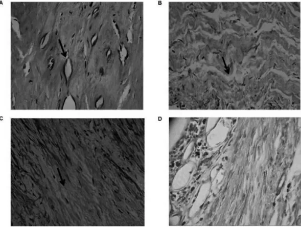
Fig. 2 - Rheumatic mitral valves present neovascularization and an increase of mononuclear iniltrates - Longitudinal cuts were done and then ixed and stained with hematoxylin and eosin (HE) and analyzed by optical microscopy (40x). A: The arrows indicate neovascularization and tissue breakdown of collagen ibers. B and C: The arrows indicate the presence of ibroblasts. D: Photomicrograph showing inlammatory iniltrate, ibrosis and some neoformed vessels.
the amplicons were present in 94.11% and 100% of the reactions, respectively, with different degrees of intensity.
All experiments were performed in duplicate to conirm
the reliability of the data obtained using this technique. In quantitative analysis, the ETr-A/GAPDH mean ratio is 33.04 ± 18.09%; while the ETr-B/GAPDH mean ratio is 114.58 ± 42.30% (Table 4). Pearson’s correlation (R) between ETr-A/GAPDH and ETr-B/GAPDH of each sample is 0.73 (P= 0.0004), which is strongly positive.
In histological analysis on mitral valves, there were
enormous amounts of ibrocytes, dense connective
tissue, type I collagen (eosinophilic) and extracellular
ground substance. The inlammatory process exhibited
high cellularity, tissue vascularization with permeating capillaries, and transformed or neoformed collagen (Figure
2). It was also possible to observe ibroblasts, lymphocytes, and dystrophic calciication areas (tissue necrosis with
deposits). Regarding histopathological individual features,
ibrosis is found in all of the 17 samples (100%), mononuclear iniltrate in 15 samples (88.23%), neovascularization in nine samples (52.94%), calciication in ten samples (58.82%) and ossiication in any sample (Table 5).
Table 4. Densitometric analysis of the amounts of the ampliied
ETr-A and ETr-B genes after normalization with the GAPDH gene (ETr-A/GAPDH and ETr-B/GAPDH ratio).
Number LAM01 LAM02 LAM03 LAM04 LAM05 LAM06 LAM07 LAM08 LAM09 LAM10 LAM11 LAM12 LAM13 LAM14 LAM15 LAM16 LAM17 Mean Std. Dev
ETr-A/GAPDH 44.94% 69.83% 18.90% 28.57% 36.06% 0.00% 24.93% 19.80% 38.89% 19.91% 61.05% 21.34% 63.41% 27.54% 32.67% 23.36% 30.47% 33.04% 18.09%
ETr-B/GAPDH 136.47% 200.24% 113.79% 107.22% 116.40% 98.45% 72.11% 59.86% 109.75% 115.43% 113.94% 83.15% 219.42%
77.93% 98.89% 80.09% 144.82% 114.58% 42.30%
Table 5. Histopathological individual features of 17 rheumatic mitral valves.
LAM01 LAM02 LAM03 LAM04 LAM05 LAM06 LAM07 LAM08 LAM09 LAM10 LAM11 LAM12 LAM13 LAM14 LAM15 LAM16 LAM17
Fibrosis x x x x x x x x x x x x x x x x x
Iniltrate
x x x 0 x x x x x x x x x 0 x x x
X represents presence of characteristic and 0 represents absence of it Neovascularization
x 0 x 0 x x x x 0 x 0 0 x 0 0 x 0
Calciication
x x 0 x 0 x 0 x x x x 0 0 0 x 0 x
Ossiication
0 0 0 0 0 0 0 0 0 0 0 0 0 0 0 0 0
DISCUSSION
Our data suggest that the expression of ET-3 has no
signiicant relation to rheumatic mitral valve disease in situ, because the ampliication of the corresponding fragment was not visible in the gel. We also visualized the ampliication
of the ETr-A and ETr-B gene fragments in almost 100% of the samples. Moura et al. [21], studying 37 mitral valves, demonstrated that endothelin receptors (ETr-A and ETr-B) were present in this type of human tissue. However there
were differences in the intensity of the ampliied bands,
which suggests that the receptors displayed different expression levels. The presence of ETr-A in the samples is interesting result but expected because in addition to being the predominant receptor in cardiac myocytes, this
receptor is involved in inlammatory processes, mitogenesis
and pathological vasoconstriction which are exuberant in rheumatic disease during the exudative and proliferative stages [16,21].
On the other hand, the presence of ETr-B in the samples in a higher proportion than ETr-A (114.58 ± 42.30% vs. 33.04 ± 18.09%) is not expected, because this receptor presents vasodilating properties, mediated by the release of nitric oxide (NO) and prostacyclin, which inhibits production of endothelin [16]. Beside vasodilator function, ETr-B, has an important role in removing excess of endothelin, being responsible for the maintenance of normal plasma levels of this peptide [22,23]. This paradoxical behavior of endothelin receptors is also found in some pathological conditions, such as chronic heart failure and myocardial ischemia [22].
Endothelin has some inluence on histopathological
features encountered in our seventeen mitral valves, such as neoangiogenesis (through the expression of ETR-A receptors that lead to increased vascular endothelial
growth factor - VEGF) and calciication (by increasing
the gene expression of osteocalcin and osteopontin)
[24,25]. Regarding ibrosis, endothelin stimulates type I
collagen production, inhibition of collagenase activity and abnormal production of extracellular matrix promoting a
reactive ibrosis [25-27]. This mechanism can be mediated for basic ibroblast growth factor (bFGF), which
up-regulates the expression of ETr-A and perhaps of ETr-B [22]. Endothelin also actives neutrophils, mast cells and stimulates monocytes to release some cytokines, such as TGF-beta and TNF-alfa [22].
The present data are preliminary but may be of great value because they may form the foundation for further investigation in this area, especially given the scarcity of studies of endothelins and its receptors in the context of rheumatic valve disease. In this study, the different patients may have different levels of intensity of expression of these receptors because they were experiencing different stages of rheumatic valve disease. Histological data are important indicators of the degree of valve involvement and suggest an interaction between neovasculogenesis and
inlammatory molecular events. The presence of receptors
REFERENCES
1. American College of Cardiology; American Heart Association Task Force on Practice Guidelines (Writing Committee to revise the 1998 guidelines for the management of patients with valvular heart disease); Society of Cardiovascular Anesthesiologists,
Bonow RO, Carabello BA, Chatterjee K, de Leon AC Jr,
Faxon DP, Freed MD, et al. ACC/AHA 2006 guidelines for the management of patients with valvular heart disease: a report of the American College of Cardiology/American Heart Association Task Force on Practice Guidelines (writing Committee to Revise the 1998 guidelines for the management of patients with valvular heart disease) developed in collaboration with the Society of Cardiovascular Anesthesiologists endorsed by the Society for Cardiovascular Angiography and Interventions and the Society of Thoracic Surgeons. J Am Coll Cardiol. 2006;48(3):e1-148.
2. Brasil. Ministério da Saúde. DATASUS: informações de saúde. Available at://www.datasus.gov.br/tabnet/tabnet.htm Accessed on: June 2010.
3. Costa LP, Domiciano DS, Pereira RMR. Características
demográicas, clínicas, laboratoriais e radiológicas da febre
reumática no Brasil: revisão sistemática. Rev Bras Reumatol. 2009;49:617-22.
4. Carapetis JR. Rheumatic heart disease in developing countries. N Engl J Med. 2007;357(5):439-41.
5. Marron K, Yacoub MH, Polak JM, Sheppard MN, Fagan D,
Whitehead BF, et al. Innervation of human atrioventricular and arterial valves. Circulation. 1996;94(3):368-75.
6. Waller BF, Howard J, Fess S. Pathology of mitral valve stenosis and pure mitral regurgitation: part II. Clin Cardiol. 1994;17(7):395-402.
7. Schmitto JD, Lee LS, Mokashi SA, Bolman RM 3rd, Cohn LH, Chen FY. Functional mitral regurgitation. Cardiol Rev. 2010;18(6):285-91.
8. Veinot JP. Pathology of inlammatory native valvular heart
disease. Card Pathol. 2006;15(5):243-51.
9. Spieker LE, Noll G, Ruschitzka FT, Lüscher TF. Endothelin receptor antagonists in congestive heart failure: a new therapeutic principle for the future? J Am Coll Cardiol. 2001;37(6):1493-505.
10. Sharma D, Singh A, Trivedi SS, Bhattacharjee J. Role of
endothelin and inlammatory cytokines in pre-eclampsia.
A pilot North Indian study. Am J Reprod Immunol. 2011;65(4):428-32.
11. Yanagisawa M, Kurihara H, Kimura S, Goto K, Masaki T.
A novel peptide vasoconstrictor, endothelin, is produced by vascular endothelium and modulates smooth muscle Ca2+ channels. J Hypertens Suppl. 1988;6(4):S188-91.
12. Schneider MP, Boesen EI, Pollock DM. Contrasting actions of endothelin ET(A) and ET(B) receptors in cardiovascular disease. Annu Rev Pharmacol Toxicol. 2007;47:731-59.
13. Rossi GP, Pitter G. Genetic variation in the endothelin system: do polymorphisms affect the therapeutic strategies? Ann N Y Acad Sci. 2006;1069:34-50.
14. Davenport AP. Endothelin converting enzyme in human tissues. Histochem J. 1998;30:359-74.
15. Kirkby NS, Hadoke PW, Bagnall AJ, Webb DJ. The endothelin
system as a therapeutic target in cardiovascular disease: great expectations or bleak house? Br J Pharmacol. 2008;153(6):1105-19.
16. Masaki T. Historical review: endothelin. Trends Pharmacol Sci. 2004;25(4):219-24.
17. Russell FD, Molenaar P. The human heart endothelin system: ET-1 synthesis, storage, release and effect. Trends Pharmacol Sci. 2000;21(9):353-9.
18. Sambrook J, Russel DW. Extraction, puriication and analysis
of mRNA from eukaryotic cells. In Sambrook J, Russel DW, eds. Molecular cloning: a laboratory manual. 3rd ed. New York: Cold Spring Harbor Laboratory Press;2001. p.610-21.
19. Yuan SM, Wang J, Hu XN, Li DM, Jing H. Transforming
growth factor-β/Smad signaling function in the aortopathies.
Rev Bras Cir Cardiovasc. 2011;26(3):393-403.
20. Yuan SM, Wang J, Huang HR, Jing H. Osteopontin expression and its possible functions in the aortic disorders and coronary artery disease. Rev Bras Cir Cardiovasc. 2011;26(2):173-82.
21. Moura EB, Gomes MR, Corso RB, Faber CN, Carneiro FP, Pacheco YG. Amplification of the genes that codify endothelin-1 and its receptors in rheumatic mitral valves. Arq Bras Cardiol. 2010;95(1):122-30.
22. Mayes, MD. Endothelin and endothelin receptor antagonists in systemic rheumatic disease. Arthritis Rheum. 2003;48(5):1190-9.
23. Chen MC, Wu CJ, Yip HK, Chang HW, Chen CJ, Yu TH, et al.
Increased circulating endothelin-1 in rheumatic mitral stenosis: irrelevance to left atrial and pulmonary artery pressures. Chest. 2004;125(2):390-6.
24. Shimojo N, Jesmin S, Zaedi S, Otsuki T, Maeda S, Yamaguchi N, et al. Contributory role of VEGF overexpression in endothelin-1-induced cardiomyocyte hypertrophy. Am J Physiol Heart Circ Physiol. 2007;293(1):H474-81.
25. Wu SY, Zhang BH, Pan CS, Jiang HF, Pang YZ, Tang CS, et al. Endothelin-1 is a potent regulator in vivo in vascular
calciication and in vitro in calciication of vascular smooth
muscle cells. Peptides. 2003;24(8):1149-56.
26. Brás-Silva C, Leite-Moreira AF. Efeitos miocárdicos da endotelina-1. Rev Port Cardiol. 2008;27(7-8):925-51.