RBCCV 44205-1430 DOI: 10.5935/1678-9741.20120105
Sanguineous normothermic, intermittent
cardioplegia, effects on hypertrophic
myocardium. Morphometric, metabolic and
ultrastructural studies in rabbits hearts
Efeitos da cardioplegia sanguínea normotérmica intermitente, em miocárdio hipertróico. Estudos
morfométricos, metabólicos e ultraestruturais em corações coelhos
Clovis Carbone Junior
1, José Eduardo de Salles Roselino
2, Valder Rodrigues Mello
3, Paulo Roberto
Barbosa Evora
4, Albert Amin Sader
51. MD. PhD Department of Surgery and Anatomy, Ribeirão Preto Faculty of Medicine, University of São Paulo - Experimental execution, data evaluation and text review; Ribeirão Preto, SP, Brazil.
2. MD, PhD Department of Biochemistry and Immunology, Ribeirão Preto Faculty of Medicine, University of São Paulo - Data evaluation and review; Ribeirão Preto, SP, Brazil.
3. BsC, PhD Department of Surgery and Anatomy, Ribeirão Preto Faculty of Medicine, University of São Paulo - Data evaluation and review; Ribeirão Preto, SP, Brazil.
4. Full Professor Department of Surgery and Anatomy, Ribeirão Preto Faculty of Medicine, University of São Paulo (Docente da Divisão de Cirurgia Torácica e Cardiovascular) - Paper review data and writing; Ribeirão Preto, SP, Brazil.
5. Full Professor Department of Surgery and Anatomy, Ribeirão Preto
Faculty of Medicine, University of São Paulo - Study design, data evaluation and text review; Ribeirão Preto, SP, Brazil.
Work carried out at Ribeirão Preto Faculty of Medicine, University of São Paulo, Ribeirão Preto/São Paulo, Brazil.
Correspondence address: Paulo Roberto Barbosa Evora
Rua Barbosa, 367 – apt. 15 – Centro – Ribeirão Preto, SP Brasil – CEP 14015-120
E-mail: [email protected]
Article received on September 5th, 2012
Article accepted on October 1st, 2012
Abstract
Objectives: The present investigation aimed to study the protective effect of intermittent normothermic cardioplegia in rabbit's hypertrophic hearts.
Methods: The parameters chosen were 1) the ratio heart weight / body weight, 2) the myocardial glycogen levels, 3) ultrastructural changes of light and electron microscopy, and 4) mitochondrial respiration.
Results: 1) The experimental model, coarctation of the aorta induced left ventricular hypertrophy; 2) the temporal evolution of the glycogen levels in hypertrophic myocardium
demonstrates that there is a signiicant decrease; 3) It was
observed a time-dependent trend of higher oxygen consumption
values in the hypertrophic group; 4) there was a signiicant time-dependent decrease in the respiratory coeficient rate in
the hypertrophic group; 5) the stoichiometries values of the ADP: O2 revealed the downward trend of the values of the
hypertrophic group; 6) It was possible to observe damaged
mitochondria from hypertrophic myocardium emphasizing the large heterogeneity of data.
Conclusion: The acquisition of biochemical data, especially the increase in speed of glycogen breakdown, when anatomical changes are not detected, represents an important
result even when considering all the dificulties inherent in
the process of translating experimental results into clinical practice. With regard to the adopted methods, it is clear
that morphometric methods are less speciic. Otherwise,
the biochemical data allow detecting alterations of glycogen concentrations and mitochondria respiration before the morphometric alterations should be detected
Resumo
Objetivos: O presente estudo teve como objetivo estudar o efeito protetor da cardioplegia normotérmica intermitente
em corações hipertróicos de coelhos.
Métodos: Os parâmetros escolhidos foram: 1) relação peso cardíaco/peso corporal; 2) níveis de glicogênio nos músculos cardíacos; 3) alterações ultraestruturais por microscopia óptica e eletrônica; e 4) respiração mitocondrial.
Resultados: 1) O modelo experimental de coarctação
da aorta induziu hipertroia ventricular esquerda; 2) a
evolução temporal dos níveis de glicogênio no miocárdio
hipertróico demonstra que há diminuição signiicativa;
3) observou-se tendência dependente do tempo para
maiores valores do consumo de oxigênio para o grupo
hipertróico; 4) houve diminuição dependente do tempo da taxa de coeiciente respiratório no grupo hipertróico; 5) os
valores estequiométricos da ADP: O2 revelou a tendência
decrescente no grupo hipertróico; 6) observaram-se lesões mitocondriais do miocárdio hipertróico, enfatizando a
grande heterogeneidade dos dados.
Conclusão: A aquisição de dados bioquímicos, principalmente o aumento na velocidade de quebra do glicogênio, quando mudanças anatômicas não são detectadas, representa um resultado importante, mesmo quando se
consideram todas as diiculdades inerentes ao processo
translacional de resultados experimentais para a prática clínica. No que diz respeito aos métodos adotados, é evidente
que os métodos morfométricos são menos especíicos. Os
dados bioquímicos permitem a detecção de alterações das concentrações de glicogênio e respiração mitocondrial antes das alterações morfométricas serem detectadas.
Descritores: Parada cardíaca induzida. Hipertroia
ventricular esquerda. Procedimentos cirúrgicos cardiovasculares. Abbreviations, Acronyms & Symbols
ADP Adenosine diphosphate
LVW/BW Left ventricular and body weight ratio
RCR Respiratory control ratio
INTRODUCTION
The current success of cardiac surgery was provided, among other relevant factors, by better understanding the myocardium protection. The timeline of this understanding includes: 1) systemic hypothermia [1]; 2) intra-operative cardiac arrest induced by potassium citrate [2]; 3) anoxic arrest of the heart [3]; 4) the use of chemicals that could rapidly determine cardiac arrest [4]; 5) Miscellaneous studies relating to the composition, temperature and mode of drug administration named cardioplegic solution [5-7], and; 6) normothermic cardioplegia [8,9].
In cardiac hypertrophy, whether it is induced in an experimental model or observed clinically, energy metabolism is compromised highlighting that Sink et al. [10] demonstrated that the hypertrophic heart must be stopped immediately emphasizing the need of additives to the cardioplegia solution. It is noteworthy that Cooley et al., in 1972, described the "Stone Heart", i.e., ischemic contracture, more frequent and severe in the hypertrophic heart [11]. This ischemia/reperfusion phenomenon allowed
the resumption of the discussion, and inal conclusion,
about the pivotal role of myocardial protection as a means of avoiding this extremely serious intraoperative complication. Therefore, even after more than 60 years, continued studies of the hypertrophic myocardium energy metabolism under the action of cardioplegia are still necessary to improve surgical procedures in this pathological condition.
Most of experimental researches on cardioplegia were
made in hearts of normal animals. Thus, speciically, the
present investigation aimed to study the protective effect of intermittent normothermic cardioplegia in rabbits hypertrophic hearts. The parameters chosen were 1) the ratio heart weight/body weight to evaluate the myocardium hypertrophy, 2) the heart muscle glycogen levels, 3) ultrastructural changes by light and electron microscopy, and 4) mitochondrial respiration. As a secondary objective, the study aimed to evaluate the adequacy of the adopted methods.
METHODS
Experimental design
New Zeland rabbits (n=76; 1.7 - 2.5 kg) were anesthetized using pentobarbital sodium (30 mg/kg intravenous). The animals underwent tracheostomy, and they were ventilated using an endotracheal tube (3.0 mm,
Rusch, Telelex Medical, Durham, NC, USA) with 100%
O2 in a pressure-controlled mode (Takaoka 600, K. Takaoka Indústria e Comércio Ltda, São Bernardo do Campo, SP, Brazil). The ear marginal vein was cannulated for volemic
reposition with saline solution (NaCl 0.9%, 10 ml/kg/h).
The Institutional Animal Care and Use Committee of the Ribeirão Preto Faculty of Medicine, University of São Paulo, Brazil reviewed and approved the procedures for animal handling, which were in accordance with the Guide for the Care and Use of Laboratory Animals published by the U.S. National Institutes of Health (NIH Publication No. 85-23, revised 1996).
normal (n=36) and Group II/hypertrophy (n=40). The number of rabbits was based on literature data, mainly involving mitochondria respiration. Also, rabbits were elected as experimental animal based on those studies. The samples were collected immediately, 60 and 90 minutes after cardioplegia infusion.
Cardiac hypertrophy was induced by coarctation of the abdominal aorta just distal to the diaphragm, according the technique of Leclercq et al. [12], using a midline laparotomy as access. The aorta was dissected and looped with cotton thread 00 (two zeros), the stenosis of the artery
was calibrated with a 2 mm needle (50% stenosis), which
was removedafter tying the artery. The wall plans were sutured, and the waiting time for the induction of cardiac hypertrophy was 14 days.
On the morning of the fourteenth postoperative day, under anesthesia, all animals were submitted to median
sternotomy, identiication and fast excision of the heart
after sectioning of its vessels (superior and inferior vena cava, aorta artery and pulmonary artery and vein). The whole heart was immediately immersed in a beaker
containing 0.9% saline solution and, ice cooler stored
to keep the temperature between 0 and 3oC. The hearts were subjected to cardiac arrest by normothermic blood
cardioplegia solution (37°C), composed of 40% blood and 60% solution of sodium chloride 0.9% to 50 mEq/L of
potassium chloride to induce cardiac arrest and 30 mEq/L for maintenance doses. After cardioplegic arrest, the hearts were subjected to a period of myocardial ischemia of 60 and 90 minutes, kept in a water bath at 37°C, using multidose intermittent antegrade blood cardioplegia every 20 minutes, the proportion of 5 ml/kg body weight. The group with zero time of ischemia was used as the control. The atria were extracted, and the ventricles were cut into small fragments of about 2 mm. During this procedure,
repeated washings were done with 0.9% saline solution at
a temperature of 0 to 3oC. For each time was carried out biochemical determination of the following parameters: analysis of mitochondrial oxygen consumption and measurement of the glycogen concentration.
Mitochondrial function
The mitochondrial fraction was isolated by the method of Bullock et al. [13] Mitochondrial function was determined by a polarographic method [14], using an OXY 5 polarograph-oxygraph (Gilson Medical Eletronics, Inc., W. Beltline Middeton, WI, USA). The respiration medium contained 0.23 M sucrose, 8 mM potassium phosphate, 9.5 mMTris, pH 7.0, 0.14 mg/ml albumin, and 1 mM EDTA. The mitochondrial fraction was assayed at 1 2 mg/ml protein concentration in the oxygraph chamber. Substrates were added as a mixture of malate, pyruvate, keto-glutarate, and b-hydroxy-butyrate, each at 48 mM
inal concentration. State III (activated respiration) was
obtained after the addition of 400 nmoles of adenosine diphosphate (ADP). State IV (basal respiration) was measured when all ADP had been converted to ATP, a condition indicated by the return to basal respiratory levels. The ratio of respiration rate after the addition of ADP (state III) to respiration rate during the basal state (state IV) corresponded to the respiratory control ratio (RCR). The parameters of oxidative phosphorylation were calculated according to Chance and Williams [15] and Estabrook and Pullman [16] and were expressed in nanoatoms of oxygen used per mg protein per minute. Mitochondrial protein content was determined by the biuret method [17].
Measurements of myocardial glycogen
Glycogen was extracted with 30% KOH from 500
mg of the left ventricle myocardium. After centrifugation at 800g for 10 min, 1 mL of supernatant solution was transferred to a tube incubated on ice and mixed with 2 mL
luid with anthrone. The mixture was boiled for 10 min and
then cooled immediately on ice, followed by incubation at room temperature for 10 min. The absorbance was read at 620 nm by a spectrophotometer and, the values were
expressed in % of humid weigh [18].
Morphologic study
A cross-sectional slice of the left ventricle chamber was obtained after the cardioplegic arrest was immersed
and ixed in Bouin solution for 24 hours, dehydrated in
ethanol and included in Paraplast. Cuts (5 µm) were stained with hematoxylin-eosin, Mallory’s trichrome and PicroSirius Red with the aid of a digitizing tablet (MINI-MOP - Kontron Elektronics). Measurements were taken directly on the blades and the values are expressed in
millimeters (inal images 800 Xs).
For transmission electron microscopy, a group of blocks was selected and processed for ultrathin sections. The areas of cytoplasm, where mitochondria predominated, were photographed under a microscope Philips EM 208 (Transmission Electron Microscopy) at original
magniication of 20.000X. The negatives were double enlargedand copied yielding the inal images at 40.000X. These ampliications were analyzed for the determination
of mitochondrial diameters (maximum and minimum) through the use of the tablet (MINI-MOP - Kontron Elektronics), and the results were expressed in micrometers
as the correction was made for the magniication factor.
Statistical analysis
For statistical analysis of the body weight and to assess the ventricular induction of myocardial hypertrophy was used the t-test to compare values with similar and different
For the biochemical parameters (glycogen and mitochondrial respiration), statistical comparisons were carried out between control and hypertrophic myocardium groups, and among times of ischemia. Initially, tests were
normal (Shapiro and Wilk) not inding suitable conditions
for the application of parametric statistics. It was decided then to apply the Kruskal-Wallis test on each group separately (normal versus hypertrophic) in order to study the effect of ischemia time on biochemical variables,
adopting the signiicance level of 5%.
The Mann-Whitney test was carried out in each subgroup of time (0, 60 and 90). For multiple comparisons among the subgroups of time (0, 60 and 90), for each biochemical variable mentioned above, it was used the method of Dunn.
RESULTS
Induction of experimental cardiac hypertrophy
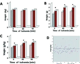
In Figure 1A, are represented the values of body weight of all animals studied during cardioplegic arrest times of 0, 60 and 90 min. It is observed a similarity between Groups
I and II (signiicance of 5%).
In Figure 1B are represented the values of ventricular myocardium weight with the statistical t-test showing
a signiicant increase (signiicance of 5%) in the
hypertrophied heart at all ischemic times.
Figure 1C shows the values of the left ventricular and body weight ratio (LVW/BW in g/kg) with the statistical
t-test showing a signiicant increase (signiicance of 5%)
in the hypertrophied heart at all ischemic times.
Figure 1D demonstrates the dispersion of the ratio values of ventricular weight/body weight in normal and hypertrophic groups, but corresponding to 0 min ischemia. This analysis aims to characterize the eventual presence of cardiac hypertrophy in the control group, when it is supposed that the intracellular edema factor was absent,
making sure that the values relected the myocardium
hypertrophy in this study group.
Glycogen and mitochondrial function
Figure 2 represents the glycogen levels found in normal and hypertrophic myocardium of rabbits when subjected to infusion of normothermic blood cardioplegia. Statistical analysis was performed between samples; the Mann-Whitney analysis of normal and hypertrophic
group revealed no signiicant differences (signiicance of 5%). However, the Kruskal-Wallis test that analyzes the
temporal evolution of the glycogen levels in hypertrophic
myocardium demonstrates that there is a signiicant decrease (signiicance of 5%) at 90 minutes compared to
time 0 (zero), whereas normal myocardium did not differ
in any time (signiicance of 5%).
Fig. 1 - A - body weight (kg), B – left ventricle weight (g), C - left ventricle weight LVW/body weight (BW) ratio (g/kg); D – values of the ratio LVW/BW (g/kg) characterizing the data dispersion. Normal myocardium (column textured) and hypertrophic (full column) underwent infusion of cardioplegia at ischemia times of 0 min, 60 min and 90 min. The columns represent the mean values and the bars above the columns the standard error of the mean. In the igure D, values in the x-axis correspond to increasing numbers of samples and the line which bisects the graph corresponds to the value LVW/BW of 2.3 g/kg. The asterisk means signiicant difference between groups, with P <0.05
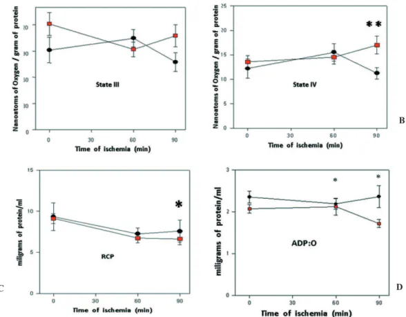
Figure 3A represents the values of the state III mitochondrial oxygen consumption, demonstrating the effect of ischemia time on the group of normal and hypertrophic myocardium. The analysis between groups by Mann-Whitney, at 90 min, showed a trend of higher values for the hypertrophic group. The Kruskal-Wallis test found no difference among samples
obtained at different times (signiicance of 5%).
Figure 3B represents the values of the state IV mitochondrial oxygen consumption, at times 0, 60 and 90 min, found in normal and hypertrophic myocardium after infusion of normothermic cardioplegic solution. The
Mann-Whitney test shows a signiicant increase in values at 90 min (signiicance of 5%) of the hypertrophic group when
compared to normal. The Kruskal-Wallis test did not show
differences among the results (signiicance of 5%).
The values of the respiratory control ratio (RCR) observed
in Figure 3C, were measured in normal and hypertrophic myocardium after infusion of cardioplegic solution at 0, 60
and 90 min. The Mann-Whitney test found no signiicant differences between groups (signiicance of 5%). However,
the analysis between the different times observed by the
Kruskal-Wallis test, showed a signiicant decrease in the RCR (signiicance of 5%), in the hypertrophic group, at 90
min of ischemia when compared with time zero.
Figure 3D represents the stoichiometric values of the ADP:O2 found in normal and hypertrophic myocardium after normothermic cardioplegic arrest. The Mann-Whitney test revealed a downward trend of the values of the hypertrophic group when compared with normal myocardium. For the hypertrophic group, the Kruskal-Wallis test showed differences between them with a
signiicant decrease (P <0.05) values for ADP:O2.
Fig. 3 - Analysis of mitochondrial respiration: A - State III, B - State IV, C - Respiratory Control Ratio (RCR), and D - mitochondrial oxygen consumption (ADP:O2). Comparison between normal hearts (black circles) and hypertrophic hearts (red squares) of rabbits submitted to the infusion of normothermic blood cardioplegic solution with cardiac arrest of 60 and 90 minutes and the control group. The mitochondria (1 mg protein/ml) were tested at 30°C using Alpha-ketoglutarate as respiratory substrates. Breathing was activated with 400 nanomoles of ADP. Values are expressed in oxygen nanoatoms O2/mg protein.min. The symbols represent the average values with the corresponding standard deviation. Group normal heart (control n = 12, n = 60 min n=13, 90 min n = 12); Group hypertrophic heart (control/n = 16; 60 min/n=12; 90 min/n = 12). A – No signiicant differences; B - The asterisks (**) refer to the signiicant increase (P <0.05) in the hypertrophic group compared with the normal at 90 min, C - The asterisk refers to a signiicant difference (P <0.05) between the time 0 and 90 min of the hypertrophic group, D - the asterisk refers to a signiicant difference (P <0.05) between 60 and 90 min and 0 and 90 min of the hypertrophic group
A B
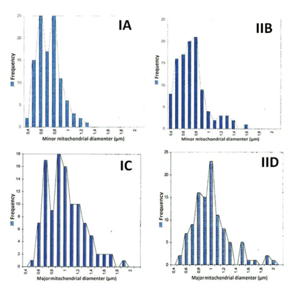
The graphs of Figure 4 represent the minor (A and B) and major (C and D) mitochondrial diameters analyzed by electron microscopy, increased 40.000x. One can observe a large heterogeneity of data, when divided by its frequency. Comparing data from Groups I and II and their respective subgroups of cardioplegic arrest in time zero, there was a higher trend in Group II of increasing the mitochondrial diameter. The data of minor mitochondrial diameters are most similar in its distribution. However, in Group II and its respective subgroup of 60 min of ischemia, there was a marked increase in both mitochondrial diameters, demonstrating intracellular edema with cardioplegic arrest.
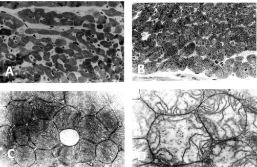
Figure 5 presents light (A, B) and electron (B, C) micrographs of transverse sections of the myocardium
of rabbits submitted to cardioplegic cardiac arrest. A - micrograph (block No. 11647b) belonging to the
subgroup 0 minutes in Group I, shows myocardial ibers
involving the interstitium where it is observed blood vessels and collagen (800X); B - micrograph (block No. 11597g) belonging to 0 minutes subgroup in Group II
reveals a signiicant increase in the transverse diameter of myocardial ibers occupying the space of interstitium
(800X); C - micrograph (block No 11647h) belonging to the subgroup 0 minutes in group I reveal various aspects of normal mitochondria (40.000X), and; D - Micrograph (block No 11692f) belonging to the subgroup 0 minutes in Group II shows mitochondria with increased diameter and loss of their morphological characteristics (40.000X).
Fig. 5 - Light (A, B) and electron (C, D) micrographs of transverse sections of the myocardium of rabbits submitted to cardioplegic cardiac arrest. A – micrograph of a specimen belonging to the subgroup 0 minutes in Group I, shows myocardial ibers sectioned transversely,and in the involved interstitium is observed blood vessels and collagen (800X); B – micrograph of a specimen belonging to 0 minutes subgroup in Group II reveals a signiicant increase in the transverse diameter of myocardial ibers occupying the space of interstitium (800X), C - micrograph belonging to the subgroup 0 minutes in group I reveals various aspects of normal mitochondria (40.000X), D - Micrograph belonging to the subgroup 0 minutes in Group II shows mitochondria with increased diameter and loss of their morphological characteristics (40.000X)
DISCUSSION
Myocardial hypertrophy is considered the most
eficient event, among the compensatory mechanisms of
heart diseases, when the muscle is exposed to overload depending on extramyocardial disease. Several mechanical and neurohormonal factors act as myocardial growth factors and change the pattern of protein synthesis, resulting in a ventricular remodeling. The several mechanisms in response to the decrease of cardiac performance, initially adaptive, became developmentally pernicious [19].
The basic data of the present investigation were: 1) the experimental model, coarctation of the aorta induced left ventricular hypertrophy; 2) the temporal evolution of the glycogen levels in hypertrophic myocardium demonstrates
that there is a signiicant decrease; 3) It was observed a
time-dependent trend of higher oxygen consumption
values for the hypertrophic group; 3) there was a signiicant time-dependent decrease in the respiratory coeficient rate
in the hypertrophic group; 4) the stoichiometric values of the ADP: O2 revealed the downward trend of the values of the hypertrophic group; 5) It was possible to observe damaged mitochondria from hypertrophic myocardium emphasizing the large heterogeneity of data.
Experimental model
Concerning the experimental model, coarctation of
the aorta induced left ventricular hypertrophy. Indeed, there has been a bandage of the aorta in order to cause a pressure overload in the proximal portion. The sustained high pressure trigger a complex process whose ultimate expression was the increased thickness of the myocardium without enlargement of the ventricular cavity as a consequence of adaptive morphological response [20]. The
data, conirming the appropriateness of the methodology are shown in Figure 1A markedly and signiicant weight of
the left ventricle of rabbits in the experimental hypertrophy
protocol can be observed. To rule out the possible inluence
and 3.9 for obvious failure. In this initial understanding of myocardial hypertrophy, it is reasonable to distinguish two types of cardiac hypertrophy: a physiological normal or increased contractility, and, a pathologically reduced contractile function [22].
Glycogen metabolism
Considering hypertrophy as a disease process, not just a physiological response, and its risk factor role for clinical complications commonly present in cardiac surgery, the biochemical approach of myocardial protection is mandatory. Thus, the present study in rabbits used intermittent antegrade blood cardioplegia which, admittedly, provides protection to the myocardium reduces the energy expenditure and decreases the rate of glycogenolysis. The normothermia experimental option experiment was adopted, based on studies performed in our laboratory showing a larger decrease glycogen levels in normothermic condition, compared with hypothermia [23]. In the present study, there was no difference in glycogen concentration (time zero x 90 minutes) within the control group samples. Surely, this observation is
due to the inluence of the variability of sample values
in an experimental situation in which there was an increase of glycogen breakdown. However, the decrease in the concentrations of the samples occurred at lower speed when compared to the drop rate observed in hypertrophic hearts. The results of the muscle glycogen content showing high variability are already referred in
the scientiic literature [24]. The statistical analysis did
not establish a difference in the levels of myocardium glycogen after ninety minutes of ischemia. The variability factor, as already mentioned in the preceding paragraph, must have contributed to the lack of difference. One data to be highlighted in this research is that it was possible
to observe signiicant differences among the values of
muscle glycogen in hypertrophic hearts, considering the values obtained after ninety minutes of cardioplegia infusions. As simplest interpretation of these results, one should consider that glycogenolysis is established more rapidly in the muscle subjected to conditions which favor hypertrophy. In order to complete the conceptualization of the study, it is necessary to emphasize that metabolic adaptation to anaerobic condition is made by the degree of the glycogenolytic pathway activation. This may explain the differences among positive results observed in normal hearts, and negative results observed in hearts subjected to conditions that promote hypertrophy.
Mitochondrial respiration
The irst detail to be discussed concerning the
preparation of mitochondria is the control samples that presented RCR close to 10, which is a valuable indicator
of the quality. The values of the state III mitochondrial oxygen consumption, which denotes the effect of ischemia time, showed at 90 min, a trend of higher values for the hypertrophic group, but, analyzing the temporal evolution it was not observed differences among the results. The
values of the state IV showed a signiicant increase of
the hypertrophic group when compared to normal, at 90 minutes, without differences in the temporal evolution. The
RCR did not show signiicant differences between normal
and hypertrophic group. However, the analysis of the
evolution time line showed signiicant RCR decrease in the
hypertrophic group, at 90 min of ischemia, when compared with time zero. The stoichiometric values of the ADP: O2 found in the normal and hypertrophic myocardium after normothermic cardioplegic arrest revealed the downward trend of the values of the hypertrophic group with the time line statistical analysis revealing decreased values. Observing the respiratory values, all together, the only possible conclusion is that the hypertrophic myocardium is more susceptible to changes elicited by prolonged ischemia.
Morphometric analysis
Electron microscopy revealed many aspects of normal
myocardial ibers, while the hypertrophic hearts showed signiicantly increased transverse diameters occupying
the interstitial space. The same occurred in relation to the diameters of the mitochondria showed an increase in their diameters and loss of their morphological characteristics (Figures 4 and 5). However, onecan observe the large heterogeneity of data, when were divided by frequency.
Demonstrating intracellular edema with cardioplegic arrest, in a study of ultrastructural analysis of hypertrophic rabbits myocardium, Goldstein et al. [25] showed a range from 7 to 142 days for clinically manifestations
be conirmed anatomically. Furthermore, these authors
reported that the pleomorphism variations make it
dificult, or almost impossible, to estimate the number or
size of normal mitochondria. Perhaps this fact explains the
dificulty in inding clearest results in relation to the values
of mitochondrial diameter during the period studied. However, the trend of population shift to higher values is clearly shown in frequency histograms.
Concluding remarks
The acquisition of biochemical data, especially the increase in speed of glycogen breakdown, when anatomical changes are not detected, represents an
important result even when considering all the dificulties
expertise, always seeking for the best. At last, regarding hypertrophied heart protection, experimental studies have shown the superiority of tepid blood cardioplegia in relation to hypothermic crystalloid solution. Cardioplegic strategies to protect the hypertrophic heart during cardiac surgery is, surely, the most controversial subject concerning cardioprotection and an eternal challenge since all tested cardioplegic techniques confer suboptimal myocardial protection. Therefore, we should always have in mind that the state-of-the-art was not achieved yet [26-30].
Limitations of the study
The investigation showed that normothermic sanguineous, intermittent cardioplegia protects the hypertrophic myocardium against the deleterious effects of ischemia followed by reperfusion. But, no comparative studies with other techniques of myocardial protection. Therefore, any attempt to clinical correlation would be speculative.
As the induction of ventricular hypertrophy was performed by an aortic coarctation for 14 days, it is likely to consider the physiological type because this is an adaptation to a pressure overload. This doubt about the type of hypertrophy can be considered a possible limitation of the study. At least two well-conducted studies by Brazilian authors proved the adequacy of myocardial hypertrophy induced by the technique of aortic coarctation [31,32].
REFERENCES
1. Bigelow WG, Lindsay WK, Greenwood WF. Hypothermia; its possible role in cardiac surgery: an investigation of factors governing survival in dogs at low body temperatures. Ann Surg. 1950;132(5):849-66.
2. Melrose DG, Dreyer B, Bentall HH, Baker JB. Elective cardiac arrest. Lancet. 1955;269(6879):21-2.
3. Cooley DA, Beall AC Jr, Grondin P. Open-heart operations with disposable oxygenators, 5 per cent dextrose prime, and normothermia. Surgery. 1962;52:713-9.
4. Kirsch U, Rodewald G, Kalmár P. Induced ischemic arrest. Clinical experience with cardioplegia in open-heart surgery. J Thorac Cardiovasc Surg. 1972;63(1):121-30.
5. Gay WA Jr, Ebert PA. Functional, metabolic, and morphologic effects of potassium-induced cardioplegia. Surgery. 1973;74(2):284-90.
6. Tyers GF, Hughes HC Jr, Todd GJ, Williams DR, Andrews EJ, Prophet GA, et al. Protection from ischemic cardiac arrest by coronary perfusion with cold Ringer’s lactate solution. J Thorac Cardiovasc Surg. 1974;67(3):411-8.
7. Bretschneider HJ, Hübner G, Knoll D, Lohr B, Nordbeck H, Spieckermann PG. Myocardial resistance and tolerance to ischemia: physiological and biochemical basis. J Cardiovasc Surg (Torino). 1975;16(3):241-60.
8. Lichtenstein SV, el Dalati H, Panos A, Slutsky AS. Long cross-clamp time with warm heart surgery. Lancet. 1989;1(8652):1443.
9. Lichtenstein SV, Ashe KA, el Dalati H, Cusimano RJ, Panos A, Slutsky AS. Warm heart surgery. J Thorac Cardiovasc Surg. 1991;101(2):269-74.
10. Sink JD, Pellom GL, Currie WD, Hill RC, Olsen CO, Jones RN, et al. Response of hypertrophied myocardium to ischemia: correlation with biochemical and physiological parameters. J Thorac Cardiovasc Surg. 1981;81(6):865-72.
11. Cooley DA, Reul GJ, Wukasch DC. Ischemic contracture of the heart: "Stone heart". Am J Cardiol. 1972;29(4):575-7.
12. Leclercq JF, Sebag C, Swynghedauw B. Experimental cardiac hypertrophy in rabbits after aortic stenosis or incompetence or both. Biomedicine. 1978;28(3):180-4.
13. Bullock G, Carter EE, White AM. The preparation of mitochondria from muscle without the use of a homogeniser. FEBS Lett. 1970;8(2):109-11.
14. Sordahl LA, Besch HR Jr, Allen JC, Crow C, Lindenmayer GE, Schwartz A. Enzymatic aspects of the cardiac muscle cell: mitochondria, sarcoplasmic reticulum and nonovalent cation active transport system. Methods Achiev Exp Pathol. 1971;5:287-346.
15. Chance B, Williams GR. The respiratory chain and oxidative phosphorylation. Adv Enzymol Relat Subj Biochem. 1956;17:65-134.
16. Estabrook RW, Pullman ME. Oxidations and phosphorylations. New York:Academic Press;1967. p.7-8.
17. Lowry OH, Rosebrough NJ, Farr AL, Randall RJ. Protein measurement with the Folin phenol reagent. J Biol Chem. 1951;193(1):265-75.
18. Colowick SP, Kaplan NO. Methods in enzymology. New York:Academic Press;1957. p.34.
19. Levy D, Garrison RJ, Savage DD, Kannel WB, Castelli WP. Prognostic implications of echocardiographically determined left ventricular mass in the Framingham Heart Study. N Engl J Med. 1990;322(22):1561-6.
20. Grossman W, Barry WH. Diastolic pressure-volume relations in the diseased heart. Fed Proc. 1980;39(2):148-55.
muscle in aortic insuficiency in the rabbit. J Mol Cell Cardiol.
1970;1(3):235-47.
22. Wikman-Coffelt J, Parmley WW, Mason DT. The cardiac hypertrophy process. Analyses of factors determining pathological vs. physiological development. Circ Res. 1979;45(6):697-707.
23. Carbone Jr C; Roselino JES, Carneiro JJ, Sader AA. Análise comparativa da reserva de glicogénio do miocardio isquémico de coelhos submetidos a cardioplegia hipotérmica ou normotérmica. Rev Bras Cir Cardiovasc. 1992;7(1):9-13.
24. Steenbergen C, Perlman ME, London RE, Murphy E. Mechanism of preconditioning. Ionic alterations. Circ Res. 1993;72(1):112-25.
25. Goldstein MA, Sordahl LA, Schwartz A. Ultrastructural analysis of left ventricular hypertrophy in rabbits. J Mol Cell Cardiol. 1974;6(3):265-73.
26. Gomes WJ, Ascione R, Suleiman MS, Bryan AJ, Angelini GD. Efeitos das cardioplegias sanguíneas hipotérmica e normotérmica nos substratos intracelulares em pacientes com corações hipertróficos. Rev Bras Cir Cardiovasc. 2000;15(2):160-8.
27. Ascione R, Caputo M, Gomes WJ, Lotto AA, Bryan AJ,
Angelini GD, et al. Myocardial injury in hypertrophic hearts of patients undergoing aortic valve surgery using cold or warm blood cardioplegia. Eur J Cardiothorac Surg. 2002;21(3):440-6.
28. Cressoni ES, Avanci LE, Braile DM, Cicogna AC, Lima-Oliveira AP, Gerez MA, et al. Myocardial protection to the hypertrophied heart: the eternal challenge. Rev Bras Cir Cardiovasc. 2008;23(1):97-107.
29. Cressoni ES, Avanci LE, Braile DM, Lima-Oliveira AP, Taboga SR, Martins AS, et al. Effects of myocardial protection in hypertrophic rabbit hearts: structural and ultra structural analysis. Rev Bras Cir Cardiovasc. 2007;22(1):24-32.
30. Suleiman MS, Hancock M, Shukla R, Rajakaruna C, Angelini GD. Cardioplegic strategies to protect the hypertrophic heart during cardiac surgery. Perfusion. 2011;26(Suppl 1):48-56.
31. Martins AS, Aguilera NW, Matsubara BB, Bregagnollo EA. Experimental myocardial hypertrophy induced by a minimally invasive ascending aorta coarctation. Braz J Med Biol Res. 2001;34(3):413-5.
32. Souza Vilarinho KA, Petrucci O, Baker RS, Vassallo J, Schenka AA, Duffy JY, et al. Early changes in contractility indices and
ibrosis in two minimally invasive congestive heart failure