RBCCV 44205-1582 DOI: 10.5935/1678-9741.20140075
Comparison of two surgical techniques for creating
an acute myocardial infarct in rats
Comparação de duas técnicas cirúrgicas para criar um infarto agudo do miocárdio em ratos
Luiz Guilherme Achcar Capriglione
1, MsC; Fabiane Barchiki
1, MsC; Gabriel Sales Ottoboni
1, MsC;
Nelson Itiro Miyague
1, PhD; Paula Hansen Suss
1, PhD; Carmen Lúcia Kuniyoshi Rebelatto
1, PhD;
Cláudia Turra Pimpão
1, PhD; Alexandra Cristina Senegaglia
1, PhD; Paulo Roberto Brofman
1, PhD
1Pontifícia Universidade Católica do Paraná (PUCPR), Curitiba, PR, Brazil.
This study was carried out at Pontifícia Universidade Católica do Paraná (PUCPR), Curitiba, PR, Brazil.
Financial support from Conselho Nacional de Desenvolvimento Cientíico e Tecnológico-CNPq
Correspondence address: Paulo Roberto Brofman
Pontifícia Universidade Católica do Paraná
Rua Imaculada Conceição, 1155 - Prado Velho, Curitiba, PR Brazil - Zip code:80215-901
E-mail: [email protected]
Article received on October 31th, 2013 Article accepted on May 26th, 2014 Abstract
Objective: To perform a comparative assessment of two surgical techniques that are used creating an acute myocardial infarc by occluding the left anterior descending coronary artery in order to generate rats with a left ventricular ejection fraction of less than 40%.
Methods: The study was completely randomized and com-prised 89 halothane-anaesthetised rats, which were divided into three groups. The control group (SHAM) comprised fourteen rats, whose left anterior descending coronary artery was not oc-cluded. Group 1 (G1): comprised by 35 endotracheally intubated and mechanically ventilated rats, whose left anterior descending coronary artery was occluded. Group 2 (G2): comprised 40 rats being manually ventilated using a nasal respirator whose left anterior descending coronary artery was occluded. Other differences between the two techniques include the method of performing the thoracotomy and removing the pericardium in order to expose the heart, and the use of different methods and suture types for closing the thorax. Seven days after surgery, the cardiac function of all surviving rats was determined by echocardiography.
Results: No rats SHAM group had progressed to death or had left ventricular ejection fraction less than 40%. Nine of the
16 surviving G1 rats (56.3%) and six of the 20 surviving G2 rats (30%) had a left ventricular ejection fraction of less than 40%.
Conclusion: The results indicate a tendency of the technique used in G1 to be better than in G2. This improvement is probably due to the greater duration of the open thorax, which reduces the pressure over time from the surgeon, allowing occlusion of left anterior descending coronary artery with higher accuracy.
Descriptors: Myocardial infarction. Echocardiography. Mod-els, Cardiovascular.
Resumo
Objetivo: Realizar uma avaliação comparativa de duas téc-nicas cirúrgicas que são usadas para criar um infarto agudo do miocárdio pela oclusão da artéria coronária descendente anterior esquerda, a im de gerar ratos com uma fração de ejeção ventri -cular esquerda inferior a 40%.
left ventricular ejection fraction (LVEF) of less than 40%. The consequent on cardiac function in these rats was assessed using echocardiography.
METHODS
Animals
This animal study and the procedures detailed herein were reviewed and approved by the Local Ethics Committee on Animal Research (Identiication numbers: PUCPR 180 and 540). The study comprised 89 male 100-day-old albino Wistar rats (Rattus norvegicus) (mean weight 348.6 grams ±24.3 (standard deviation (SD)]. The rats were obtained from the central animal facility of the Pontifícia Universidade Católica do Paraná, Curitiba, Brazil, which has an in-house breeding programme. The rats were housed in open-top poly-propylene cages (41cm x 34cm x 16cm (height)) in groups of three or four rats/cage in a temperature- (18-21C°) and hu-midity-controlled (55-65% relative humidity) environment with a 12-hour light-dark cycle, and had ad libitum access to a standard rodent chow (NUVITAL®, Colombo, Paraná,
Bra-zil) and water. The bedding (pine wood shavings, Inbrasfa-ma, São José dos Pinhais, Paraná, Brazil) in each cage was changed daily.
A comparative experimental study was performed. After a 2-day acclimatization period, the rats were randomly divided by lot of cages into three groups according to the surgical procedure that they underwent. An AMI was created in 35 rats of the Group 1 (G1) and 40 rats of the Group 2 (G2) by two different surgical methods (see later). Group 3 (SHAM) comprised fourteen rats, used as control of the experiment, in which ones the AMI was not created. The methods of
anaes-Abbreviations, acronyms & symbols
AMI Acute myocardial infarction
EDA End diastolic area
EDV End diastolic volume
ESA End systolic area
ESV End systolic volume
G1 Group 1
G2 Group 2
HR Heart rate
IM Intramuscular IP Intraperitoneal LADCA Left anterior descending coronary artery LVEF Left ventricular ejection fraction
SD Standard deviation
SHAM Control group
INTRODUCTION
Cell therapy has been proposed as a future therapy for
myocardial diseases, and the eficacy of the different types
of cell therapy for cardiomyopathies has been investigated in rats with experimental acute myocardial infarction (AMI)[1]. Two different surgical techniques for creating an AMI in rats have been described in the medical science literature. Both techniques have four common steps: a lateral thoracotomy through the left fourth intercostal space, removal of the peri-cardium, permanent occlusion of the left anterior descending coronary artery (LADCA), and closure of the thorax. However, the techniques differ in terms of (a) the need for endotracheal intubation, (b) the type of ventilatory support, (c) the duration of the open-chest surgery, and (d) visualisation of the
myocar-dial infarction after occlusion of the LADCA artery. The irst
technique, which was initially described by Johns & Olson[2], is simple, does not require sophisticated equipment, and is still widely used by other investigators[3-6]. In this technique, the animal can be nasally ventilated using a respirator because of the duration of open thorax is short, thereby eliminating the need for endotracheal intubation. In the second technique, which is also commonly used by investigators[1,7], the animals are obligatorily intubated and are mechanically ventilated in the positive end-expiratory pressure mode because the duration
of open thorax is longer than that of the irst technique. Other
differences between the two techniques include the method of performing the thoracotomy and removing the pericardium in order to expose the heart, and the use of different methods and suture types for closing the open thorax.
The aim of this study was to compare two different surgical methods for creating an AMI in order to generate rats with a
endotraquealmente e ventilados mecanicamente, cuja artéria coronária descendente anterior esquerda foi ocluída. Grupo 2 (G2): constituído por 40 ratos sendo ventilados manualmente
uti-lizando um respirador nasal, cuja artéria coronária descendente anterior esquerda foi ocluída. Outras diferenças entre as duas técnicas incluem o método de realizar a toracotomia e remover o pericárdio, a im de expor o coração, e o uso de diferentes métodos e tipos de sutura para fechar o tórax. Sete dias após a cirurgia, a função cardíaca de todos os ratos sobreviventes foi determinada por ecocardiograia.
Resultados: Nenhum rato do grupo SHAM foi a óbito ou teve fração de ejeção ventricular esquerda menor que 40%. Nove dos 16 ratos sobreviventes do G1 (56,3%) e seis dos 20 ratos sobre-viventes do G2 (30%) tiveram uma fração de ejeção ventricular esquerda inferior a 40%.
Conclusão: Os resultados indicam uma tendência da técnica utilizada no G1 ser melhor do que a do G2. Esta melhora deve-se provavelmente à maior duração do tórax aberto, o que reduz a pressão de tempo sobre o cirurgião, permitindo uma oclusão da ar-téria coronária descendente anterior esquerda com maior acurácia.
thesia, thoracotomy, and exposure of the heart and LADCA in these fourteen SHAM rats were identical to those that were done in the G2 rats (see later). In the SHAM rats, the LADCA was not occluded after placing 4-0 silk thread around the exposed vessel.
Anaesthesia
Full details of the anaesthetic protocols for the two groups
of rats are presented in Table 1. For G1 rats, the rats were irst
pre-medicated by intraperitoneal (IP) injections of 1.25mg/ kg diazepam (Valium®, 5 mg/ml, Teuto, Goiás, Brazil) and 12.5 mg/kg ketamine (Vetanarcol®, 50 mg/ml, Laboratórios König S.A., Avellaneda, Argentina), and an intramuscular (IM) injection of 5 mg/kg meperidine (Dolosal®, 50 mg/ml, Cristália, São Paulo, Brazil). Five minutes after the injections, anaesthesia was induced by ~4% halothane (Tanohalo®, Cris-tália, São Paulo, Brazil) in 100% oxygen in a glass induction chamber (Chiarorn, Brazil). Each rat was then endotracheally intubated, and their anaesthesia was maintained by ~2% halothane vaporized in 100% oxygen (~150 ml/minute) in a semi-closed breathing circuit. Halothane delivery to the anaesthetised rats was not continuous: it was stopped at the time of LADCA occlusion or when the rat was at the desired depth of anaesthesia. Each rat was mechanically ventilated using a ventilator (Harvard model 683 small animal ventilator, Harvard Apparatus, MA, USA), which was set 70-80 breaths/ minute and a minute volume of 175-200 ml/min.
For G2 rats, different pre-medication and anaesthesia protocols were used because the duration of the surgery was
shorter than that of the G1 rats. In this group, the rats were irst
pre-medicated by an IM injection of 5 mg/kg meperidine. Five minutes after the injection, anaesthesia was then induced by
~4% halothane in 100% oxygen in a glass induction chamber (Chiarorn. Brazil). The rats were not endotracheally intubat-ed, and the anaesthesia was maintained by ~2% halothane in 100% oxygen (~350 ml/minute) using a facial mask. Halothane delivery to the anaesthetised rats was not continuous: it was stopped at the time of LADCA occlusion or when the rat was at the desired depth of anaesthesia. The rats were manually ventilated at ~70-80 breaths/minute and a minute volume of ~175-200 ml/min during the thoracotomy and in the imme-diate postoperative period using a nasal respirator that was adapted[7] (Figure 1).
Fig. 1 -Nasal respirator for manual ventilation of the G2 rats during the thoracotomy and occlusion of the left anterior descending branch of the left coronary artery.
*Group 2 (G2)
Table 1. Details of the pre-medication and anaesthetic protocols and the peri-and post-operative medications of the two groups of rats in whom an acute myocardial infarction (AMI) was created.
Anaesthesia Protocol Inhalation Anaesthesia Dissociative Anaesthesia Muscle Relaxant Intraoperative Analgesic Postoperative Analgesics
Antibiotic
Anticholinergic* Diuretic* Positive Inotrope*
Group 1 (n =35) Halothane (vaporizer) Ketamine 12.5mg/kg IP Diazepam 1.25mg/kg IP Meperidine 5mg/kg IM
Morphine 1mg/kg SC, three times per day
for 48 hours Flunixin 2.5mg/kg SC, once a day for 48 hours
Enroloxacin 10mg/kg IM,
once daily for 4 days Atropine 40µg/kg IM Furosemide 1-4mg/kg IM Adrenaline 0.04-0.2mg/kg IM
Group 2 (n=40) Halothane (facial mask)
not given not given Meperidine 5mg/kg IM
Morphine 1mg/kg SC, three times per day
for 48 hours Flunixin 2.5mg/kg SC, once a day for 48 hours
Enroloxacin 10mg/kg IM,
once daily for 4 days Atropine 40µg/kg IM Furosemide 1-4mg/kg IM Adrenaline 0.04-0.2mg/kg IM
The body temperature of the rats during the surgery and while they recovered from anaesthesia was maintained by keeping them on a heated table (MASTER DIGITAL SA-300®, Ch@mpion/Electronics, France). Post-operative pain
was relieved for 48 hours after surgery using lunixin and
morphine, whose doses and route and frequency of admin-istration are given in Table 1. Rats which developed adverse cardio-respiratory and renal effects following creation of the AMI were treated with atropine, furosemide, adrenaline, if
necessary, and the speciic details of each drug’s dose and
route of administration are also listed in Table 1.
Surgical creation of an AMI
Before surgery, the surgical site was prepared by pre-surgical shaving and skin antisepsis using 95% alcohol and 2% chlorhexidine gluconate. In the G1 rats, the thoracotomy was performed through the left fourth intercostal space after surgically separating the latissimus dorsi and pectoral mus-cles. The intercostal space was kept open using a 7-cm Alm self-retaining retractor in order to visualize the beating heart.
The pericardium was then removed using a sterile lexible
cotton-tipped rod. After exposing the heart, which was not
exteriorized, the LADCA was irst identiied, and then occlud -ed 2 mm from its origin between the left atrial -edge and the pulmonary artery sulcus using 7-0 polypropylene thread. The thorax was then closed in two layers with simple interrupted
4-0 monoilament nylon sutures.
In the G2 rats, the latissimus dorsi and pectoral muscles
were irst separated prior to thoracotomy, which was performed
through either the left fourth intercostal space. A continuous cotton thread was then placed around the surgical incision before opening the chest and exposing the heart in order to facilitate rapid closure of the open thorax immediately following occlu-sion of the LADCA (Figure 2). The thorax was opened using 16-cm thoracic Crile forcepsand in the sequence it was applied a lateral compression to the right thorax to physically tear the pericardium exteriorizing the heart. After exteriorizing the heart, the LADCA was occluded 2 mm from its origin by ligating the artery between the pulmonary artery and the left atrial auricle with 4-0 silk thread. Silk thread was used to occlude the artery because it has a higher tensile strength than 7-0 polypropylene thread, thereby making it less likely to break when occluding the artery. The entire procedure was rapidly performed in order to ensure a high survival rate of the rats. The thorax was closed immediately after occlusion of the LADCA. Each rat was re-turned to its home cage when it was fully recovered from the anaesthesia and surgery, and kept under the identical conditions that were described in the “Animals” subsection.
Echocardiography
Echocardiographic examination was performed by an ex-perienced professional, without the knowledge of the groups formed. An echocardiographic examination of the fourteen
SHAM rats and all surviving G1 and G2 rats was performed seven days after surgery. For this purpose, the rats were sedated by an IM injection of 50 mg/kg ketamine and 5mg/kg xylazine (Rompun®, 20 mg/ml, Bayer S.A., São Paulo, Brazil). When sedated, their thoracic hair was removed by shaving, and they were placed in the dorsal decubitus position with the body slightly inclined to the left. Two-dimensional transthoracic echocardiography was performed using a multi-frequency linear-array ultrasound transducer (15L6, bandwidth 15 MHz, Philips Ultrasound, USA) whose output was recorded on a Hewlett Packard Sonos 5500 Ultrasound System. Ejection fraction (LVEF), end systolic volume (ESV), end diastolic volume (EDV), end systolic area (ESA), and end diastolic area (EDA) of the left ventricle were determined from the
images using Simpson’s method[8]. The heart rate (HR) of these rats was simultaneously measured by an electrocardio-graph that was incorporated into the ultrasound system. All echocardiographic measurements were performed using the same equipment and were repeated three times by the same examiner. The results are represented as the mean of three independent measurements.
After the echocardiographic examination, the rats were then returned to their home cages, where they were kept for thirty days under the identical conditions that were described in the “Animals” subsection. The rats were also followed-up daily for clinical signs of illness and behavioural problems, such as aggression or stereotypic behaviours. After 30 days, the rats were humanely killed without the presence of other
Fig. 2 - The route of the continuous cotton thread that is placed around the surgical incision before opening the chest in the G2 rats.
rats by an overdose of halothane after being placed in the glass induction chamber that was used to induce anaesthesia. After
conirmation of death, each rat was necropsied.
Histopathology of the hearts
After euthanasia, the heart of each rat from the G1, G2, and SHAM groups was removed for histopathological
anal-ysis. For this purpose, the hearts were ixed in a 10% neutral
buffered formalin solution (Biotec, Labmaster, Pinhais-PR) for 24 hours. Histological sections of the heart were prepared for haematoxylin and eosin (H&E) and Masson trichrome staining by standard procedures using a commercial kit (Easy Path®, Bio-Optica Milano S.P.A., Milan, Italy). Briely, the formalin-maintained samples were washed in tap water, dehydrated using an ascending alcohol series, and then
em-bedded in parafin blocks. Sections (5-µm thick) were cut,
mounted on glass slides, hydrated using distilled water, and then stained. H&E staining was performed in order to locate the infarction and Masson trichrome staining was used to assess collagen deposition in the infarct.
Statistical analysis
The echocardiographic measurements of all surviving G1 and G2 rats, whose LVEF was less than 40% and the rats SHAM group were compared and statistically analysed using a computerized statistical software programme (Prism version 5.0 for Windows, GraphPad Software Inc., CA, USA).
The normal distribution of the samples was assessed by the Kolmogorov-Smirnov test. Means of echocardiographic parameters as well as the duration of anesthesia and surgery were compared using the test of ONE-WAY (ANOVA) fol-lowed by Bonferroni test.
The chi-square test for proportions was used to determine whether the mortality rates in the SHAM, G1 and G2 groups were different from each other. The Fisher exact test was used to determine differences in proportions between groups SHAM, G1 and G2 for the occurrence of LVEF less than 40%. Data are presented as mean±SD, the level of statistical
signiicance was set at 5% (α=0.05).
RESULTS
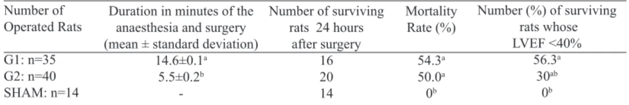
The entire duration of anaesthesia and surgery was signii -cantly longer (P<0.0001) in the G1 rats than in the G2 rats (Table 2). None of the fourteen SHAM rats died after surgery, and each SHAM rats had a LVEF that was greater than 40%
(Table 2). Speciically, the values of the ive echocardiograph -ic measurements in these rats were 61.67±7.23% for LVEF, 0.59±0.07 ml for EDV, 0.23±0.05 ml for ESV, 1.09±0.08 cm2 for EDA, and 0.59±0.08 cm2 for ESA, statistically different in the G1 and G2 (Table 3).
Nineteen G1 rats (54.3%) and 20 G2 rats (50%) died during
the irst 24 hours after surgery, and the mortality rates in the two groups were not signiicantly different (Table 2).
Table 3. Comparison of the means of echocardiographic measurements between the groups SHAM and G2, G1.
Variables G1 (n=9)
G2 (n=6)
SHAM (n=14)
P value
LVEF (%) 28,0 ±9,2a 26,5 ±6,1a 61.6 ±7.2b 0,0001 ESV (mL) 0,54 ±0,12a 0,55 ±0,12a 0.23 ± 0.05b 0,0001
LVEF=ejection fraction of the left ventricle; ESV=end systolic volume; EDV=end diastolic volume; ESA=end systolic area; EDA=end diastolic area; HR=heart rates; bpm=beats per minute. Different letters between rows: signiicant difference (P<0.05)
EDV (mL) 0,74 ±0,09a 0,74 ±0,13a 0.59 ±0.07b 0,0007
ESA (cm2) 1,12 ±0,35a
1,01 ±0,12a
0.59 ± 0.08b 0,0001
EDA (cm2)
1,23 ±0,1a 1,23 ±0,11a 1.0 ±0.08b 0,0022 HR (bpm) 258,4 ±41,6a 255,2 ±40,2a 227,3 ± 34,8a
>0,05 Table 2. Comparison of the duration of the anesthetic-surgical procedure and the percentage of rats with ejection fraction of the left ventricle (LVEF) less than 40% between G1, G2 and SHAM groups.
Number of Operated Rats
G1: n=35 G2: n=40 SHAM: n=14
Duration in minutes of the anaesthesia and surgery (mean ± standard deviation)
14.6±0.1a 5.5±0.2b -Mortality Rate (%) 54.3a 50.0a 0b
n=sample size; different letters between rows = P<0.05, and is the signiicance of the difference between the groups
Number (%) of surviving rats whose LVEF <40%
56.3a 30ab
0b Number of surviving
rats 24 hours after surgery
The propensity for complications during surgery, namely pulmonary atelectasis and haemorrhage, was higher in the G2 rats than in the G1 rats. In contrast, the propensity for post-op-erative complications, namely respiratory depression, heart failure, and pulmonary oedema, was higher in the G1 rats than in the G2. The main causes of death in the G1 and G2 rats were cardiac dilation, heart failure, and pulmonary oedema. Nine of the 16 surviving G1 rats (56.3%) and six of the 20 surviving G2 rats (30%) had a LVEF of less than 40%. The proportion of rats with a LVEF of less than 40% in the groups G1 and G2
was not signiicantly different from each other (P=0.176). The
proportion of rats with LVEF less than 40% was signiicantly
higher in G1 compared to SHAM group (P=0.014), but there
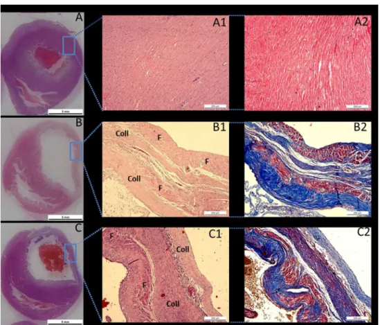
Fig. 3 - Panoramic photomicrographs of the cross-sections of the heart from a SHAM rat (A), a G1 rat (B), and a G2 rat (C).
*In the G1 and G2 rats, a transmural infarct is observed on the free wall of the left ventricle. Haematoxylin and eosin staining of cardiac tissue from a SHAM rat (A1) in which no myocardial infarct was observed, the myocardium is preserved and no histological alterations in cardiac microarchitecture were found. Haematoxylin and eosin staining of cardiac tissue in a G1 rat (B1) and a G2 rat (C1) shows an organized area of cicatricial collagen (Coll)
in the transmural infarct of remanescent muscle ibres (F). Masson trichrome staining of
cardiac tissue from a SHAM rat (A2), a G1 rat (B2), and a G2 rat (C2) in which the muscle
ibres are stained red and the collagen is stained blue
was no difference in this rate between the G2 and SHAM group (P=0.0743), (Table 2). Overall, 25.7% of all G1 rats and 15% of all G2 rats had a LVEF of less than 40%. Table 3 shows the comparison of the means of echocardiographic measurements between the groups SHAM and G2, G1. The mean values of each echocardiographic measurement in the nine G1 rats were
not signiicantly different from those in the six G2 rats.
Histopathological analysis of hearts from the ten SHAM rats revealed preserved cardiac muscle tissue (Figure 3). Histological analysis of hearts from both the G1 and G2 rats revealed the presence of an organized transmural infarct with
ibrosis and intense collagen deposits in the free wall of the
DISCUSSION
The mortality rate in the G1 and G2 during the irst 24 hours
following surgery in this study was between 50-55%, and this rate is similar to that reported (40-65%) in other studies[3,9]. Zomoff et al.[10] reported that the cardiac remodelling that occurs after AMI is associated with a high prevalence of cardiac rupture, arrhythmias, and the formation of aneurysms. Other investiga-tors have reported that arrhythmias, such as sustained ventricular
tachycardia and ventricular ibrillation, are the principal cause
of death in rats with an experimentally-induced AMI[9,11]. Although there was no statistical difference between groups, we found that the number of G1 rats with a LVEF of less than 40% was greater than found in the group G2. We attribute this increased to the duration of the surgical procedure that was used to create the AMI. The resultant number of rats with a LVEF of less than 40% is also determined by several intrinsic and extrinsic factors. The intrinsic determinants
include the rat’s age and lineage, individual variations in the
anatomy of the coronary artery, and individual physiological factors. On the age of the rat, spontaneous improvement in systolic function and ventricular volume can occur within 30 days after creation of an AMI in young rats[12].Hence, adult rats whose ages of the approximately 150 days and have a body weight of approximately 350 grams are preferred for creating the experimental AMI[12].The extrinsic determinants include
the site of the occlusion, which is dependent of the surgeon’s
experience and skill in occluding the LADCA. If the LADCA is occluded close to its origin, the size of the infarct will be large and the mortality rate can be as high as 100%[13].
Kissin et al.[14] has also reported that the prolonged duration
(three hours) of halothane-induced anaesthesia can inluence
the onset of postoperative deleterious effects and could cause postoperative death in rats with an experimentally-induced
AMI. Speciically, they showed that increasing the duration
of halothane anaesthesia after creation of the AMI causes pro-longed hypotension and increases the size of the myocardial infarction. Then, it is possible that, beyond the surgery, the duration of halothane-induced anaesthesia accounted for the higher propensity for a severe left ventricular dysfunction in the G1 rats than in the G2 rats. However, in a mild form, as in this study the difference in anaesthesia time between groups was only nine minutes.
In our study, histopathological examination of the hearts of all rats with a LVEF of less than 40% revealed that these hearts had histopathological characteristics that were similar to those that have been reported by others[3,4]. Speciically, we found that the infarcted hearts were dilated, and this dilation was associated with a reduced thickness of the free wall of the left ventricle with intense collagen deposits. According to Fishbein et al.[4], these cardiac alterations in the G1 and G2 rats probably occur about 21 days after creation of the exper-imental AMI. Consequently, cardiac muscle tissue loss results
in a decrease in LVEF with an increase in end systolic and end diastolic volume of the left ventricle. This process results in ventricular dilatation and an increase in diastolic tension[10,12].
We found no signiicant differences in the echocardiographic
measurements of the G1 and G2 rats whose LVEF were less than 40%.
However, when comparing the two surgical techniques for creating the AMI, the technique that was used in the G2 rats is more advantageous than that used in the G1 rats in terms of (a) the duration of the surgical procedure, (b) the need for
speciic rodent equipment or instrumentation, (c) the healing process, as measured by the low incidence of suture break-down, and the reduced time of the complete closure of the surgical wound and removal of the sutures, and (d) the extent of acoustic shadowing in the echocardiographic evaluation. Nonetheless, the surgical technique that was used for creating the AMI in the G1 rats does have some advantages over that used for creating the AMI in the G2 rats. Firstly, the rate of operative complications, such as the frequency of pulmonary atelectasis and haemorrhage, is lower. Secondly, the use of a
lexible cotton-tipped rod enables easier removal of the peri -cardium. Thirdly, the risk of cardiac rupture is lower because the heart is not exteriorized as in the G1. Finally, the duration of the open thorax is longer, which reduces the time pressure on the surgeon.
Our study has several limitations. The techniques showed are the most practiced, but the low rate of severely infarcted rats remains a limitation of this experimental model. Due to this limitation, the power of the statistical test used to analyze the proportion of LVEF less than 40% between the groups was low (0.35). To increase the power of the test, we would have a greater number of samples in each group, making the statistical results of this analysis more reliable, but for ethical reasons this was not done. A second limitation of our study was the necessity of sedate the rats with ketamine and xylazine for echocardiographic examination. This sedative mixture not only affects the HR, but also decreases cardiac contractility[15]. Although the doses of ketamine and xylazine were identical for all rats, we used minimal doses in order to immobilize the rats for the examination. Echocardiographic measurements were also performed in the fourteen SHAM rats, which were also sedated by the identical doses of ketamine and xylazine. We found that the values of these measurements were higher than those in the rats with an AMI. Accordingly, we concluded that the changes in the echocardiographic measurements that were found in the rats with an AMI are due to the presence of the AMI and not to the sedatives.
CONCLUSION
be better than G2. This improvement is probably due to the greater duration of the open thorax, which reduces the time pressure on the surgeon, allowing a LADCA occlusion with greater accuracy.
Authors’ roles & responsibilities
LGAC Study design, anesthetic and surgical procedures, handling and care of animals, interpretation of results and writing FB Histopathological analysis, handling and care of animals,
interpretation of results
GSO Anesthetic and surgical procedures and interpretation of results NIM Echocardiographic examination
PHS Preparation of images and discussion
CLKR Histopathological analysis and interpretation of results CTP Statistical analysis
ACS Study design, revision of the manuscript and inal approval PRB Orientation for study design and inal approval
REFERENCES
1. Senegaglia AC, Barboza LA, Dallagiovanna B, Aita CA, Hansen
P, Rebelatto CL, et al. Are puriied or expanded cord
blood-derived CD133+ cells better at improving cardiac function? Exp Biol Med (Mywood). 2010;235(1):119-29.
2. Johns TN, Olson BJ. Experimental myocardial infarction. I. A method of coronary occlusion in small animals. Ann Surg. 1954;140(5):675-82.
3. Zornoff LA, Paiva SA, Minicucci MF, Spadaro J. Experimental myocardium infarction in rats: analysis of the model. Arq Bras Cardiol. 2009;93(4):434-40.
4. Fishbein MC, Maclean MB, Maroko PR. Experimental myocardial infarction in the rat: qualitative and quantitative changes during pathologic evolution. Am J Pathol. 1978;90(1):57-70.
5. Pimentel EB, de Moraes AC, Forechi L, Machado RC, Baldo MP, Mill JG. Kinetics of the electrocardiographic changes after permanent coronary occlusion in rats: Relationship with infarct size. Pathophysiology. 2012;19(4):277-81.
6. Louzada RA, Oliveira PF, Cavalcanti-de-Albuquerque JP, Cunha-Carvalho L, Baldanza MR, Kasai-Brunswick TH,
et al. Granulocyte-colony stimulating factor treatment of
chronic myocardial infarction. Cardiovasc Drugs Ther. 2010;24(2):121-30.
7. Yu Y, Zhan ZH, Wei SG, Chu Y, Weiss RM, Heistad DD, et al. Central gene transfer of interleukin-10 reduces hypothalamic
inlammation and evidence of heart failure in rats after myocardial
infarction. Circ Res.2007;101(3):304-12.
8. Schiller NB, Foster E. Analysis of left ventricular systolic function. Heart. 1996;75(6 Suppl 2):17-26.
9. Opitz CF, Mitchell GF, Pfeffer MA, Pfeffer JM. Arrhythmias and death after coronary artery occlusion in the rat. Continuous telemetric ECG monitoring in conscious, untethered rats. Circulation. 1995;92(2):253-61.
10. Zornoff LA, Paiva SA, Duarte DR, Spadaro J. Ventricular
remodeling after myocardial infarction: concepts and clinical implications. Arq Bras Cardiol. 2009;92(2):150-64.
11. Li Y, Kloner RA. Is there a gender difference in infarct size and arrhythmias following experimental coronary occlusion and
reperfusion? J Thromb Thrombolysis.1995;2(3):221-5.
12. Pabis FC, Miyague NI, Francisco JC, Woitowicz V, Carvalho KA,
Faria-Neto JR, et al. Echocardiographic assessment of myocardial
infarction evolution in young and adult rats.Arq Bras Cardiol.
2008;91(5):321-6.
13. Pfeffer MA, Pfeffer JM, Fishbein MC, Fletcher PJ, Spadaro J, Kloner RA, et al. Myocardial infarct size and ventricular function in rats. Circ Res. 1979;44(4):503-12.
14. Kissin I, Stanbridge R, Bishop SP, Reves JG. Effect of halothane on myocardial infarct size in rats. Can Anaesth Soc J. 1981;28(3):239-43.
15. Stein AB, Tiwari S, Thomas P, Hunt G, Levent C, Stoddard MF,
et al. Effects of anesthesia on echocardiographic assessment of