RBCCV 44205-1580 DOI 10.5935/1678-9741.20140058
Risk factors of atheromatous aorta in
cardiovascular surgery
Fatores de risco de ateromatose da aorta em cirurgia cardiovascular
Fernando A. Atik
1, MD; Isaac Azevedo Silva
1, MD; Claudio Ribeiro da Cunha
1, MD
1Instituto de Cardiologia do Distrito Federal (IC-DF), Brasília, DF, Brazil.
This study was carried out at Instituto de Cardiologia do Distrito Federal (IC-DF), Brasília, DF, Brazil.
No inancial support.
Correspondence address: Fernando Antibas Atik
Instituto de Cardiologia do Distrito Federal
Estrada Parque Contorno do Bosque s/n - Cruzeiro Novo – Brasília, DF, Brasil – Zip Code: 70658-700
E-mail: [email protected]
Article received on October 3rd, 2013 Article accepted on March 24th, 2014
Abstract
Objective: To determine the prevalence and proile of ascend
-ing aorta or aortic arch atheromatous disease in cardiovascular surgery patients, its risk factors and its prognostic implication early after surgery.
Methods: Between January 2007 and June 2011, 2042 con
-secutive adult patients were analyzed, with no exclusion criteria. Atheromatous aorta diagnosis was determined intraoperatively by surgeon palpation of the aorta. Determinants of atheroma
-tous aorta, as well as its prognostic implication were studied by multivariate logistic regression.
Results: Prevalence of atheromatous aorta was 3.3% (68 pa
-tients). Determinants were age > 61 years (OR= 2.79; CI95%= 2.43 - 3.15; P<0.0001), coronary artery disease (OR=3.1; CI95%=2.8 - 3.44; P=0.002), hypertension (OR=2.26; CI95%=1.82 - 2.7; P=0.03) and peripheral vascular disease (OR=3.15; CI95%= 2.83 - 3.46; P=0.04). Atheromatous aorta was an independent predictor of postoperative cerebrovascular accident (OR=3.46; CI95%=3.18 - 3.76; P=0.01).
Conclusion: Although infrequent, the presence of athero
-matous aorta is associated with advanced age, hypertension, coronary artery disease and peripheral vascular disease. In those patients, a more detailed preoperative and intraoperative assess
-ment of the aorta is justiied, due to greater risk of postoperative cerebrovascular accident.
Descriptors: Atherosclerosis. Aorta. Thoracic Surgery. Cardio
-vascular Surgical Procedures.
Resumo
Objetivo: Determinar a prevalência e as características de ateromatose da aorta ascendente e/ou arco aórtico em cirurgia cardiovascular, os fatores de risco de sua ocorrência e a implica
-ção prognóstica imediata da mesma.
Métodos: No período de janeiro de 2007 a junho de 2011, 2042 pacientes adultos consecutivos foram analisados, sem critérios de exclusão. A detecção de ateromatose da aorta foi realizada por meio de palpação durante o ato operatório. Os fatores de risco de ateromatose da aorta e a sua implicação prognóstica foram determinados por regressão logística multivariada.
Resultados: A prevalência de ateromatose da aorta foi de 3,3% (68 pacientes). Os fatores de risco foram a idade > 61 anos (OR= 2,79; IC95%= 2,43 - 3,15; P<0,0001), doença arterial coronária (OR=3,1; IC95%=2,8 - 3,44; P=0,002), hipertensão arterial sistê
-mica (OR=2,26; IC95%=1,82 - 2,7; P=0,03) e doença vascular pe
-riférica (OR=3,15; IC95%= 2,83 - 3,46; P=0,04). A ateromatose da aorta foi preditor independente da ocorrência de acidente vascular cerebral no pós-operatório (OR=3,46; IC95%=3,18 - 3,76; P=0,01).
Conclusão: Embora infrequente, a presença de ateromatose da aorta tem maior ocorrência de acordo com a idade, com a presença de hipertensão arterial sistêmica, doença arterial coronária e doença vascular periférica. Nestas situações, é justiicada investigação pré e intraoperatória mais detalhada, pois a presença de ateromatose deter
-mina maior chance de acidente vascular cerebral no pós-operatório.
Descritores: Aterosclerose. Aorta. Cirurgia Torácica. Proce
the Research Ethics Commission under the protocol number
069883/2013, in accordance with the Helsinki rules.
The surgeries were isolated coronary artery bypass grafting (CABG) in 911 patients (44.6%), isolated valve surgeries in 561 (27.5%) and combined surgeries in 400 patients (19.6%), and other procedures in 170 (8, 3%). Other procedures were composed of congenital disease in adult surgeries in 84 pa
-tients (4.1%), isolated aorta surgeries in 45 pa-tients (2.2%)
and miscellaneous surgeries in the rest.
Of the patients that we identiied the atheromatous aorta, 47 (69%) underwent isolated CABG, 5 (7%) valve operation and the rest of the combined operations (24%). Cardiopulmo
-nary bypass was not used in 15 (32%) patients of those who
underwent isolated CABG. On the other hand, it was used in
all submitted to other operations.
The decision regarding the performance of surgery from
diagnosis of atheromatosis was the surgeon’s discretion. The
aorta is no longer handled entirely in 16% of patients. Changes in cannulation site occurred in 79% of patients, change of clamping site in 69% and change in proximal anastomoses or aortotomy in 59%. The aortic replacement under deep hypo
-thermic circulatory arrest was performed in 16% of patients.
Statistical analysis
Categorical variables were expressed as frequencies and percentages and continuous by mean and standard deviation.
Continuous variables with heterogeneous distribution were
expressed by medians and conidence intervals relating to one standard deviation. When comparing the pre- and intraopera -tive characteristics of morbidity and mortality events between
the groups, the chi-square test, Fisher exact test, Student’s t test
were used when indicated. Multivariate logistic regression was
used to determine the risk of atheromatosis and its prognostic implications factors in the occurrence of death, stroke and acute renal failure. Variables signiicantly (P <0.05) related to each of
the events by univariate analysis were retained. Then, stepwise
backward logistic regression was used in the construction of multivariate models. Calibration and discrimination of the models were determined by the Hosmer-Lemeshow test and
the analysis of the ROC curve (receiver-operating character
-istic), respectively.
RESULTS
Prevalence and descriptive atheromatosis
The atheromatous ascending aorta and/or aortic arch were diagnosed by the surgeon during surgery in
68 patients, which corresponds to 3.3% of the total. The intraoperative indings were isolated calciied plate bounded by 35%, extensive or multiple plate calciied in 33% and porcelain aorta by 32%. Given these indings, various types of impact emerged: impossibility of any manipulation of the aorta in 33.3%, cannulation failure
Abbreviations, acronyms and symbols
CABG Coronary artery bypass grafting
LC Left coronary
LV Left ventricle
NYHA New York Heart Association
INTRODUCTION
The presence of atheromatous disease of the thoracic aorta is a known complicating factor in patients undergoing cardio
-vascular surgery, since it determines changes in intraoperative planning, and increases the risk for increased morbidity and
mortality[1].
The correlation between atherosclerosis in the coronary
arteries and other arterial sites have been extensively docu -mented[2,3], especially in the carotid arteries[4]. In turn, patients
with atherosclerosis of the carotid arteries also have a higher rate of atheromatous thoracic aorta[5]. On the other hand,
ath-eromatosis of the thoracic aorta is common in the elderly, and
population studies[6,7] found that these patients have a higher prevalence of cardiovascular events and stroke.
Although this evidence demands a more careful
monitor-ing in risk groups, there are no national data on the subject. Knowledge of the prevalence and atheromatous aorta risk factors may provide greater predictability of its occurrence and prognosis. They can also lead to therapeutic changes that aim to minimize the operative risk.
The aim of this study are to determine the prevalence
and characteristics of atheromatous ascending aorta and/or
aortic arch in patients undergoing cardiovascular surgery in
a Brazilian center, the risk factors for its occurrence and its
immediate prognostic implications.
METHODS
From January 2007 to June 2011, 2042 consecutive adult
patients underwent cardiovascular surgery. The mean age was 57.4±15 years (range 16 years to 87 years) and 1168 (57.2%) were male. All patients were studied, with no exclusion cri
-teria. Pre-, intra- and post-operative of the patients were pro
-spectively collected and stored in an electronic database. The diagnosis of atheromatous aorta was performed by its palpa -tion during surgery by the surgeon, and the collec-tion of data took into account the full account of the surgeon in relation
in 9.7%, clamping impossibility 4.2% and impossibility of cannulation and clamping 2.8%. In the remaining half of the patients, there was possibility of cardiopulmonary bypass with changes in local cannulation, clamping and proximal anastomoses.
Risk factors of ateromatose
In Table 1 are described the differences found between
the control group and atheromatosis by univariate analysis as regards demographics, comorbity and risk factors widely
recognized for cardiovascular surgery. Thus, the older age in
the atheromatosis group was particularly signiicant (65.8±9.7
years versus 54.3±15 years, P<0.0001). Still, all of atheroscle-rosis risk factors occurred more frequently in atheromatosis
group compared to the control. In addition, there was a higher
frequency of involvement of other arterial territories in
ath-eromatous aorta group, for example, coronary artery disease, especially when associated with left main coronary artery obstruction and peripheral vascular disease. There was an
association trend between obstructive carotid artery disease and aortic atheromatosis.
Multivariate analysis identiied independent risk factors for atheromatous disease of the aorta in the study population (Table 2), as the age older than 61 years (OR=2.79; 95% CI
2.43 to 3.15; P<0.0001), the presence of coronary artery dis
-ease (OR=3.1; 95% CI 2.8 to 3.44; P=0.002), hypertension
(OR=2.26; 95% CI 1.82 to 2.7; P=0.03) and peripheral vascular
disease (OR=3.15; 95% CI 2.83 to 3.46; P=0.04).
Table 1. Preoperative characteristics of patients with and without atheromatous aorta who underwent
cardiovascular surgery. Demographics Age (years) Male Weight (kg) Height (cm)
Previous cardiac surgery Functional class (NYHA) I
II III IV
Coronary artery disease Acute coronary syndrome Myocardial infarction <30d Trunk lesion LC
Number of affected vessels 1
2 3
LV ejection fraction Pulmonary hypertension
Carotid disease Risk factors
Arterial hypertension
Diabetes mellitus Dyslipidemia Smoking Comorbidities Prior stroke Atrial ibrillation
Peripheral vascular disease
Prognostic implications
Table 3 shows the impact of atheromatous aorta in hos
-pital mortality and major morbidity outcomes by univariate analysis. 1There was a higher hospital mortality and higher incidence of stroke, sepsis, myocardial infarction, acute renal failure, prolonged mechanical ventilation and longer hospital stay in patients with atheromatous aorta compared to the
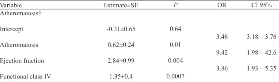
control. Regarding the multivariate analysis, the presence of atheromatous aorta was an independent predictor of the occurrence of stroke in the postoperative period (OR=3.47; 95% CI 3.18 to 3.76; P=0.01), as documented in Table 4. There was no correlation in the analysis of risk factors, including the
presence of atheromatous aorta and hospital mortality or acute renal failure in the postoperative period.
Table 2. Multivariate analysis of risk factors for atheromatous aorta in patients undergoing cardiovascular
surgery.
Variable Atheromatosis†
Intercept
Age > 61 years
DAC
SAH
Peripheral vascular disease
Estimate±SE
3.24±0.33
0.51±0.13
0.57±0.19
0.41±0.19
0.57±0.28
P
<0.0001
<0.0001
0.002
0.03
0.04
OR
2.79
3.1
2.26
3.15
CI 95%
2.43 – 3.15
2.8 – 3.44
1.82 – 2.7
2.83 – 3.46
† Hosmer-Lemeshow test, P=0.09; C-statistic 0.77, CAD=coronary artery disease, Hypertension. CAD=coronary artery disease; SH=systemic arterial hypertension
Table 3. Morbidity and mortality data in patients with and without aortic atheromatosis undergoing
cardiovascular surgery.
Hospital mortality
Stroke
Myocardial infarction
Sepsis
Acute renal failure
Prolonged mechanical ventilation Intensive care unit stay (days) Length of stay (days)
Atheromatosis (N=68)
12 (17.6%) 7 (10.4%) 10 (14.7%) 13 (19.1%) 12 (17.6%) 6 (8.9%)
3 (2; 5) 15 (8; 25)
Control (N=1974)
114 (5.7%) 44 (2.2%) 96 (4.8%) 136 (6.9%)
81 (4.1%) 44 (2.2%)
2 (2; 4) 11 (7; 19)
P
<0.0001 <0.0001 0.0003 0.0001 <0.0001
0.0005 0.24 0.03
Table 4. Multivariate analysis of the occurrence of stroke in patients undergoing cardiovascular surgery.
Variable Atheromatosis†
Intercept
Atheromatosis
Ejection fraction
Functional class IV
Estimate±SE
-0.31±0.65
0.62±0.24
2.84±0.99
1.35±0.4
P
0.64
0.01
0.004
0.0007
OR
3.46
9.42
3.86
† Hosmer-Lemeshow test, P=0.9; C-statistic 0.7
CI 95%
3.18 – 3.76
1.98 – 42.6
It was not identiied a greater risk of stroke according to the type of the atheromatous disease in the aorta. Patients with porcelain aorta had the same risk as other patients with delimited atherosclerotic plaques (12.5% vs. 9.5%, P=0.87).
Also, avoiding manipulation of the aorta did not prevent the occurrence of cerebral complications (9% vs. 10.2%, P=0.91), as well as changing the cannulation site (P=0.61), clamping (P=0.12) or using circulatory arrest (P=0.9).
DISCUSSION
This observational study aimed at exploring atheromatous ascending aorta and/or aortic arch in a group of consecutive adult patients undergoing cardiovascular surgery. Despite the prevalence is relatively low in this population, it is consistent
with other[8] literature series. The prevalence could be higher if our population was older, since there was direct relationship between age and the presence of atheromatosis. Davila-Ro -man et al.[9] found important atheromatous ascending aorta in approximately one third of patients older than 80 years who underwent cardiovascular surgery. The increase in life expec
-tancy of the Brazilian population will certainly determine a higher incidence of patients with this problem being treated
for cardiovascular surgery.
In addition to age, it was identiied that the main athero -sclerosis risk factors[5] are the same related to atheromatous aorta by univariate analysis. However, only hypertension was considered an independent risk factor in the multivariate
analysis. In the latter, other factors found in our series were
coronary artery disease and peripheral vascular disease, re
-inforcing the coexistence of atherosclerotic involvement in
different arterial territories.
Most studies in the [10-12] literature considers as risk factors
for aortic atheromatosis the obstructive carotid artery disease, abdominal aortic aneurysm, left main coronary artery
ob-struction, diabetes mellitus and hypertension. The calciied
aortic stenosis and chronic renal failure are still other risk factors involved[13]. Looking critically, preoperative patient characteristics have little predictive value, since most of the patients referred to the cardiovascular surgery has such fea -tures, which therefore requires more accurate assessments. In
our experience, such evaluations are performed infrequently and dependent on the doctor’s suspicion level.
The importance of completing the diagnosis in order to enable the best possible surgical planning. Traditional image
tests that can identify the atheromatous aorta are inaccurate,
as the chest X-ray or cardiac catheterization. Transesopha
-geal echocardiography[14] is better than the previous ones, however, presents image acquisition limitations in the distal ascending aorta and proximal aortic arch, frequently used sites for cannulation and clamping. Computed tomography[15] is an excellent test to detect atheromatous aorta, however,
it has disadvantages in relation to the use of radiation and
its use is associated with high cost for the health system. It
can, however, be used in the groups at the most risk prior
to surgery[13].
During surgery, the surgeon palpates traditionally the
aorta to diagnose aortic atheromatosis. However, Wareing et al.[10] studied more than 500 patients and found palpation be imprecise and it was detected only 38% of positive diagnoses on ultrasound. The best imaging method is the epiaortic ul -trasound[16,17] performed intraoperatively with high-resolution
transducers which measure the wall and the lumen of the aorta in the various segments.
Precise informations regarding the pathological features of the aorta have implications for the management of cardio
-pulmonary bypass, myocardial and cerebral protection and surgical planning, in order to minimize unwanted embolism microparticle atheroma, especially to the brain. Our study
and others[1,18] showed that the presence of atheromatous aor -ta is an independent risk factor in the development of stroke postoperatively, with absolute risk by about 10%, a risk con -sistent in several series.
Blauth et al.[12] reviewed the autopsy indings of 221 pa
-tients undergoing cardiac surgery. It was revealed that
cere-bral embolism occurred in 37% of patients with atheroscle
-rosis of the aorta, compared to only 2% of those who had not.
Van der Linden et al.[19] showed that the kind of impairment of the aorta has a differential risk of stroke. In our experience, we could not corroborate the indings of this latest study of higher risk related to the type of involvement, including as regards the porcelain aorta.
The various types of surgical strategy are possible to be performed, as shown in our experience, and there is no
consensus in the literature regarding the best strategy. These
include the revascularization with composite arterial grafts under cardiopulmonary bypass without aortic clamping, changes on the site of cannulation, clamping and proximal anastomoses, up to the aortic replacement under total circu
-latory arrest with deep hypothermia.
However, none of these approaches is capable of prevent
-ing the risk of displacement of atheromatous particles. It’s intuitive to state that avoiding any manipulation of the aorta is advised in the presence of atheromatosis. However, this strategy is not possible in all cases, particularly the need for
combined surgeries involving the heart valves and thoracic aorta. There are also some data from observational stud-ies[1,8,10-12,14] suggesting that the change in operative tactics to minimize the risk of complications, although the morbidity
remain higher than usual.
We could not ind in our superiority experience any ap
reduce this risk. Neither the replacement of the aorta under total circulatory arrest with deep hypothermia reached the same beneit, although not carrying additional risk.
Cardiac surgery with aortic manipulation guided by epiaortic ultrasound has uncertain clinical value. Clini -cal studies have detected low cerebral embolization by
transcranial Doppler[20] and improved neuropsychological performance[21,22], but it has not been demonstrated lower
stroke rates.
In a patient with porcelain aorta, carrier or aorta valve disease requiring surgery, most surgeons hesitate to perform complicated surgical procedures involving total circulatory arrest and aortic replacement in elderly patients who often already has other operative risk factors, which opens the possibility of transapical aortic valve replacement when in -dicated[23]. On the other hand, if the proposed surgery would be isolated CABG, performing it without cardiopulmonary bypass and without manipulation of the aorta seems to be a more appropriate strategy when possible[24].
It should be noted that our experience does not corroborate
such conduct, nor there is strong evidence in the literature to
support them up to this point.
Study limitations
This is an observational study, with the limitations inherent
to its design. We used palpation of the aorta as a diagnostic method of atheromatosis, which certainly reduced the preva
-lence. The use of epiaortic ultrasound should be the standard of our service. The limited number of patients and the very different approaches adopted by surgeons in the presence of atheromatous hamper determining the impact of the change of intraoperative tactic in the results of morbidity and mortality. For this reason, this was not listed as objective of this study. A multicenter study of more robust number of patients facing this purpose would be interesting to be developed.
CONCLUSION
Although infrequent, the presence of atheromatous
aorta has higher incidence according to age, with the
presence of hypertension, coronary artery disease and pe
-ripheral vascular disease. In these situations, it is justiied pre- and intraoperative more detailed research, because the presence of atheromatous determines greater chance of stroke postoperatively.
Authors' roles & responsibilities
FAA Data collection, statistical analysis, data interpretation and writing of the manuscript
IAS Manuscript writing CRC Manuscript review
REFERENCES
1. Gillinov AM, Lytle BW, Hoang V, Cosgrove DM, Banbury MK, McCarthy PM, et al. The atherosclerotic aorta at aortic valve
replacement: surgical strategies and results. J Thorac Cardiovasc Surg 2000;120(5):957-63.
2. Yamamoto H, Shavelle D, Takasu J, Lu B, Mao SS, Fisher H, et
al. Valvular and thoracic aortic calcium as a marker of the extent and severity of angiographic coronary artery disease. Am Heart J. 2003;146(1):153-9.
3. Budoff MJ, Nasir K, Mao S, Tseng PH, Chau A, Liu ST, et al.
Ethnic differences of the presence and severity of coronary atherosclerosis. Atherosclerosis. 2006;187(2):343-50.
4. Folsom AR, Kronmal RA, Detrano RC, O’Leary DH, Bild DE,
Bluemke DA, et al. Coronary artery calciication compared with carotid intima-media thickness in the prediction of cardiovascular disease incidence: the Multi-Ethnic Study of Atherosclerosis (MESA). Arch Intern Med. 2008;168(12):1333-9.
5. Takasu J, Budoff MJ, O’Brien KD, Shavelle DM, Probstield JL, Carr JJ, et al. Relationship between coronary artery and descending thoracic aortic calciication as detected by computed tomography: the Multi-Ethnic Study of Atherosclerosis. Atherosclerosis. 2009;204(2):440-6.
6. Witterman JC, Kannel WB, Wolf PA, Grobbee DE, Hofman A,
D’Agostino RB, et al. Aortic calciied plaques and cardiovascular disease (the Framingham Study). Am J Cardiol 1990;66(15):1060-4.
7. Hollander M, Hak AE, Koudstaal PJ, Bots ML, Grobbee
DE, Hofman A, et al. Comparison between measures of atherosclerosis and risk of stroke: the Rotterdam study. Stroke. 2003;34(10):2367-72.
8. Zingone B, Gatti G, Spina A, Rauber E, Dreas L, Forti G, et al. Current role and outcomes of ascending aortic replacement for
severe nonaneurysmal aortic atherosclerosis. Ann Thorac Surg.
2010;89(2):429-34.
9. Dávila-Román VG, Kouchoukos NT, Schechtman KB, Barzilai
B. Atherosclerosis of the ascending aorta is a predictor of renal dysfunction after cardiac operations. J Thorac Cardiovasc Surg. 1999;117(1):111-6.
10. Wareing TH, Davila-Roman VG, Barzilai B, Murphy SF,
Kouchoukos NT. Management of the severely atherosclerotic
ascending aorta during cardiac operations. A strategy for detection and treatment. J Thorac Cardiovasc Surg. 1992;103(3):453-62.
11. Mills NL, Everson CT. Atherosclerosis of the ascending aorta
and coronary artery bypass. Pathology, clinical correlates, and operative management. J Thorac Cardiovasc Surg. 1991;102(4):546-53.
An emerging problem in cardiac surgery. J Thorac Cardiovasc Surg. 1992;103(6):1104-11.
13. Nishi H, Mitsuno M, Tanaka H, Ryomoto M, Fukui S, Miyamoto
Y. Who needs preoperative routine chest computed tomography for prevention of stroke in cardiac surgery? Interact Cardiovasc Thorac Surg. 2010;11(1):30-3.
14. Katz ES, Tunick PA, Rusinek H, Ribakove G, Spencer FC, Kronzon I. Protruding aortic atheromas predict stroke in elderly patients undergoing cardiopulmonar bypass: experience with intraoperative transesophageal echocardiography. J Am Coll Cardiol. 1992;20(1):70-7.
15. Takeda Y, Hoshiga M, Tatsugami F, Morinaga I, Takehara K, Hotchi J, et al. Clinical significance of calcification in ascending aorta as a marker for the requirement of coronary
revascularization. J Atheroscler Thromb. 2009;16(4):346-54.
16. Zingone B, Rauber E, Gatti G, Pappalardo A, Benussi B, Dreas L, et al. The impact of epiaortic ultrasonography scanning on the risk of perioperative stroke. Eur J Cardiothorac Surg. 2006;29(5):720-8.
17. Davila-Roman VG, Barzilai B, Wareing TH, Murphy SF, Kouchoukos NT. Intraoperative ultrasonography evaluation of the ascending aorta in 100 consecutive patients undergoing cardiac surgery. Circulation. 1991;84(5 Suppl):III47-53.
18. Djaiani G, Fedorko L, Borger M, Mikulis D, Carroll J, Cheng
D, et al. Mild to moderate atheromatous disease of the thoracic
aorta and new ischemic brain lesions after conventional coronary
bypass graft surgery. Stroke. 2004;35(9):e356-8.
19. van der Linden J, Hadjinikolaou L, Bergman P, Lindblom D. Postoperative stroke in cardiac surgery is related to the location and extent of atherosclerotic disease in the ascending aorta. J Am Coll Cardiol; 2001;38(1):131-5.
20. Borger MA, Taylor RL, Weisel RD, Kulkarni G, Benaroia M, Rao V, et al. Decreased cerebral emboli during distal aortic arch
cannulation: a randomized clinical trial. J Thorac Cardiovasc Surg. 1999;118(4):740-5.
21. Goto T, Baba T, Yoshitake A, Shibata Y, Ura M, Sakata R. Craniocervical and aortic atherosclerosis as neurologic risk
factors in coronary surgery. Ann Thorac Surg. 2000;69(3):834-40.
22. Hammon JW Jr, Stump DA, Kon ND, Cordell AR, Hudspeth AS, Oaks TE, et al. Risk factors and solutions for the development of neurobehavioral changes after coronary artery bypass grafting. Ann Thorac Surg. 1997;63(6):1613-8.
23. Kempfert J, Van Linden A, Linke A, Schuler G, Rastan A, Lehmann S, et al. Transapical aortic valve implantation: therapy of choice for patients with aortic stenosis and porcelain aorta? Ann Thorac Surg. 2010;90(5):1457-61.