CRANIOCEREBRAL INVOLVEMENT IN LYMPHOMA
JORGE D. CORREALE * — DAVID A. MONTEVERDE * — JOSÉ A. BVERI * EDGARDO G. REICH * — NÉSTOR LUCATELLI **
SUMMARY — Nine-hundred-eighty-nine patients with lymphoma were studied. Fifty-three cases (5.3%) had lymphomatous craniocerebral infiltration. The principal factors of risk for this complication were: advanced stage of the lymphoma (III or IV), diffuse histiocytic, diffuse poorly differentiated lymphocytic, or mixed cellularity lymphoma histological type, bone marrow involvement, and previous systemic chemotherapy. Thirty-two per cent of the cases of meningeal lymphomatous infiltration were asymptomatic and represented autopsy findings. CT-scan was an useful test to detect brain focal parenchymatous infiltration, as opposed to meningeal infiltration. Mean survival time in patients with lymphomatous meningeal infiltration was 4.3 months, following the combined use of systemic chemotherapy, radiation therapy and intrathecal methotrexate. Two cases had primary cerebral lymphoma, although without associated immunodeficiency Twenty patients (2%) had intracranial hemorrhage, in clear relationship with platelet alterations. Fifteen patients (1.5%) had CNS infection, caused by common bacteriae or opportunistic agents. In 7 cases, the diagnosis was made at autopsy. Thirty-six autopsies were performed. In 8 cases (22%), pathologic findings such as, demyelination, microcalcificat ons, coagulative necrosis, or gliosis, suggested complications from treatment.
Infiltración linfomatosa craniocerebral.
RESUMEN — Fueron estudiados 989 pacientes con linfoma. Tuvieron infiltración linfomatosa craneocerebral 53 casos (5.3%). Los principales factores de riesgo para esta complicación fueron: a. estado avanzado del linfoma (III o IV); b. las formas difusas histiocíticas, difusa pobremente diferenciada o celularidad mixta; c. el compromiso de la medula osea y de la quimioterapia sistêmica previa. En el 32% de los casos la infiltración meníngea linfomatosa fué asintomática y represento hallazgos de autópsia. La tomografía cerebral fué de utilidad para detectar infiltraciones parenquimatosas focales, no así para Ias infiltraciones meníngeas. El tiempo medio de sobrevida en pacientes con infiltración meníngea linfomatosa fué de 4.3 meses, siguientes al uso combinado de terapia radiante a craneo total, quimioterapia sistêmica y/o intratecal con methotrexate. Dos casos con linfoma cerebral primário no estuvieron asociados con inmunodeficiencia. Hemorragias intracraniales se observaron en 20 pacientes (2%), en relaciõn con alteraciones plaquetarias. En 15 casos hubo infección del SNC (1.5%), causada uor bactérias comunes o por agentes oportunistas. En 7 de esos casos el diagnóstico se hizo por autópsia. En 8 de 36 casos autopsiados (22%) se observaron desmielinización, microcalcificaciones, necrosis coagulativa o gliosis, sugestivas de complicaciones por los tratamientos efectuados.
In recent years, advances in the treatment of lymphoma have permitted a longer life expectancy, and consequently the appearance of neurological complications in more advanced stages of the disease. Most of the drugs used in the treatment of lymphoma have difficulties penetrating the blood-brain barrier. For that reason, lymphomatous cells localized in the central nervous system ( C N S ) may not be reached by
chemo-From the Division of Neurology (*) and Division of Pathology (**), José Maria Ramos Mejia Hospital, Buenos Aires.
therapeutic agents, and may proliferate at that level. Bone marrow involvement from lymphomatous invasion, medication toxicity, or both, can determine abnormalities in the number and function of platelets, with subsequent occurrence of hemorrhagic phenomena. The use of chemotherapeutic agents and radiation therapy ( R T ) has also brought about new complications. Chemotherapy or RT can have direct toxic effects on the CNS which, in turn, can produce necrotic lesions, demyelinating micro-angiopathy, or a subacute necrotizing leukoencephalopathy. Secondly, treatment can produce immunodepression superimposed to the immunological deficiency caused by the lymphoma, thus creating conditions for the development of opportunistic infections.
In the present study, we assess the different craniocerebral lesions caused by the above mentioned mechanisms in patients with lymphoma.
SUBJECTS AND METHODS
Four-hundred-twenty-four inpatients were studied prospectively between 1984 and 1987 in the Neurology and Hematology Departments of the José Maria Ramos Meiia Hospital of Buenos Aires. They all had a diagnosis of lymphoma. In a small proportion of patients, initial neurological symptoms led to the subsequent diagnosis of lymphoma. At the same time, 565 outpatient charts were reviewed, all of them with diagnosis of lymphoma. This made a total of 989 cases. Five-hundred-sixty-three cases corresponded to non-Hodgkin's lymphoma (NHL,), while the remaining 426 to the Hodgkin's type (HL). In our population, 73 patients (7.4%) had evidence of craniocerebral involvement consisting of lymphomatous infiltrations, hemorrhages, infections or complications from treatment. The diagnosis of CNS lymphomatous infiltration was based on either: (a) the presence of signs and symptoms of craniocerebral involvement in patients with diagnosis of lymphoma, in whom there was evidence of structural abnormalities shown by ancillary methods such as CT scan of the head, brain radionuclide scan or plain X-Rays of the skull, and in whom there was improvement or disappearance of the pathological images and of the clinical symptoms after treatment with chemotherapy, RT, or both; or (b) cerebrospinal fluid (CSF) cytology, brain biopsy, or autopsy.
Analysis of the clinical picture included age of the patient, sex, duration of the disease, histological type of lymphoma, clinical stage, bone marrow involvement, CSF study, ancillary methods, treatment administered, and response. Treatment included RT at doses from 2400 to 5000 Rads over 15 to 20 sessions, systemic chemotherapy (SCh), intrathecal metho-trexate administration (IT Mtx) followed by leucovorin rescue, or a combination of them. The only patients who underwent surgery were those who had neurological involvement as the initial clinical picture, i.e., a space-occupying mass, and in whom diagnosis of lymphoma was unknown yet. Hemorrhagic phenomena were demonstrated by means of CT scan or autopsy, when the latter was performed. Diagnosis of CNS infection was based on Hooper's criteria for immunosuppressed patients 26. Complications from treatment were included when verified by pathological studies in patients that received RT, SCh, or IT Mtx. A total of 36 autopsies were performed. NHLs were classified according to the histological criteria of Rappaport 51. For the HLs, Luke's criteria were used 41. Clinical stage was established according to the recommendations of the Ann Arbor Conference 5. One case of Burkitt's lymphoma was also included.
RESULTS
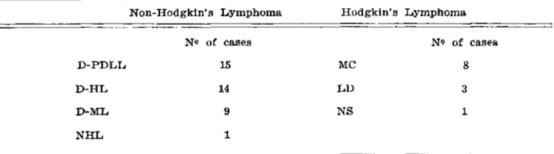
INFILTRATIONS — Meningeal, brain, or skull lymphomatous infiltrations were seen in 53 patients (5.3%). There were 39 cases with NHL, 12 with HL, 1 with Burkitt's lymphoma, and 1 with a T-cell type lymphoma. In the NHL group, there were 25 men and 14 women. Their mean age was 48 ± 17.3 years. Duration of the disease, from the time of diagnosis of lymphoma until the appearance of the first neurological symptom, was 26.4 months (range 1-108 months). In 5 patients (13%), the neurological picture was the first mani-festation of the disease. In the HL group, there were 7 men and 5 women. Their mean age was 37 ± 16 years. Duration of the illness, at the time of the neurological complication, was 52.9 months (range 1-84 months). The lymphoma histological types are summarized in Table 1. There was a clear relationship between the clinical stage of the lymphoma and the occurrence of CNS lymphomatous infiltration. Eleven per cent of the cases with lymphomatous infiltrations were on stages II A or B, 22.7% on stages III A or B, and 66% on stages IV A or B.
to the orbit or extending to adjacent areas. Finally, four patients had infiltration of the skull. In 11 cases, there was a combination of brain parenchymatous lymphomatous infil-tration with either meningeal or orbital involvement. Ten case® of lymphomatous meningeal infiltration were diagnosed at autopsy, without any prior indication of such an involvement. Two cases corresponded to primary cerebral lymphomas (PCL) of the diffuse histiocytic type. Their age at onset was 57 and 52 years, and their survival time was 8 and 11 months, respectively. None of them had associated immunodeficiency. Eight patients with brain meningeal infiltration had concomittant spinal cord involvement. Forty-five patients with craniocerebral infiltration underwent a bone marrow biopsy, which showed lymphomatous infiltration in 70% of them. However, when considered separatedly, patients with HL and craniocerebral infiltration had bone marrow involvement in all cases. The commonest symptoms and signs are summarized in Table 2.
In all cases with brain parenchymatous or skull lymphomatous infiltration, positive findings were seen on the CT scan. In 55% of the cases, there were single hyperdense images, which enhanced homogeneously after contrast administration. In the remaining cases, the images were isodense but enhanced after contrast administration. In all cases, they had varying degrees of surrounding edema and mass effect, depending on their size. They were contiguous to either the cortex or the ependymum. On these studies images either correlated with the pathological findings or disappeared after specific treatment. Nevertheless, CT scans of the head were negative in patients with lymphomatous meningitis except for one case, where there was periependimal contrast enhancement suggesting meningeal involvement.
Fourteen patients with CT scan images consistent with focal brain infiltration were treated with RT. There was subsequent disappearance of the lesions in 9 patients (64%). Twenty-one cases of lymphomatous meningeal infiltration were treated with a combination of RT at doses of 2400 to 4000 Rads, SCh, and IT Mtx. None of them showed a satisfactory response to treatment. Their survival time had a mean of 4.3 months from the onset of symptoms.
HEMORRHAGES — Twenty patients (2%) had intracranial hemorrhages. Among them, 18 had abnormalities in the platelet count or function. Two cases had an elevated platelet count, and they had intracranial hemorrhages secondary to platelet function abnormalities. The types of the intracranial hemorrhages were: diffuse suffusions (60%), petechiae (26%), or parenchimatous hematomas (26%). In 12% of cases, two or more of these findings occurred combined.
INFECTIONS — Fifteen patients (1.5%) had CNS infections. In most cases, the etiological agent was a bacteria or a virus, either in the HL or the NHL group (Table 3). All the infections occurred in patients on advanced stages of the lymphoma. The clinical pictures corresponded to meningoencephalitis or meningitis in 7 cases. One patient had a brain abscess caused by Staphylococcus aureus. In the remaining patients, the clinical picture consisted of confusion and focal findings without meningeal irritation signs. Diagnosis was made at autopsy in these cases.
COMPLICATIONS FROM TREATMENT — Thirty-six autopsies were performed. Lesions that can be related to treatment, i.e., circumscribed or diffuse demyelination areas, micro-calcifications, coagulative necrosis or gliosis, were seen in 8 patients (Table 4). One patient, who had been treated with RT alone, had cerebral microcalcifications. In this case, lesions can be ascribed to RT. In the other cases, it was not possible to single out a definitive relationship between the pathological findings and treatment, since more than one of the therapeutic agents used could have caused demyelination.
COMMENTS
Different series reported varying incidences of brain and meningeal lymphoma-tous infiltration in patients with lymphoma. Figures varied between 2 and 29% in patients with NHL4,2i,29,34,40,53,66. in the case of HL, there have been either few reports or series with a small number of patients 8,43,56,61; Sapozinck and Kaplan reported an incidence of 0.5% 56. in our series, 6.9% of the NHL and 2.8% of the HL patients had
evidence of craniocerebral lymphomatous infiltration. In the HL group, this complication occurred at a 10-year younger age than in the NHL group, perhaps reflecting the earlier age of onset of HL. However, the duration of the hematological disease, at the time of the CNS infiltration, w a s twice as long in the HL group (52.9 v s 26.4 months, respecti-vely). Neurological involvement w a s the first manifestation of the disease in 13% of cases of NHL. On the contrary, no case of HL had neurological involvement at the onset, in agreement with other series 33,56,60. Generally, brain lymphomatous infiltration is seen on advanced stages of the lymphomas, and is associated with histological types like diffuse histiocytic lymphoma (DHL) and diffuse poorly differentiated lymphocytic lymphoma (DPDLL)4,2i,34,37. Brain infiltration is infrequent in patients with HL or NHL of nodular type 4,17,21,34,37,40, although it may occur in the latter after trans-formation to a diffuse form 34,37,66. Meningeal lymphomatous involvement has been reported in pathological studies of up to 76% of patients with Burkitt's lymphoma with early relapse of the disease after treatment (within 3 m o n t h s )2 7
.3 6
-6 7
. Sixty-five per cent of the patients with brain infiltration were on advanced stages of the disease, i.e., IV A or B. In patients with NHL, DPDLL accounted for 3 8 % of the cases and, DHL for 36%, in agreement with other authors 34,37,66. in the group of patients with HL, we found a greater incidence of histological types of mixed cellularity (66.6%). There have been controversial results in this regard in the different pu-blished series 8,11.56. Between 9.5 and 7 3 % of the lymphomatous meningeal infiltra-tions are asymptomatic and are diagnosed at a u t o p s y1 7
.2 4
-6 6
. In our series, 3 2 % of meningeal infiltrations fell into this category.
infiltration 17,37,40. T h i s association s u g g e s t s dissemination from the bone marrow to the dura, subarachnoid space, and subsequent invasion to the Virchow-Robin space. The incidence of bone marrow infiltration in patients with early DHL is significantly lower than in patients with nodular forms (15 vs 40%, respectively). However, meningeal involvement in the former is frequent 17. There is no clear explanation for this fact 4. W e found bone marrow involvement in 7 0 % of the cases of lymphoma with craniocerebral infiltration. In the case of HL with brain and meningeal infil-tration, the bone marrow w a s involved in all cases. In the cases of brain and menin-geal infiltration without bone marrow involvement, the initial lymphomatous infiltration occurred in the nasal cavum, orbit, or spinal nerve roots, from where the disease disseminated to the CNS.
Lymphoma cells could be demonstrated in 78% of the CSF studies of patients with craniocerebral infiltration. According to different series 2,21,42,66, lymphoma cells can be seem in the CSF study in 70 to 88% of the patients with craniocerebral infil-tration when these studies are performed with cytocentrifugation techniques. However, these figures can drop to 50%, if only one CSF sample is drawn 42.66. Most authors recommend the study of at least three samples of CSF when looking for lymphoma cells. The study of surface markers, the identification of deoxynucleotidyltransferase, and the use of immunocytochemical techniques on CSF lymphocytes decrease the number of false positive results due to viral or mycotic infections 13.14,16,25,39. The CT scan of the head is not a very useful test to detect meningeal lymphomatous infiltrations. Even using larger doses of contrast, enhancement similar to that occurring in patients with infectious or carcinomatous meningitis is not frequently seen 3,12,48. On the contrary, brain focal parenchymatous infiltrations tend to appear as iso or hyperdense masses in contact with the cortical surface or the ependymum. T h i s fact would suggest dissemination to the brain parenchyma from the dura and CSF 48. in patients with orbital infiltrations, enlargement of the optic nerve can be seen on the CT scan 3. In our series CT scan findings were similar to those reported by others. In only one case of meningeal infiltration, w a s periependymal contrast enhancement observed on the CT scan. Conversely, the CT scan w a s very useful in revealing brain focal parenchymatous infiltrations.
Different conditions have been considered as risk factors predisposing to brain involvement in lymphomas. Specifically, the presence of a DPDLL, a diffuse indiffe-rentiated histological type, prior treatment with SCh, bone marrow involvement, age below 35 years, or extranodal involvement, have been singled out as risk factors 34,40. The combination of these factors permits the prediction of the risk of CNS infiltration for each patient through a multivariable regression model. With the above mentioned data, Litam et al.4
<> proposed a table that would group patients a s having low, medium, or high risk of infiltration of the CNS. In our series, 95% of the patients met the criteria for inclusion in the high risk group for craniocerebral infiltration.
The lack of a uniform protocol for the treatment of these patients, as well as the different clinical forms observed, did not allow us to draw valid conclusions on the efficacy of any single therapeutic approach. However, there are two important observations. In the first place, when using combined SCh, RT, and ITMtx for the treatment of meningeal lymphomatous infiltrations, we observed a mean survival time of 4.3 months. In other series it is r e p o r t e d ^ a mean survival time of 8 months
with the use of Ommaya reservoirs for the intrathecal administration of chemothe-rapeutic agents, along with RT in the same situation. Therefore, this would suggest the possible usefulness of the administration of chemotherapy with this technique. Secondly, consideration should be given to chemotherapeutic prophylactic treatment in patients at high risk for brain and meningeal lymphomatous infiltrations 29,40,49 since 80% of these occur within 5 months following treatment. Thus, prophylaxis should start early in the course of treatment, e.g., in the first three months 40.
patients with lymphoma is related to bone marrow involvement from lymphomatous infiltration or from chemotherapy toxicity. A s opposed to intracranial hematomas, brain suffusions and petechiae are more frequently an autopsy finding, as previously suggested 9. Patients with lymphoma are well-known for their predisposition to infections. Opportunistic agents, the association of different infectious agents com-promising the same host, or the appearance of several infectious foci, are a frequent occurrence in these patients 2 6
> 54
. According to Hooper et a l .2 6
, 2.7% of patients with HL and 0.6% of those with NHL develop some kind of CNS infectious cation in the course of their disease. In our series, the incidence of such compli-cation w a s 1.9 and 1.2% in each group, respectively. Because of their poor sympto-matology, early diagnosis of the CNS infection is frequently difficult in these patients 54. In 7 cases ( 4 6 % ) , diagnosis of ONS infection w a s made at autopsy, in agreement with other s e r i e s7
. Common bacteriae, as well a s opportunistic agents, are capable of causing CNS infections in immunocompromised patients at varying frequencies 6,7,26. In our series, w e observed a predominance of bacterial and viral agents, even though we also found Trypanosoma cruzi, a parasite, causing one case of meningoencephalitis. This last entity was initially described in 1 9 1 16 2
, and w a s subsequently seen in patients with immunodeficiency from different etiologies 30.4 5
.
Previous work has reported neurotoxicity from IT Mtx, RT, and less frequently, from SCh 10,15,19,31,44,47,50,55,57,58,63-65. Lesions are much more frequent in the white matter of the brain and consist of foci of oligodendrocyte loss with severe reactive astrocyte proliferation, coagulation necrosis of the vascular endothelium, fibrinoid degeneration, and vascular thrombosis 57,58,63. Occasionally, there are associated calci-fications 15,19,47,55. T h e clinical picture corresponds to a progressive encephalopathy with confusion, seizures, spasticity, ataxia, coma, and death 10,31,44. \n 22% of the
autopsies, w e found elements suggesting lesions secondary to treatment, i.e., focal or diffuse demyelination, microcalcifications, coagulative necrosis, or gliosis. In only one case, could the use of RT as the only treatment, be held responsible for the observed demyelinating lesions. In the remaining cases, the lesions could have been caused by the combination of different therapeutic agents, while being difficult to single out the predominance of anyone of them in the origin of the process.
The current longer life expectancy in patients with lymphoma determined an increase in the appearance of the different types of craniocerebral involvement, most of which carry a severe prognosis. Awareness can lead to an early diagnosis and prompt treatment, which may provide relief and a longer survival time. Therefore, patients at high risck for lymphomatous infiltration should be considered for early prophylatic treatment. However, this point awaits further definition.
REFERENCES
1. Bennett CL, Putterman A, Bitran JD — Staging and therapy of orbital lymphomas. Cancer 57:1204, 1986.
2. Billingham ME, Rawlinson DG, Berry PF, Kempson RL — The cytodiagnosis of malignant lymphomas and Hodgkin's disease in cerebrospinal, pleural and ascitic fluids. Acta Cytol 19:547, 1975.
3. Brandt-Zawadski M, Enzmann DR — Computed tomographic brain scanning in patients with lymphoma. Radiology 129:67, 1978.
4. Bunn PA Jr, Schein PS, Banks PM, DeVita VT Jr — Central nervous system complications in patients with diffuse histiocytic and undifferentiated lymphoma: leukemia revisited. Blood 47:3, 1976.
5. Carbone PP, Kaplan HS, Musshoff K, Smiters DW, Tubiana M — Report of the Committee on Hodgkin's Disease Staging Classification. Cancer Res 31:1860, 1971. 6. Chernik NL, Armstrong D, Posner JB — Central nervous system infections in patients
with cancer. Medicine 52:563, 1973.
7. Chernik NL, Armstrong D, Posner JB — Central nervous system infections in patients with cancer. Cancer 40:263, 1977.
8. Cuttner J, Meyer R, Huang YP — Intracerebral involvement in Hodgkin's disease: a report of six cases and review of the literature. Cancer 43:1497, 1979.
9. Davies Jones GAB, Preston FE, Timperley WR — Neurological Complications in Clinical Hematology. Blackwell, Oxford, 1981, pg 98.
10. DeVivo DC, Malas D, Nelson JJ — Leukoencephalopathy in childhood leukemia. Neurology 27:609, 1977.
ÍL Dillman RO, Mueh J, Greco CM, Green MR — Leptomeningeal Hodgkin's disease. Ann Inter Med 92:714, 1980.
REFERENCES
1. Bennett CL, Putterman A, Bitran JD — Staging and therapy of orbital lymphomas. Cancer 57:1204, 1986.
2. Billingham ME, Rawlinson DG, Berry PF, Kempson RL — The cytodiagnosis of malignant lymphomas and Hodgkin's disease in cerebrospinal, pleural and ascitic fluids. Acta Cytol 19:547, 1975.
3. Brandt-Zawadski M, Enzmann DR — Computed tomographic brain scanning in patients with lymphoma. Radiology 129:67, 1978.
4. Bunn PA Jr, Sehein PS, Banks PM, DeVita VT Jr — Central nervous system complications in patients with diffuse histiocytic and undifferentiated lymphoma: leukemia revisited. Blood 47:3, 1976.
5. Carbone PP, Kaplan HS, Musshoff K, Smiters DW, Tubiana M — Report of the Committee on Hodgkin's Disease Staging Classification. Cancer Res 31:1860, 1971. 6. Chernik NL, Armstrong D, Posner JB — Central nervous system infections in patients
with cancer. Medicine 52:563, 1973.
7. Chernik NL, Armstrong D, Posner JB — Central nervous system infections in patients with cancer. Cancer 40:263, 1977.
8. Cuttner J, Meyer R, Huang YP — Intracerebral involvement in Hodgkin's disease: a report of six cases and review of the literature. Cancer 43:1497, 1979.
9. Davies Jones GAB, Preston FE, Timperley WR — Neurological Complications in Clinical Hematology. Blackwell, Oxford, 1981, pg 98.
10. DeVivo DC, Malas D, Nelson JJ — Leukoencephalopathy in childhood leukemia. Neurology 27:609, 1977.
12. Enzmann LR, Krikorian J, Yorke C, Hayward R — Computed tomography in leptome¬ ningeal spread of tumor. J Comput Assist Tomogr 2:448, 1978.
13. Erenerudh J, Olsson T, Berlin G, Gustafsson B, Karlsson H — Cell surface markers for diagnosis of central nervous system involvement in lymphoproliferative disease. Ann Neurol 20:610, 1986.
14. Ezrin-Waters C, Klein M, Deck J, Lang AE — Diagnostic importance of immunological markers in lymphoma involving the central nervous system. Ann Neurol 16:668, 1984. 15. Flament-Durand J, Ketelbant-Balase P, Maurus R, Regnier R, Spehl M — Intracerebral
calcifications appearing during the course of acute lymphocytic leukemia treated with methotrexate and X-rays. Cancer 35:319, 1975.
16. Goodson JD, Strauss GM — Diagnosis of lymphomatous leptomeningitis by cerebrospinal fluid lymphocyte cell surface markers. Am J Med 66:1057, 1979.
17. Griffin JW, Thompson RW, Mitchinscn MJ, Kiewiet JC, Welland FH — Lymphomatous leptomeningitis. Am J Med 51:200, 1971.
18. Hanto DW, Frizzera G, Purtilo DT — Clinical spectrum of lymphoproliferative disorders in renal transplant recipients and evidence for the role of Epstein-Barr virus. Cancer
Res 41:4253, 1981.
19. Harwood-Nash MB, Reilly BJ — Calcification of the basal ganglia following radiation therapy. Am J Radiol 108:392, 1970.
20. Helle TL, Britt RH, Colby TV — Primary lymphoma of the central nervous system: clinicopathological study and experience at Stanford. J Neurosurg 60:94, 1984.
21. Herman TS, Hammond N, Jones SE, Butler JJ, Byrne GE, McKelvey E — Involvement of the central nervous system by non-Hodgkin's lymphoma. Cancer 43:390, 1979. 22. Hochberg FH, Miller G, Schooley RT — Central nervous system lymphoma related to
Epstein-Barr virus. N Engl J Med 309:745, 1983.
23. Hochberg FH, Miller DC — Primary central nervous system lymphoma. J Neurosurg 68:835, 1988.
24. Höch D, Herold M, Kästner R, Anger G, Schreider D — Die Meningosis neoplastica bei unreifzelligen Leukosen und malignen Lymphomen. Folia Haematol 112:515, 1985. 25. Hooijkaas H, van Dongen JJM, HHhlen K, van Zanen GE — Immunological characterisation
of cells in cerebrospinal fluid from patients with lymphoid malignancies. Lancet 1:518, 1984.
26. Hooper DC, Pruitt AA, Rubin RH — Central nervous system infection in the chronically immunosuppressed. Medicine 61:166, 1982.
27. Janota I — Involvement of the nervous system in malignant lymphoma in Nigeria. Br J Cancer 20:47, 1966.
28. Jiddane M, Nicoli F, Diaz P — Intracranial malignant lymphoma: report of 30 cases and review of the literature. J Neurosurg 65:592, 1986.
29. Johnson GJ, Oken MM, Anderson JR, O'Conell MJ, Glick JH — Central nervous system relapse in unfavourable histology non-Hodgkin's lymphoma: is prophylaxis indicated? Lancet 2:685, 1984.
30. Jost L, Turin M, Etchegoyen F, Leiguarda R, Torcuato A, Iotti R — Meningoencefalitis chagásica en paciente con tratamiento inmunosupresor por transplante renal. Rev Neurol Arg 3:425, 1977.
31. Kay HEM, Knapton PJ, O'Sullivan JP — Encephalopathy in acute leukemia associated with methotrexate therapy. Arch Dis Child 47:344, 1972.
32. Knowles DM II, Jakobiec FA — Orbital lymphoid neoplasms: a clinicopathologic study of 60 patients. Cancer 46:576, 1980.
33. Lascalles R, Burston J — Hodgkin's disease presenting with symptoms of cranial nerve involvement. Arch Neurol 7:359, 1962.
34. Law IP, Dick FR, Blom J, Berquein PR — Involvement of central nervous system in non-Hodgkin's lymphoma. Cancer 36:225, 1975.
35. Lazzarino M, Morra E, Rosso R — Clinicopathologic and immunologic characteristics of non-Hodgkin's lymphoma presenting in the orbit: a report of eight cases. Cancer 55:1907, 1985.
36. Levine PH, Kamaraiu LS, Lieberman PH — The American Burkitt's lymphoma registry: eight years' experience. Cancer 49:1016, 1982.
37. Levitt LJ, Dawson DM, Rosenthal DS, Moloney WC — CNS involvement in non-Hodgkin's lymphoma. Cancer 45:545, 1980.
38. Levy RM, Bredesen DE, Rosemblum ML — Neurological manifestations of the acquired immunodeficiency syndrome (AIDS): experience at UCSF and review of the literature. J Neurosurg 62:475, 1985.
40. Litani JP, Cabanillas F, Smith TL, Bodey GP, Freireich EJ — Central nervous system relapse in malignant lymphomas: risk factors and implications for prophylaxis. Blood 54:1249, 1979.
41. Lukes RJ, Butler JJ — The pathology and nomenclature of Hodgkin's disease. Cancer Res 26:1063, 1966.
42. Mackintosh FR, Colby TV, Podolsky WJ — Central nervous system involvement in non-Hodgkin's lymphoma: an analysis of 105 cases. Cancer 45:586, 1982.
43. Marshall G, Roesman V, van den Noort S — Invasive Hodgkin's disease of brain: report of two new cases and review of American and European literature with clinical pathologic correlations. Cancer 22:621, 1968.
44. Mcintosh S, Klatskin EM, O'Brien RT — Chronic neurologic disturbance in childhood leukemia. Cancer 37:853, 1976.
45. Monteverde DA, Taratuto AL, Lucatelli N — Meningoencefalitis chagásica aguda en pacientes inmunosuprimidos. Rev Neurol Arg 2:260, 1976.
46. Moover R, Fraumeni JF Jr — Risk of cancer in renal transplant recipients. Lancet 2:55, 1973.
47. Mueller S, Bell W, Seibert J — Cerebral calcifications associated to intrathecal metho-trexate therapy in acute lymphocytic leukemia. J Pediatr 88:650, 1976.
48. Pagani JJ, Libshitz HI, Wallace S, Hayman LA — Central nervous system leukemia and lymphoma: computed tomographic manifestations. Am J Radiol 137:1195, 1981. 49. Perez-Soler R, Smith TL, Cabanillas F — Central nervous system prophylaxis with
combined intravenous and intrathecal methotrexate in diffuse lymphoma of aggresive histologic type. Cancer 57:971, 1986.
50. Peyland-Ramu N, Poplack DG, Pilzo DA, Adaraato BT, DiChiro G — Abnormal CT scans of the brain in asymptomatic children with acute leukemia after prophylactic treatment of the central nervous system with radiation and intrathecal chemotherapy. N Ensrl J Med 298:815, 1978.
51. Rappaport H — Tumors of hematopoietic system. In Atlas of Tumor Pathology. Armed Forces Institute of Pathology, Washington DC, 1966, pg 91.
52. Raz I, Siegal T, Siegal T, Polliack A — CNS involvement by non-Hodgkin's lymphoma: response to a standard therapeutic protocol. Arch Neurol 41:1167, 1984.
53. Rosemberg SA, Diamond HD, Jaslowitz B, Craver LF — Lymphosarcoma: a review of 1269 cases. Medicine 40:31, 1961.
54. Rubin RH, Hooper DC — Central nervous system infection in the compromised host. In Molavi A, LeFrock JL (eds): Med Clin North Am. Saunders, Philadelphia, 1985, pg 281.
55. Rubinstein LJ — Radiation changes in intracranial neoplasms and the adjacent brain. In Tumours of the CNS. Atlas of Tumor Pathology. Armed Forces Institute of Pathology, Washington DC, 1972, pg 349.
56. Sapozink MD, Kaplan HJ — Intracranial Hodgkin's disease: a report of 12 cases and review of the literature. Cancer 52:1301, 1983.
57. Shapiro WE, Chernik NL — Necrotizing encephalopathy following intraventricular instillation of methotrexate. Arch Neurol 28:96, 1973.
58. Smith B — Brain damage after intrathecal methotrexate. J Neurol Neurosurg Psychiat 38:810, 1975.
59. Spillane JA, Kendall BE, Moseley IF — Cerebral lymphoma: clinical radiological corre-lation. J Neurol Neurosurg Psychiat 45:199, 1982.
60. Steinherz PG, Walker R, Kroll G — Lymphomatous leptomeningitis as a presenting symptom of Hodgkin's disease. Ann Intern Med 99:342, 1983.
61. Thompson RW, DeNardo GL — Therapeutic response of intracranial Hodgkin's disease documented by brain scanning. Cancer 24:981, 1969.
62. Vianna G — Contribuição para o estudo da anatomia patológica de moléstia de Chagas. Mem Inst Osw Cruz 3:276, 1911.
63. Weiss HD, Walker MD, Wiernik PH — Neurotoxicity of commonly used antineoplastic agents: first of two parts. N Engl J Med 291:75, 1974.
64. Weiss HD, Walker MD, Wiernik PH — Neurotoxicity of commonly used antineoplastic agents: second of two parts. N Engl J Med 291:127, 1974.
65. Winkelman MD, Hines JD — Cerebellar degeneration caused by high dose cytosine arabinoside: a clinicopathological study. Ann Neurol 14:520, 1983.
66. Young RC, Howser DM, Anderson T — Central nervous system complications of non--Hodgkin's lymphoma. Am J Med 66:435, 1979.