THE CENTRAL NERVOUS SYSTEM LEUKEMIA
zyxwvutsrqponmlkjihgfedcbaZYXWVUTSRQPONMLKJIHGFEDCBAA C L I N I C A L A N D P A T H O L O G I C A L STUDY zyxwvutsrqponmlkjihgfedcbaZYXWVUTSRQPONMLKJIHGFEDCBA
MARIA INÊS VILHENA LANA-PEIXOTO * MARCO AURÉLIO LANA-PEIXOTO *
An increasing number of patients with central nervous system leukemia
(CNSL) are now being observed. This has been attributed to lenghtening
survival since the advent of effective antileukemic therapy
4,5,26,27.CNSL may
occur as an initial manifestation of systemic leukemia
1 0. Most often, however,
it appears during either the active or the hematologic remission
2 5. The
development of CNS involvement usually represents a poor p r o g n o s i s
2 6.
The present communication reports on clinical and pathological findings
in 18 patients with CNSL.
M A T E R I A L S A N D M E T H O D S
Clinical data on 18 patients from Saint Louis University Hospital with diagnosis of CNSL w e r e reviewed including age, sex, date of diagnosis, morphological type oi leukemia, neurological signs and symptoms, hemogram, myelogram, cerebrospinal fluid findings, status of the systemic leukemia at onset of the CNS involvement (28); interva between the onset of the disease and CNS involvement; total survival and survival after CNS involvement. Autopsy descriptions and histologic slides of CNS were reviewed in each case. Special emphasis was placed on amount and location of leukemic cells and hemorrhage. Other lesions including edema, demyelination, infarct, infection and calcification w e r e also evaluated. One or m o r e slides were generally examined from each of the following sites: frontal, parietal and occipital c o r t e x ; internal capsule and basal ganglia; hippocampus, midbrain, pons, medulla and cerebellum. Sections form at least three levels of spinal cord were also examined in most instances.
Intracranial hemorrhage was classified according to Phair (22) i n : A. Intracerebral — 1. petechial and of no clinical significance; 2. focal, either single or multiple b u t less
than 2.0 cm in greatest diameter and so located as to probably not to be related to the patient's death; 3. diffuse or massive, either single or multiple, but greater then 2.0 cm in greatest diameter and probably related to the patient's death. B. Subarachnoid — 1. p r i m a r y : a. petechial; b. focal, c. diffuse; 2. secondary. C. Subdural.
Leukostasis was defined as the degree of clogging of vascular lumina b y leukemic
cells (17,22) and was graded as (22): 0 normal; 1zyxwvutsrqponmlkjihgfedcbaZYXWVUTSRQPONMLKJIHGFEDCBA- f equivocal; 2-(- slight; 3-f- moderate; 4-{- marked. Perivascular and parenchymal infiltrates were similarly rated. Meningeal
infiltrates were considered of leukemic origin when consisted of less than 100 cells/high power field in the most concentrated regions. The term meningeal infiltrates indicates involvement of the leptomeninges since the dura matter was not always available.
R E S U L T S
The incidence of CNSL for the four main morphological groups of leukemia is shown in table 1. The same incidence (22.2%) was seen in acute and chronic lymphocytic leukemia (ALL. and C L L respectively), a slightly lower (16.6%) was found in acute myelogenous leukemia ( A M L ) and a notably higher (38.8%) incidence in chronic
myeloge-nous leukemia ( C M L ) . There were 10 males and 8 females. The age ranged from 10 to 87 years with a median age of the 52 years. The interval from diagnosis of leukemia to recognition of CNS involvement ranged from 2 to 2,239 days (6 years and 49 days) with a median of 147 days. A t the time of CNS manifestation minimal clinical evidence
The principal CNS signs and symptons are recorded in table 3. Disturbances of the mental status as lethargy, disorientation, delirium, stupor and coma were the most common signs. Cranial nerves dysfunctions were next in frequency, most commonly
anisocoria, ptosis, diplopia and facial paralysis. Ocular pain, upward gazezyxwvutsrqponmlkjihgfedcbaZYXWVUTSRQPONMLKJIHGFEDCBA pal3y, dysphagia and dysarthria were also seen. Other signs and symptoms included retinal
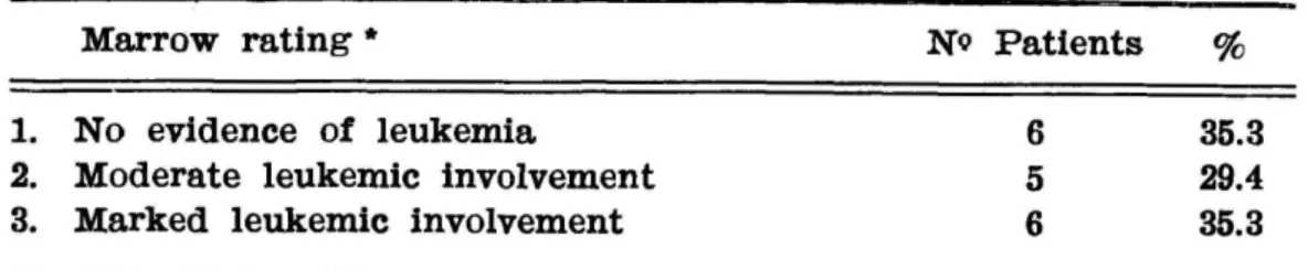
were below 10,000/cu.mm. In three patients platelet counts were available. Bone marrow findings (28) at the onset of CNS signs and symptoms suggested no evidence of leukemia in 6 patients, moderate leukemic involvement in 5 and marked involvement in 6 (Table 5). Bone marrow was not examined in one patient whose death occurred within 24 hours after admission. Lumbar puncture was performed in 8 patients. T h e cerebrospinal
fluid (CSF) was normal in three cases. Initial pressure higher than 150 mm of water was seen in four cases. Pleocytosis of mononuclear cells was found in t w o cases (18 and 726 cells/cu.mm respectively) and of hematias in three cases (30, 40 and 371 cells/cu.mm respectively). Protein levels exceeded 50 m g % in three cases, the highest value being 330 m g % whereas glucose levels were normal (45-60 m g % ) in all patients. T h e total survival time ranged from 8 to 2,250 days with a median of 300 days. The survival time after CNS involvement ranged from 1 to 180 days with a median of 21 days. zyxwvutsrqponmlkjihgfedcbaZYXWVUTSRQPONMLKJIHGFEDCBA
Table 5 — Bone marrow findings at onset of CNS involvement. (*) Aooording to Sullivan et al. 28.
COMMENTS
This autopsy study reveals that the involvement of the CNS is more
commonly found in chronic myelogenous leukemia (CML) than in the other
types of leukemia. In some clinical s e r i e s
4»
1 5»
3 1acute lymphocytic leukemia
(AAL) has been found responsible for the great majority of cases with CNS
lesions. This discrepancy might be attributed to a lower clinical suspiction
of CNSL in CML si. There was no significant difference of CNSL for the
two sexes as seen in other series
15,20,22,29,30although some authors
25,31have
found a higher incidence in males. CNS involvement was found to occur
throughout the course of the disease from early as two days after the clinical
diagnosis of systemic leukemia to as late as 6 years. At the time of diagnosis
of CNSL most patients were in status 111 (moderate disease). A similar finding
was observed by Hyman et a l .
1 5. In other series
2 3«
2 r» CNS manifestations
of leukemia occurred mostly while the systemic disease was apparently under
therapeutic control (stage I and II). Bone marrow examination at the time
of diagnosis of CNSL has shown no evidence of leukemia in 33.3% of the
cases further indicating the CNS lesions may occur in other than the terminal
phase of the systemic disease. A wide variation of symptoms and signs was
present in our cases avoiding the formulation of any specific diagnostic criteria.
CSF examination is frequently helpful in establishing a diagnosis of CNS
involvement. Changes in CSF pressure, number of cells and protein content
were found in about 63 per cent of the patients in whom lumbar puncture
was performed. In the remaining patients no CSF abnormality was detected,
confirming the finding of other authors that CSNL may be associated with
normal C S F
1 5>
2 3. As leukemic cells may often be seen in the CSF when
appropriate tecniques of cytology are employed
1 1a reducing number of normal
lumbar punctures may be expected as those techniques become routinely used.
The pathogenesis of C N S L
1 9is related to the diffuse leukemic cell
infiltration of the arachnoid
zyxwvutsrqponmlkjihgfedcbaZYXWVUTSRQPONMLKJIHGFEDCBA 2 1, 2 3 , 2 0 . Leukemic involvement initially becomesDemyelinated plaques are thought to represent progressive multifocal
leucoencephalopathy, a common pathological finding in lymphoproliferative
disorders. Papova-like virus has been demonstrated in the CNS in these
conditions
3 2and correspond to the eosinophilic nuclear inclusions seen in
oligodendrocytes. Wether the disease results from activation of virus residing
in a latent form in the brain or invasion of the brain by virus normally
residing in a harmless manner in extraneural tissues, or a primary infection
by Papova-virus is not known. Cerebral calcifications have been described
previously in CNS
2>°>
2 1and found to represent complications of X-ray therapy
and intrathecal methothrexate treatment.
The development of CNSL is usually thought to indicate a poor
progno-sis
zyxwvutsrqponmlkjihgfedcbaZYXWVUTSRQPONMLKJIHGFEDCBA4,12,13,23,25.
Evans et al.
4
showed that the median survival predicted for
those who develops CNS symptoms is 8 months; for those who do not, it is 24
months. However H y m a n
1 5suggests that there is no shortening of the survical
time when CNS leukemia is early and correctly treated. In an attempt to prevent
or delay involvement of CNS by leukemia cranial irradiation and intrathecal
methotrexate are usually added to combination chemotherapy (vincristine,
prednisone, 6-mercaptopurine, methotrexate, cyclophosphamide)
1.3,9,14,15,18,25.
Dearth et a l .
3still recommend additional utilization of l e u k a p h a r e s i s i n order
to reduce the blast cell population to safe levels.
SUMMARY
Post-mortem clinical and pathological study of 18 cases of central nervous
system leukemia showed that this complication occurred mostly in chronic
myelogenous leukemia ( 3 8 . 8 % ) . No diagnostic criteria was found. The great
majority of signs and symptoms were related to either d sturbances of the
mental status or cranial nerves dysfunction. Cerebrospinal fluid may be found
normal. CNS involvement may occur at any time during the course of systemic
leukemia, when the disease is under apparently good therapeutic control as
well as during relapse. Pathological findings in order of decre sing frequency
were: parenchymal hemorrhage ( 6 1 % ) ; subarachnoid hemorrhage ( 5 5 % ) ;
meningeal infiltrates ( 4 4 % ) ; leukostasis ( 2 8 % ) ; edema and herniation ( 2 8 % ) ;
parenchymal infiltrates ( 2 2 % ) ; ischemic infarcts ( 1 7 % ) ; progressive multifocal
leucoencephalopathy ( 1 0 % ) ; calcifications ( 5 % ) ; meningitis ( 5 % ) . Total
survival time ranged from 8 to 1980 days a median of 300 days. Survival
time after CNS involvement ranged from 1 to 180 days with a median of
21 days.
RESUMO zyxwvutsrqponmlkjihgfedcbaZYXWVUTSRQPONMLKJIHGFEDCBA
Leucemia do sistema nervoso central: estudo clinico e patológico.
houve um quadro clínico típico, mas na grande maioria dos casos os sinais
e sintomas estavam relacionados ou a alterações do estado mental ou à
disfunção de nervos cranianos. O líquor pode ser normal. Os achados
pato-lógicos em ordem decrescente de frequência foram: hemorragia intraparenqui¬
matosa ( 6 1 % ) ; hemorragia subaracnóidea ( 5 5 % ) ; infiltrados meníngeos ( 4 4 % ) ;
leucostase ( 2 8 % ) ; edema e herniação ( 2 8 % ) ; infiltrado n parênquima ( 2 2 % ) ;
infarto isquêmico ( 1 7 % ) ; leucoencefalopatia progressiva multifocal ( 1 0 % ) ; cal¬
cificações ( 5 % ) ; meningite ( 5 % ) . O tempo total de sobrevida variou de 8
a 1.980 dias, com uma mediana de 300 dias. O tempo de sobrevida após
acometimento do sistema nervoso central variou de 1 a 180 dias com uma
mediana de 21 dias. O acometimento do sistema nervoso central pod ocorrer
em qualquer época durante o curso de leucemia sistêmica, mesmo quando a
doença está aparentemente sob bom controle terapêutico, mas é mais comum
durante os seus relapsos.
R E F E R E N C E S
1. A U R , R . J . A . ; SIMONE, J . ; HUSTU, H . O . ; W A L T E R S . T . ; B A R E L L A , L . ; P R A T T , C. & P I N K E L , D . — Central nervous system therapy, and combination c h e m o -therapy of childhood lymphocytic leukemia. Blood 37:272, 1971.
2. CROSLEY, C.J.; R O R K E , L . B . ; EVANS, A. & N I G R O , M. — Central nervous system lesions in childhood leukemia. Neurology (Minneapolis) 28:678, 1978.
3. D E A R T H , J . C . ; FOUNTAIN, K . S . ; SMITHSON, W . A . ; B U R G E T , E.O. & G I L C H R I S T , G.S. — Extreme leukocytosis (blast crisis) in childhood. M a y o Clin. Proc. 53:207, 1978. 4. EVANS, A . E . ; G I L B E R T , E.S. & SANDSTRA, R . — The increasing incidence of
central nervous system leukemia in children. Cancer 26:404, 1970.
5. F I T Z P A T R I C K , H . ; L I E B E R M A N , N. & SINKS, L . F . — Staging of acute leukemia and the relationship to CNS involvement Cancer 33:1376, 1974.
6. F L A M E N T - D U R A N T , J . ; K E T E L B A N T - B A L A S S E , P . ; MAURUS, R . ; R E G N I E R , R. & S P E H L , M. — Intracerebral calcification appearing during the course of acute lymphocytic leukemia treated with methotrexate and X - r a y s . Cancer 35:319, 1975. 7. F R E I R E I C H , E.J.; THOMAS, L . B . ; F R E I , E . ; F R I T Z , R . D . & F O R K N E R , C.E.
— A distinctive type of intracerebral hemorrhage associated with blastic crisis in patients with leukemia. Cancer 13:146, 1980.
8. F R I T Z , R . D . ; F O R K N E R , C.E., J r ; F R E I R E I C H , E . H . ; F R E I , E. & THOMAS, L.B.B. — The association of fatal intracranial hemorrhage and «blastic crisis» in patients with acute leukemia. N e w Engl. J. Med. 261:59, 1959.
9. G A R W I C Z , S.; ARONSON, A.S., ELMQVIST, D. & L A N D B E R G , T. — Postirradiation syndrome and EEG findings in children with acute lymph blastic leukemia. Acta pediatr. scand. 64:399, 1975.
10. G I L B E R T , E.F. & R I C E , E.C. — Neurologic manifestation of leukemia. Pediatrics 19:801, 1957.
11. GONDOR, B. & KING, E.B. — Cerebrospinal fluid c y t o l o g y : diagnostic accuracy and comparison of different techniques. Acta cytol. 20:542, 1976.
12. GREYDANUS, D . ; B U R G E R T , E.O. & G I L C H R I S T , G.S. — Hypothalamic syndrome in children with acute lymphocytic leukemia. Mayo Clin. P r o c . 53:217, 1978.
13. H A R D I S T Y , R.M. & NORMAN, P.M. — Meningeal leukemia. Arch. Dis. Childh. 42:441, 1967.
14. H O A G L A N D , H.C. — Acute leukemia and its complications. Mayo Clin. Proc. 53: 260, 1978.
to systemic leukemia and description of clinical and laboratory manifestations. J. Hematol. 25:1, 1965.
16. L O W E N T H A L , R . M . ; B U S K A R D , N.A.; GOLDMAN, J . M . ; SPIERS, A . S . D . ; B E R ¬ G I E R , N . ; G R A U B N E R , M. & GAI/TON, D.A.G. — Intensive leukapharesis as initial therapy for chronic granulocytic leukemia. Blood 46: 835, 1975.
17. M c K E E , C. & COLLINS, R . D . — Intravascular leukocyte trombi and aggregates as a cause of morbidity and mortality in leukemia. Medicine 53:463, 1974.
IS. M E L L O R , D. — Encephalitis and encephalopathy in childhood leukemia. Develop. Med. Child. Neurol. 18:90, 1976.
19. M I L L E R , R . W . — Etiology of childhood leukemia. Ped. Clin. N. Amer. 13:267, 1966. 20. MOORE, E . W . ; THOMAS, L . B . ; S H A W , R . K . & F R E I R E I C H , E.J. — The central
nervous system in acute leukemia. Arch. int. Med. 105:142, 1960.
21. M U E L L E R , S., B E L L , W . & S E I B E R T , J. — Cerebral calcifications associated with intrathecal methotrexate therapy in acute lymphocytic leukemia. J. Ped. 88:650, 1976.
22. P H A I R , J . P . ; ANDERSON, R . E . & N A M I K I , H. — The central nervous system in childhood leukemia. Ann. int. Med. 61:863, 1964.
23. P I E R C E , M.I. — Neurologic complications in acute leukemia in children. Ped. Clin. N. Amer. 9:425, 1962.
24. P R I C E , R.A. & JOHNSON, W . W . — The central nervous system in childhood leukemia. I. The arachnoid. Cancer 31:520, 1973.
25. S H A W , R . K . ; M O O R E , E . W . ; F R E I R E I C H , E.J. & THOMAS, L.B. — Meningeal leukemia. Neurology (Minneapolis) 10:823, 1960.
26. S T E F F E Y , J.M. — The central nervous system manifestations of leukemia. J. P e d . 60:183, 1962.
27. SULLIVAN, M.P. — Intracranial complications of leukemia in children. Pediatr. 5:751, 1957.
28. SULLIVAN, M . P . ; B A T T Y , E.C.; HYMAN, C.B.; M U R P H Y , L . M . ; P I E R C E , M..i. & SEVERO, N.C. — A comparison of the effettiveness of standard dose of 6-mer¬ captopurine, combination 6-mercaptopurine and DON, and high loading, 6-mercapto¬ purine therapies. Cancer Chemotherapy Reports 18:83, 1962.
29. W E L L S , C.E. & S I L V E R , R . T . — The neurologic manifestations of the acute leukemias: a clinical study. Ann. int. Med. 46:439, 1957.
30. W E S T , R . J . ; G R A H A M - P O L E , J . ; H A R D I S T Y , R.M. & P I K E , M.C. — Factors in pathogenesis of central nervous system leukemia. Brit. med. J. 3:311, 1972.
31. W O L K , R . W . ; MASSE, S.R.; CONKLIN, R . & F R E I R E I C H , E.J. — The. incidence of central nervous system leukemia in adults wich acute leukemia. Cancer 33: 863, 1974.
32. ZU R H E I N , G. & CHAN, S. — Particles resembling papovaviruses in human cerebral
demyelinating disease. Acta neuropath. 8:57, 1967. zyxwvutsrqponmlkjihgfedcbaZYXWVUTSRQPONMLKJIHGFEDCBA