C-banding and fluorescent
in situ
hybridization with rDNA sequences in
chromosomes of
Cycloneda sanguinea
Linnaeus (Coleoptera, Coccinellidae)
Eliane Mariza Dortas Maffei1, Silvia das Graças Pompolo2and Eduard Petitpierre3
1
Universidade Estadual do Sudoeste da Bahia, Departamento de Ciências Naturais, Vitória da Conquista, BA, Brazil.
2
Universidade Federal de Viçosa, Departamento de Biologia Geral, Viçosa, MG, Brazil. 3
Institut Mediterrani d’ Estudis Avançats, Department de Recursos Naturals, Palma de Mallorca, Spain.
Abstract
The aim of this study was to describe mitotic and meiotic chromosomes ofCycloneda sanguinea using C-banding, fluorescentin situ hybridization (FISH) rDNA probes, and sequential FISH/Ag-NOR staining. The chromosome number was 2n = 18 + XX for females and 2n = 18 + Xy for males. The X chromosome was metacentric and the Y chromosome was very small. During meiosis, the karyotypic meioformula was n = 9 + Xyp, and sex chromosomes
configured a parachute at metaphase I. At the beginning of pachytene, bivalents were still individualized, and sex chromosomes were associated end-to-end through the heteropycnotic region of the X chromosome. Later in pachytene,further condensation led to the formation of a pseudo-ring by the sex bivalent. All chromosomes showed
pericentromeric heterochromatin. FISH and sequential FISH/Ag-NOR staining evidenced the location of the nucleolar organizer region in one pair of autosomes (at spermatogonial metaphase). During meiosis, these genes were mapped to a region outside the sex vesicle by FISH, although Xypwas deeply stained with silver at metaphase I.
These results suggest that these argyrophilic substances are of a nucleolar protein nature, and seem to be synthesized by a pair of autosomes and imported during meiosis (prophase I) to the sex pair, during the association of the sex chromosomes.
Key words: in situhybridization, rDNA sequences, sequential FISH/Ag-NOR staining, C-banding,Cycloneda sanguinea.
Received: March 19, 2003; Accepted: November 14, 2003.
Introduction
The lady beetleCycloneda sanguineabelongs to the family Coccinellidae, a cosmopolitan taxon that comprises 5000 species, 2000 of which are Neotropical species (Iabhlokoff-Khuzorian, 1982). Many species of this family are highly active predators and are intensively used for bio-logical control (Borror and De Long, 1988; De Bach, 1964; Gordon, 1985; Levins and Wilson, 1980).
Cytogenetic analyses of the family Coccinellidae are scarce, and the karyotypes of only 4% of the species are known. All of them are characterized by meiosis with an n = 9 + Xypin males. Sex chromosome association during
metaphase shows the typical “parachute” shape that charac-terizes Coleoptera. This peculiar type of association has been traditionally explained as being due to the presence of the nucleolar organizer region (John and Lewis 1960;
Smith and Virkki 1978; Virkkiet al. 1991), and fluorescent
in situ hybridization (FISH) using 18S-28S rDNA gene probes has corroborated this theory for some Australian species ofCicindela(Galián and Hudson, 1999) and for
Olla v-nigrum(Maffeiet al., 2001a). However, the nucleo-lar theory was questioned by other investigators, who found nucleoli in a pair of autosomes in other species (Dretset al., 1983; Postiglioni and Brum- Zorrilla 1988; Postiglioniet al. 1991).
Banded karyotypes of Coccinellidae are almost un-known, except for C-banding in six species of the genus Chilocorus, Epilachna paenulata, Eriopis connexa and seven species of lady-birds from Central Europe (Ennis, 1974; Drets et al., 1983; Maffeiet al, 2000; Rošek and Holecová, 2002).
The aim of the present study was to describe the mi-totic and meiotic chromosomes in females and males of
Cycloneda sanguinea, establishing their C-banding pattern and physical mapping of rDNA sequences using FISH and sequential FISH/Ag-NOR staining.
www.sbg.org.br
Send correspondence to E.M.D. Maffei. Universidade Estadual do Sudoeste da Bahia, Departamento de Ciências Naturais, Estrada do Bem Querer, km 4, Vitória da Conquista, 45050-200 Bahia, Ba, Brazil. E-mail: maffeiemd@hotmail.com.
Material and Methods
The Cycloneda sanguinea L. specimens were col-lected on the campus of the Universidade Federal de Viçosa (UFV). Mitotic and meiotic analyses were carried out in 12 prepupal larvae and 40 male adults, with a sample size of 10 mitotic metaphases per individual. Cytogenetic prepara-tions of mitotic metaphase chromosomes (cerebral gan-glion) and meiosis analysis were carried out as described by Imaiet al. (1988) and adapted for Coccinellidae (Maffeiet al., 2000).
C-banding was performed as described by Sumner (1972) with modifications (Maffeiet al.,2000). Slides were subjected to hydrolysis with 0.2 N HCl for 4 min at room temperature, washed in distilled water and incubated with 5% barium hydroxide at 60 °C for 8 min, then washed in 0.2 N HCl at room temperature for 30 s, incubated with 2xSSC at 60 °C for 10 min, and stained with 6.6% Giemsa in Sörensen buffer pH 6.8 for 50 min.
Sequential FISH/Ag-NOR staining followed the technique described by Maffeiet al. (2001a). One drop of 2% gelatin (2 g gelatin diluted in 100 mL distilled water and 0.5 mL formic acid) and four drops of aqueous 50% Ag-NO3solution (Merck) were added onto slides already
hybridized with rDNA probes. The slides were then cov-ered with a coverslip and incubated in a moist chamber at 38% humidity for 25 min. Finally, the material was thor-oughly rinsed with a squirt bottle for approximately 5 min, air-dried and mounted with Entellan (Merck).
Ribosomal DNA sequences were mapped using FISH (Viegas-Péquignot, 1992). The rDNA probes (pDm 238) containing the 18S, 28S and 5.8S genes were labeled with bi-otin by nick translation using the Bionick kit (Gibco). Slides with fixed meiotic chromosomes were incubated with RNase (100µg/mL) for 1 h in a moist chamber containing
70% formamide at 37 °C, followed by three incubations in 2xSSC for 3 min each, and dehydrated in 50%, 75% and 100% alcohol for 3 min each. After denaturation of the chro-mosomal DNA in 70% formamide for 2 min at 70 °C, the slides were dehydrated in ice-cold 2xSSC for 2 min and im-mersed in 50%, 75% and 100% alcohol for 2 min each. In parallel, the probe was incubated at 100 °C for 2 min and maintained on ice. Ten microliters of the probe (diluted in hybridization mixture) per hybridization area were then added onto the slides, which were incubated in a 70% formamide moist chamber at 37 °C for 36 h. After incuba-tion, the slides were immersed twice in 50% formamide at 37 °C for 2 min each, and washed with 2xSSC at 37 °C for 2 min each, followed by two washes with PBT for 2 min each.
Immunological detection
The slides were incubated with anti-biotin (Vector SP3000) and anti-goat IgG-FITC (Vector FI5000). The DNA was counterstained with 100µL propidium iodide for
1 min and washed with PBS. The slides were mounted in
13 µL Vectashield and photographed under an Olympus
BX60 photomicroscope using appropriate fluorescence fil-ters.
Results
Meiosis and mitosis
C. sanguineacells in meiosis showed a large, single regular heteropycnotic chromocenter at prophase I (zygotene). At the beginning of pachytene, the bivalents were individualized and the sex chromosomes showed lin-ear end-to-end association. As pachytene progressed, DNA condensation led to the formation of a pseudo-ring by the sex bivalents (Figure 1A). At metaphase I, the meioformula was n = 9 + Xyp(Figure 1B), followed by normal
chromo-some segregation at anaphase I.
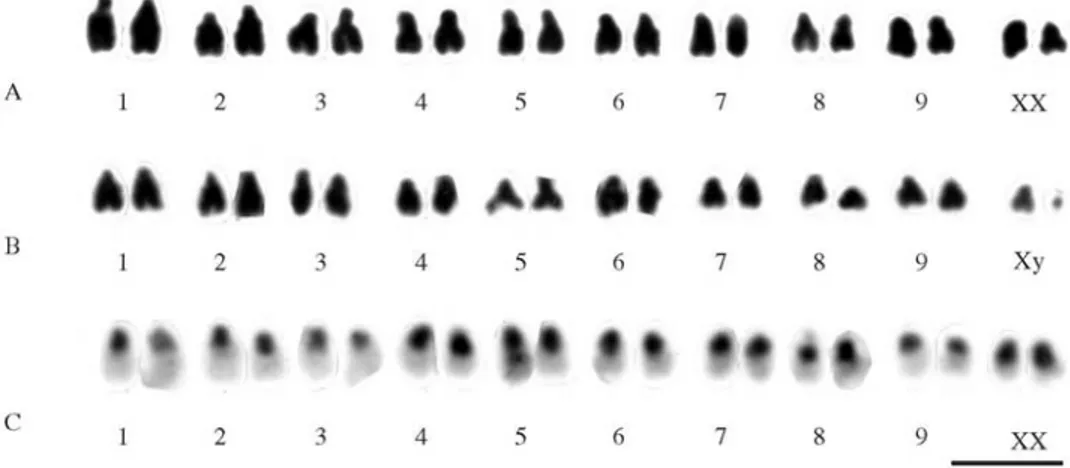
The analysis of mitotic cells with standard staining showed a chromosome number of 2n = 18 + XX in 10C. sanguineafemales and of 2n = 18 + Xy in two males. The chromosomes of this species were mainly submetacentric and the y chromosome was very small (Figures 2A and 2B). C-banding revealed heterochromatin in the pericentromeric regions of all chromosomes, including the short arms (Figure 2C).
Location of rDNA genes and Ag-NOR banding
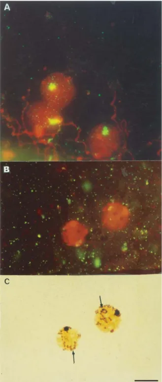
The location of rDNA sequences was evidenced by FISH as a large block at prophase I (early zygotene)
(Figure 3A). Sequential FISH/Ag-NOR staining showed that the rDNA sequences were located outside the sex vesi-cle (Figures 3B and 3C). Sequential FISH/Ag-NOR treat-ment showed a marked silver staining of both the sex vesicles (prophase I) and the lumen of the sex chromo-somes associated in a parachute format (metaphase I).
Discussion
Mitosis and meiosis
The lady-beetleCycloneda sanguineahas a chromo-some number of 2n = 18 + XX for females and a meioformula of n = 9 + Xypfor males, characteristic of
most Coleopteran species. This may also represent the ba-sic (ancestral) karyotype, especially in the suborder Polyphaga (Smith, 1950; Smith and Virkki, 1978). As re-cently reported forEriopis connexa(Maffeiet al., 2000) andOlla v-nigrum(Maffeiet al., 2001a), all members of the family Coccinellidae (42%) apparently share the same chromosomal behavior: at prophase I (pachytene) of the male meiosis, sex chromosomes appear associated end-to-end, with the short arm of the X chromosome join-ing the y chromosome in a linear fashion followed by a pseudo-ring and, finally, a parachute is visible at metaphase I, with the expanded chromatin of the short arm of the X chromosome contributing to the parachute. These results are in agreement with the model of Xypsex chromosome
association proposed by Dretset al. (1983) forEpilachna paenulata (Coleoptera, Coccinellidae). In this species, C-banding analysis revealed a sex chromosome association different from other Coleopteran species studied, due to the extensive role played by constitutive heterochromatin seg-ments in chromosome association and Xypformation. The
authors proposed a model where the parachutes consist of three different segments: two corpuscles showing intense heterochromatin upon C-banding and a euchromatic seg-ment that forms the parachute. The euchromatic segseg-ment is
acknowledged as the long arm of the X chromosome in the parachute, and the formation of the parachute itself is the result of the association between the X and y chromosomes. It has been suggested that this association is not mediated by the nucleolar organizer region, since no nucleolar mate-rial associated with the sex chromosomes was detected. The observations made by Virkkiet al. (1991) on the be-havior of Xypassociation in six Coleopteran species by
sil-ver staining supported the interpretation of Drets et al. (1983).
In the present study, the heterochromatin of C. sanguineawas mainly located in the pericentromeric re-gion of all chromosomes and in the short arms, as observed for most Coleopterans studied with C-banding technique (Ennis, 1974; Angus, 1982, 1983; Dretset al., 1983; Juan and Petitpierre, 1989; Holecováet al., 1997; Rošek and Holecová, 2000; 2002).
Location of rDNA sequences (FISH) and gene activity
Meiotic chromosomes (prophase I) and NOR-banding analysis in spermatogonial metaphase of C. sanguinea are described for the first time in the present work. In an earlier study using NOR-banding techniques in mitotic cells of adult males, Maffeiet al. (2001b) reported active regions as restricted to one pair of autosomes. They also indicated that rDNA sequences occur in a region out-side the sex vesicle (prophase I). Sequential FISH/Ag-NOR confirmed the result obtained by silver staining regarding the Xypbivalent. FISH using rDNA probes has been little
applied in Coleoptera. Juan et al. (1993) applied rDNA FISH toTenebrio molitor andMisolampus goudoti, both bearing 2n = 20 chromosomes and an Xypsex chromosome
system. InT. molitor, mitotic metaphases showed that the nucleolar organizer regions were located on two pairs of autosomes and on one pair of sex chromosome, thus sup-porting the classic hypothesis of the nucleolar origin of the
Xypassociation. In contrast, inM. goudoti, the rDNA
se-quences were restricted to one pair of autosomes. In
Cicindela melancholica, FISH mapped the rDNA se-quences to one of three X chromosomes and to the y chro-mosome (multiple sex system). However, in Cicindela
paludosa(a related species), which has an X0 sex determi-nation system, the rDNA sequences were restricted to one pair of autosomes (Galián et al., 1995). FISH for the 18S-28S rDNA genes was applied to otherCicindela spe-cies (Cicindelini tribe), and in three spespe-cies of them,
Cicindela cardinalba,Cicindelasp. (saetigera group) and
Cicindela gillesensis, these genes were mapped to two of the four sex chromosomes that contributed to the sex vesi-cle. On the other hand, inMegacephala whelani, fluores-cent hybridization during meiosis and mitosis revealed that the rDNA sequences were located on three pairs of autosomes (Galián and Hudson, 1999). This result does not support the classic nucleolar theory. The detection of argyrophilic substances inC. sanguineaduring mitosis and meiosis by FISH and sequential FISH/Ag-NOR suggests that these substances are nucleolar proteins synthesized by a pair of autosomes and imported at prophase I, during sex chromosome association.
Acknowledgements
We are grateful to Dr. Shirlei M. Recco-Pimentel, Klélia Santos, and Dr. Luciana B. Lourenço for providing the labeledDrosophilarDNA probe and for technical assis-tance, and to Dr. Lúcia Massutti de Almeida (Departa-mento de Zoologia, Universidade Federal do Paraná, Curitiba, PR) for identifying the species used in the present study. This research was supported by Fundação de Amparo à Pesquisa do Estado de Minas Gerais (FAPE-MIG), Coordenação de Aperfeiçoamento de Pessoal de Ensino Superior (CAPES) and Universidade Federal de Viçosa, Viçosa, MG, Brazil.
References
Angus RB (1982) Separation of two species standing as
Helophorus aquaticos(L.) (Coleoptera), Hydrophilidae) by banded chromosome analysis. Syst. Entomol 7:256-281. Angus RB (1983) Separation ofHelophorus grandis maritimus
andoccidentalissp. (Coleoptera, Hydrophilidae) by banded chromosome analysis. Syst. Entomol 8:1-13.
Borror DJ and De Long DM (1988) Introdução ao estudo dos insetos. Blucher, São Paulo, 653 pp.
De Bach P (1964) Biological control of insect pests and weeds. Chapman & Hall, London, 844 pp.
Drets ME, Corbella E, Panzera F and Folle GA (1983) C-banding and nonhomologous associations. II The “parachute” Xyp
sex bivalent and behavior of heterochromatic segments in
Epilachna paenulata. Chromosoma 88:249-255.
Ennis TJ (1974) Chromosome structure in Chilocorus
(Coleoptera: Coccinellidae). In: Fluorescent and Giemsa banding patterns. Can J Genet Cytol 16:651-661.
Galián J and Hudson P (1999) Cytogenetic analysis of Australian tiger beetles (Coleoptera: Cicindelidae). Chromosome num-ber, sex-determining system and localization of rDNA genes. J Zool Syst and Evol Res 37:1-6.
Galián J, Serrano J, De La Rúa P, Petitpierre E and Juan C (1995) Localization and activity of rDNA genes in tiger beetles (Coleoptera, Cicindelinae). Heredity 74:524-530.
Gordon RD (1985) Coccinellidae (Coleoptera) of America North of Mexico. J New York Entomol Society 93:1-912. Holecová M, Rozek M and Lachowska D (1997) C-banded
karyo-type ofOtiorhynchus corvusBoheman 1843 (Coleoptera, Curculionidae). Cytologia 62:209-212.
Iabhlokoff-Khuzorian SM (1982) Les Coccinelles Coleópteres Coccinellidae. Tribu Coccinellini des Regions Paléartique et Orientale. Boubeé, Paris, 568 pp.
Imai HT, Taylor RW, Crosland MWJ and Crozier RH (1988) Modes of spontaneous chromosomal mutation and karyo-type evolution in ants with reference to the minimum inter-action hypothesis. Jpn J Genet 63:159-85.
John B and Lewis KR (1960) Nucleolar controlled segregation of the sex chromosomes in beetles. Heredity 15:431-39. Juan C and Petitpierre E (1989) C-banding and DNA content in
seven species of Tenebrionidae (Coleoptera). Genome 32:834-839.
Juan C, Pons J and Petitpierre E (1993) Localization of tandemly repeated DNA sequences in beetle chromosomes by fluores-centin situhybridization. Chromosome Res 1:167-174. Levins R and Wilson M (1980) Ecological theory and pest
man-agement. Ann Rev Entomol 25:287-308.
Maffei EMD, Gasparino E and Pompolo SG (2000) Karyotypic characterization by mitosis, meiosis and C-banding of
Eriopis connexa Mulsant (Coccinellidae: Coleoptera: Polyphaga), a predator of insect pests. Hereditas 132:79-85. Maffei EMD, Pompolo SG, Campos LAO and Petitpierre E
(2001a) Sequential FISH analysis with rDNA genes and Ag-NOR banding in the lady-beetle Olla v-nigrum
(Coleoptera: Coccinellidae). Hereditas 135:13-18. Maffei EMD, Pompolo SG, Silva-Junior JC, Caixeiro APA,
Rocha MP and Dergam JA (2001b) Silver staining of
nucle-olar organizer regions (NORs) in some species of Hymenoptera (bees and parasitic wasp) and Coleoptera (lady-beetles). Cytobios 104:119-125.
Postiglioni A and Brum-Zorrilla N (1988) Non-relationship be-tween nucleolus and sex chromosome system Xyp in
Chelymorpha variabilis Boheman (Coleoptera: Chrysomelidae). Genetica 77:137-41.
Postiglioni A, Stoll M and Brum-Zorrilla N (1991) Haploid karyotype analysis of Chelymorpha variabilis Boheman (Coleoptera, Chrysomelidae) with microspreading tech-niques. Rev Brasil Genet 14:653-60.
Rošek M and Holeková M (2000) C-banding patterns in chromo-somes and sperm of Strophosoma capitatum (De Geer, 1775) (Coleoptera: Curculionidae, Brachyderinae). Folia Biologica (Kraków) 48:33-35.
Rošek M and Holeková M (2002) Chromosome number, C-banding and sperm of some ladybird species from Central Europe (Coleoptera, Coccinellidae). Folia Biologica (Kraków) 50:17-21.
Smith SG (1950) The cytotaxonomy of Coleoptera. Can Entomol 82:58-68.
Smith SG and Virkki N (1978) Animal cytogenetics. Coleoptera 1-365.
Sumner AT (1972) A simple technique for demonstrating centro-meric heterochromatin. Exp Cell Res 75:304-306. Viegas-Péquinot E (1992)In situhybridization to chromosomes
with biotinylated probes. In: Willernson D (ed)In situ Hy-bridization: A Practical Approach. Oxford University Press, IRL, Press, pp 137-158.
Virkki N, Mazzella C and Denton A (1991) Silver staining of the coleopteran Xypsex bivalent. Cytobios 67:45-63.