Karyotype differentiation of four
Cestrum
species (
Solanaceae
)
based on the physical mapping of repetitive DNA
Jéferson Nunes Fregonezi
1, Thiago Fernandes
1, José Marcelo Domingues Torezan
2, Ana Odete S. Vieira
2and André Luís Laforga Vanzela
11
Universidade Estadual de Londrina, Centro de Ciências Biológicas, Departamento de Biologia Geral,
Londrina, PR, Brazil.
2
Universidade Estadual de Londrina, Centro de Ciências Biológicas, Departamento de Biologia Animal
e Vegetal, Londrina, PR, Brazil.
Abstract
We studied the karyotypes of four BrazilianCestrumspecies (C. amictum,C. intermedium,C. sendtnerianumandC. strigilatum) using conventional Feulgen staining, C-Giemsa and C-CMA3/DAPI banding, induction of cold-sensitive
regions (CSRs) and fluorescentin situhybridization (FISH) with rDNA probes. We found that the karyotypes of all four species was 2n = 2x = 16, with, except for the eighth acrocentric pair, a predominance of meta- and submetacentric chromosomes and various heterochromatin classes. Heterochromatic types previously unreported inCestrumas neutral C-CMA3
0
/DAPI0
bands, CMA3 +
bands not associated with NORs, and C-Giemsa/CSR/DAPI
-bands were found. The heterochromatic blocks varied in size, number, position and composition. The 45S rDNA probe preferentially located in the terminal and subterminal regions of some chromosomes, while 5S rDNA appeared close to the centromere of the long arm of pair 8. These results suggest that karyotype differentiation can occur mainly due to changes in repetitive DNA, with little modification in the general composition of the conventionally stained karyotype.
Key words: Cestrum, chromosome banding, CSRs, karyotype evolution, rDNA probes. Received: August 24, 2004; Accepted: May 31, 2005.
Introduction
Along withVestia (Willd) andSessea(RuizetPav) the genus Cestrum (L.) is part of the tribe Cestreae (G. Don) of theCestroideae-Solanaceae). The genusCestrum
contains about 250 species distributed in the tropical and subtropical regions of the Americas (D’Arcy, 1986) and in-cludes herbs, shrubs and trees up to 12 m in height. Mem-bers of this genus are recognized by their actinomorphic flowers with tubular-infundibuliforme corolla, five sta-mens attached to the corolla tube up to the middle or higher, bilocular ovary with berry-like fruits, few compressed seeds and a straight embryo (Cronquist, 1988). The karyo-type ofCestrum species contains 2n = 2x = 16 chromo-somes of similar size and shape, most of which are meta- or submetacentric except for one subtelocentric pair (Berg and Greilhuber, 1992, 1993a,b). In general,Solanaceaespecies
such as those of the generaSolanumandCapsicum(2n = 2x = 24) exhibit small chromosomes (1.2-2.4mm forSolanum
and 3.5-4.0mm forCapsicum) and constancy in the
distri-bution of meta- and submetacentric chromosomes (Ber-nardello and Anderson, 1990, Ber(Ber-nardello et al., 1994, Moscone, 1993). Nevertheless, the chromosomes of the
Cestreaeare the largest in the family, reaching up to 14mm
and containing 1.36 pg of DNA per chromosome (Berg and Greilhuber, 1992, 1993a,b, Sykorováet al., 2003a).
Berg and Greilhuber (1992, 1993a,b) studied five species of Cestrum (C. aurantiacum, C. elegans, C. fasciculatum,C. parqui, andC. strigilatum) using several banding techniques, and found four different heterochro-matic types: (i) large blocks unassociated with nucleolar or-ganizing regions (NORs); (ii) numerous smaller blocks forming intercalary, proximal or distal dots; (iii) CMA3+
(chromomycin A3) bands associated with NORs; and (iv)
DAPI+(4’-6-Diamidino-2-phenylindole) bands associated with cold-sensitive regions (CSRs). The CSRs were first described by Dyer (1963) and at present have been reported in 11 genera of monocotyledons but only three dicotyle-dons.
www.sbg.org.br
Send correspondence to André L.L. Vanzela. Universidade Esta-dual de Londrina, Centro de Ciências Biológicas, Departamento de Biologia Geral, Laboratório de Biodiversidade e Restauração de Ecossistemas, Caixa Postal 6001, 86051-990 Londrina, PR, Brazil. E-mail: [email protected].
Members ofSolanaceaeshow large diversity in hete-rochromatin accumulation and dispersion. Species of -Capsicum exhibited heterochromatic blocks in all the centromeres and secondary constrictions, besides a vari-able number of large and small bands distributed along the chromosomes (Mosconeet al., 1993). Similarly,Nicotiana
species show localized DNA repetitive segments (micro-satellites, ribosomal DNAs and telomeric sequences), while other (virus-associated) segments are dispersed along the chromosomes (Yoong Limet al., 2000), and the litera-ture suggests thatCestrumcan also follows this model.
During the research reported in this paper we com-pared the karyotypes of four BrazilianCestrumspecies (C. amictum (Schlecht.), C. intermedium (Sendtn.), C. sendtnerianum(Mart. & Sendtn.) andC. strigilatum(Ruiz & Pav.)) using Feulgen staining and chromosomal markers produced by C-Giemsa and C-CMA3/DAPI banding, CSRs
and fluorescencein situhybridization (FISH) with 45S and 5S rDNA probes. The results are discussed considering the possible mechanisms of dispersion and distribution of heterochromatic segments and of rDNA cistrons in the karyotypes of the four species.
Materials and Methods
Seeds of the fourCestrumspecies cited above (eight populations) were obtained from different localities and cultivated in tubes in the seedling nursery of the Laboratory for Biodiversity and Restoration of Ecosystems (LABRE), Biology Science Centre, State University of Londrina, Pa-raná, Brazil. Vouchers were deposited at the FUEL herbar-ium (Table 1).
Slides were prepared of root tips pre-treated with 0.05% colchicine, fixed in Carnoy solution (ethanol/acetic acid 3:1, v:v), for up to 24 h and stored at -20 °C. Conven-tional staining was carried out by the Fuelgen method. The cover slips were removed after freezing slides in liquid ni-trogen and the slides permanently mounted in Entellan. To obtain CSRs, roots were collected and pre-treated in Bristol nutritive solution (Bold, 1949) for 24 h at 0 °C and fixed in Carnoy solution. Roots were hydrolyzed in 1 M HCl, dis-sected in a drop of 45% (v/v) aqueous acetic acid and
squashed. The cover slips were removed after freezing in liquid nitrogen and the preparations stained with 2% (w/v) Giemsa and the cells photographed using Kodak Imagelink HQ ASA 25 film.
For chromosomal banding, roots were digested in an enzyme solution composed of 4% cellulase and 40% pectinase (both w/v) at 37 °C and dissected in a drop of 45% acetic acid. After removal of the cover slips, the slides were aged for three days, and then incubated in 45% acetic acid, 5% barium hydroxide and 2xSSC, pH 7,0 (Schwarzacheret al., 1980, with modifications). The materials were stained in two different ways, either with 2% Giemsa or with the fluorochromes 0.5 mg/mL CMA3for 1.5 h and 2 mg/mL
DAPI for 30 min. Slides stained with Giemsa were mounted in Entellan and those stained with the fluorochro-mes mounted with a medium composed of glycerol/ McIlvaine buffer (pH 7.0) 1:1 plus 2.5 mM MgCl2. The
cells were photographed using Kodak Imagelink HQ ASA 25 and T-max ASA 100 film.
Fluorescent in situ hybridization (FISH) was per-formed according to the procedures described by Heslop-Harrisonet al.(1991) and Cuadrado and Jouve (1994), with minor modifications. Slides were prepared as described for banding. The wheat probes pTa71 containing the 45S rDNA sequence (Gerlach and Bedbrook 1979) and pTa794 containing the 5S rDNA sequence (Gerlach and Dyer 1980) were both labeled with biotin-14-dATP by nick translation and used for FISH. Each slide was treated with 30mL of
hy-bridization mixture containing 100-150 ng of labeled probe (4mL), 50% formamide (15mL), 50% polyethylene glycol
(6mL), 20xSSC (3mL), 100 ng of calf thymus DNA (1mL),
and 10% SDS (1mL). The material was denatured at 90 °C
for 10 min, and hybridization was performed overnight at 37 °C in a humidified chamber. Post-hybridization washes were carried out in 2xSSC, 20% formamide in 0.1xSSC, 0.1xSSC and 4xSSC/0.2% Tween 20, all at 42 °C. The probes were detected with avidin-FITC conjugated and the post-detection baths were conducted in 4xSSC/0.2% Tween 20 at room temperature. Slides were mounted in 26mL of a solution composed of antifade (12.5mL) and
50% glycerol in McIlvaine buffer (pH 7) with 2.5 mM
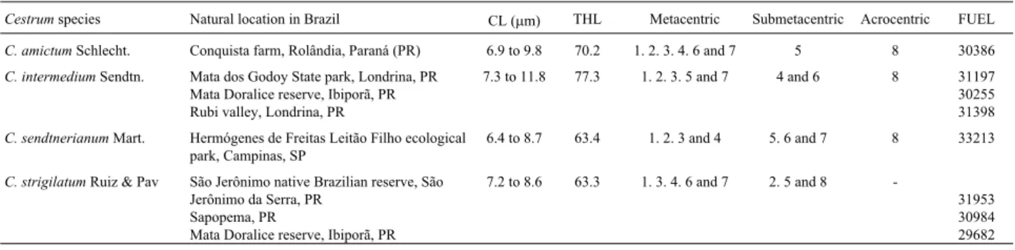
Table 1- Chromosome measurements and morphology of theCestrumspecies and populations analyzed. CL = Length of large and small chromosome pairs; THL = Total length of haploid set.
Cestrumspecies Natural location in Brazil CL (mm) THL Metacentric Submetacentric Acrocentric FUEL
C. amictumSchlecht. Conquista farm, Rolândia, Paraná (PR) 6.9 to 9.8 70.2 1. 2. 3. 4. 6 and 7 5 8 30386
C. intermediumSendtn. Mata dos Godoy State park, Londrina, PR Mata Doralice reserve, Ibiporã, PR Rubi valley, Londrina, PR
7.3 to 11.8 77.3 1. 2. 3. 5 and 7 4 and 6 8 31197 30255 31398
C. sendtnerianumMart. Hermógenes de Freitas Leitão Filho ecological park, Campinas, SP
6.4 to 8.7 63.4 1. 2. 3 and 4 5. 6 and 7 8 33213
C. strigilatumRuiz & Pav São Jerônimo native Brazilian reserve, São Jerônimo da Serra, PR
Sapopema, PR
Mata Doralice reserve, Ibiporã, PR
7.2 to 8.6 63.3 1. 3. 4. 6 and 7 2. 5 and 8
MgCl2(12.5mL), plus 1mL of 50mg/mL propidium iodide.
Photographs were taken using Kodak Proimage color ASA 100 film.
In general, 15 specimens were analyzed for each of the eight populations and idiograms constructed based on the analyses of about 10 metaphases and/or pre-metaphases obtained from each treatment. The chromosomes were classified based on the nomenclature proposed by Guerra (1986).
Results
All fourCestrumspecies studied had a diploid chro-mosome number of 2n = 2x = 16, with chrochro-mosomes vary-ing gradually in size from about 6.4mm to 11.8mm (Table
1), althoughC. intermediumexhibited slightly larger chro-mosomes. The predominant chromosome forms were meta- and submetacentric, but pair 8 ofC. strigilatumwas submetacentric while in the other species this pair was acrocentric (Table 1). Satellites were rarely seen, and the interphase nuclei were always reticulate. Karyotypical dif-ferences among the populations were not detected, even when the chromosomes were studied using several tech-niques (conventional staining, banding and FISH).
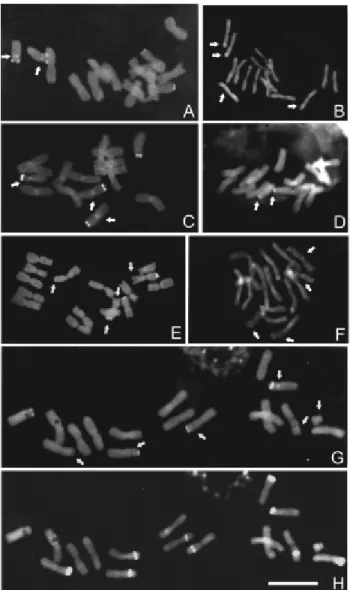
We found that C.amictumexhibited six metacentric pairs, one submetacentric and one acrocentric (Figure 1A, Table 1). C-Giemsa banding revealed heterochromatic blocks in all the chromosomes, with the intercalary bands being smaller and tenuous (or inconstant) in some prepara-tions while the terminal bands were larger and more evi-dent. The tenuous bands were distributed equidistantly on the long arms of almost all pairs except for pairs 2 and 8. Large C-Giemsa blocks were visualized mainly in the ter-minal/subterminal regions of the short arms of the minor chromosome pairs (Figures 1E and 5A) but fluorochrome banding showed large C-CMA3+ blocks associated with
NORs in the terminal regions of the short arms of pairs 6 and 8, (Figures 2A and 5A). The C-DAPI+bands were tenu-ous and slightly fluorescent, located in the terminal/subter-minal regions of the short arms of pairs 6 and 7, and on the long arms of pairs 4 and 6 (Figures 2B and 5A). Neutral bands (C-CMA30/DAPI0) appeared in the terminal regions
of the short arm of pair 3 and proximal on the long arm of pair 8 (Figures 2A, 2B and 5A). Cold-sensitive regions (CSRs) were visualized in the intercalary region of the short arm of chromosome pair 4 ofC. amictum(Figures 3A and 5A). Terminal signals on the short arms of pairs 6 and 8 were detected with FISH with the 45S rDNA probe, while FISH with the 5S rDNA probe gave a proximal signal on the long arm of pair 8 (Figures 4A, 4B and 5A).
The karyotype ofC. intermediumwas composed of five metacentric pairs, two submetacentrics and one acro-centric pair (Figure 1B and Table 1). After C-Giemsa band-ing, heterochromatic regions were visible in all the chromosomes (Figures 1F and 5B). Large C-Giemsa bands
were found in the intercalary/terminal positions of the long arms of pairs 4 and 5, in the terminal/subterminal regions of the short arm of pair 7 and 8 (both associated with NORs) and in the terminal regions of the short arms of pairs 2 and 3. More tenuous bands were detected at intercalary/termi-nal positions on the long arms of pairs 3 and 6, as well as in the intercalary part of the short arm of pair 6. Weak and in-constant Pericentromeric C-Giemsa bands occurred in pairs 1, 2, 5, 6 and 7 (Figure 5B). Fluorochrome banding re-vealed C-CMA3+ NOR-associated blocks adjacent to
C-DAPI+bands in the terminal and subterminal regions of the short arms of pairs 7 and 8 plus a tenuous neutral C-CMA30/DAPI0band in the pericentromeric region of pair 8.
Weak DAPI+ staining was also found in the peri-centromeric region of pair 7 (Figures 2C, 2D and 5B). In some preparations cold-treatment (CSR) produced evi-dence of intercalary sites on the long arms of pairs 4 and 5 and C-Giemsa banding indicated that these sites were asso-ciated with large heterochromatic blocks (Figures 3B and 5B). Fluorescentin situhybridization with the 45S rDNA probe revealed hybridization sites in the terminal region of the short arm of pair 7 and the subterminal region of pair 8 in addition to a weaker C-CMA3negative terminal site on
the long arm of pair 3. Hybridization sites with the 5S rDNA probe were found near the centromere of the long arm of pair 8 (Figures 4D and 5B).
The karyotype ofC. sendtnerianumconsisted of four metacentric pairs, three submetacentrics and one acro-centric pair (Figure 1C and Table 1). After C-Giemsa band-ing, large blocks were observable in the terminal region of the short arm of pair 1 and in the proximal region of the short arm of pair 8. Large NOR-associated terminal blocks were revealed by C-CMA3banding in both arms of pair 3
and on the short arm of pair 6. Some of these blocks also oc-curred in the intercalary regions of the short arms of pairs 1, 2, 3 and 4, on the long arm of pair 6, in the proximal region of the long arm of chromosome 7 and in both subterminal regions of pair 8. Equidistantly distributed tenuous and in-constant staining was found in the pericentromeric regions (mainly of the long arms) and intercalary/distal position of the long arms of all the chromosomes (Figure 1G). Fluoro-chrome banding showed NOR-associated terminal C-CMA3+bands on both arms of pair 3 and the short arm of
pair 6, with another intercalary band (unassociated with NORs) also occurred on the short arm of pair 6. Tenuous C-DAPI+bands were found at the subterminal regions of the short arms of pairs 3 and 7. One neutral (C-CMA30/DAPI0) band occurred in the pericentromeric
re-gion of pair 8 (Figures 2E, 2F and 5C). A single CSR was found at the intercalary position on the short arm of pair 7, coinciding with a C-Giemsa/DAPI positive band (Figures 3C and 5C). Fluorescentin situhybridization with the 45S rDNA probe revealed terminal signals in both arms of pair 3, plus a terminal site on the short arm of pair 6. In these three cases, the hybridization sites coincided with C-Giem-sa and C/CMA3 positive bands. A CMA3-negative 45S
rDNA hybridization site occurred in the terminal region of the long arm of pair 4. Hybridization with the 5S rDNA probe showed a proximal signal on the long arm of pair 8 (Figures 4C and 5C).
The karyotype ofC. strigilatumis composed of five metacentric and three submetacentric pairs (Figure 1D and Table 1). After C-Giemsa banding, large intercalary blocks were observed on both arms of pair 1, on the long arms of pairs 3 and 7 and in the subterminal regions of the short arms of pairs 3 and 6. Smaller blocks were visualized in the intercalary regions of both arms of pair 5, in the peri-centromeric regions of pairs 5 and 6 and in the subterminal regions of pairs 6, 7 and 8. Except for pair 7, all the pairs showed equidistantly distributed tenuous or inconstant
Figure 2- Chromosome banding with fluorochromes inCestrumspecies. (A) C-CMA3banding inC. amictum, arrows indicate the minor and
proxi-mal band in the pair 8. (B) C-DAPI banding inC. amictum, arrows indicate minor and interstitial bands. (C) C-CMA3banding inC. intermedium,
ar-rows indicate inconstant centromeric-pericentromeric bands and minor and proximal band in the pair 8. (D) C-DAPI banding inC. intermedium, arrows indicate major terminal bands. (E). C-CMA3 banding in C.
sendtnerianum, arrows indicate minor and terminal bands. (F) C-DAPI banding inC. sendtnerianum, arrows indicate some proximal, terminal and interstitial bands. (G) C-CMA3banding inC. strigilatum, arrows
indi-cate interstitial, proximal (pair 8) bands and a B-chromosome. (H) C-DAPI banding inC. strigilatum, arrows indicate minor interstitial bands and a B-chromosome. Bar represents 10mm.
Figure 3- Cold sensitive regions (CSRs) inCestrum. (A)C. amictum, (B)
bands in their intercalary, proximal and distal regions (Fig-ures 1H and 5D). Distal C-CMA3+bands, coinciding with
NORs and C-Giemsa bands, were found on the short arms of pairs 7 and 8 (Figure 2G) and C-Giemsa associated C-DAPI+bands were intercalary on the short arms of pairs 2, 3, 6 and 7, subterminal in pairs 4 and 8 and the peri-centromeric in pair 7 (Figure 2H). A single neutral band was detected in the proximal region of the long arm of pair 8 (Figures 2G, 2H and 5D). Cold sensitive regions were consistently observed in association with the C-DAPI+ bands of pairs 4, 6, 7 and 8, although the block observed on
the short arm of pair 2 did not constantly exhibit a CSR and the C-DAPI+block in pair 3 was not induced by the cold (Figures 3D and 5D). We found thatin situhybridization with the 45S rDNA probe gave a very similar distribution to that found inC. intermedium. There was a terminal signal on the short arm of pair 7 and subterminal in pair 8 and also a weaker C-CMA3negative terminal site on the long arm of
pair 3. Fluorescentin situhybridization with the 5S rDNA probe located a site next to the centromere on the long arm of pair 8 (Figures 4E, 4F and 5D).
Discussion
The family Solanaceae shows karyotypical unifor-mity within the same genus mainly with relation to chromo-some numbers and shapes. Illustrative examples are the species of the generaSolanum, with 2n = 24 (Bernardello and Anderson 1990, Bernardello et al. 1994), Lycium
(Stiefkens and Bernardello 2002) andCapsicum(Moscone 1993). However, variation in the chromosome base num-ber, with x = 14 and 17, has also been reported for
Hyoscyamus(Masoudet al.1999). Species ofCestrum ex-hibit uniformity in chromosome number (2n = 2x = 16) and type, including our new chromosome numbers for C. amictumand C. sendtnerianum.In general, the Cestrum
karyotypes present meta- to submetacentric chromosome
morphology for pairs 1 to 7 and subtelocentric for pair 8, as previously reported by Berg and Greilhuber (1992, 1993a,b). TheCestrumkaryotypes accumulate numerical alterations related only to the occurrence of B chromo-somes (Sykorováet al.2003b, Fregoneziet al.2004), ex-cept for a few structural changes probably associated with amplification and loss of repetitive DNA (Greilhuber 1992, 1993a,b).
The fourCestrumspecies studied by us showed hete-rochromatic blocks dispersed throughout all the chromo-somes. The heterochromatic blocks varied in size, number, position and composition, this variation being most appar-ent when comparing the 14 large blocks presappar-ent in 7 chro-mosome pairs ofC. strigilatumwith the 9 to 15 medium or large-sized blocks of C. intermedium, C. sendtnerianum
andC. amictum. The same profile appeared when compar-ing the distribution of the smaller blocks and the inconstant dots within and between the species, these dots correspond-ing to very small C-Giemsa+bands that were not detected in all the preparations and were probably due to small varia-tions in the banding and/or fixation condivaria-tions. The larger and smaller heterochromatic blocks preferentially occupied the intercalary and subterminal regions equidistantly. Five or more dots were found in some cases (e.g. C. amictum
pairs 1, 2, 3, 4 and 7, C. intermedium pair 6 and C. strigilatumpair 5) while in the other chromosomes of the four species there were at most three or four dots. The pref-erential localization of particular heterochromatic seg-ments in terminal and subterminal regions was also very evident for the C-CMA3+, C-CMA3+/NOR, C-DAPI+ and
C-DAPI+/CSRs bands, except for a few, most often interca-lary, bands found in C. amictum (pair 4) and C. intermedium(pairs 6, 4 and 5),C. sendtnerianum(pairs 6 and 7) andC. strigilatum(pairs 2, 3, 6 and 7). Similar varia-tion has been reported by Berg and Greilhuber (1992, 1993a,b) in other fiveCestrumspecies. According to Guer-ra (2000), quantitative and qualitative variation in hetero-chromatin could be common for different populations and species. Mosconeet al.(1993) reported that the amount of heterochromatin inCapsicumvaried by up to a factor of 10, with about 4% of the total length ofCapsicum chacoense
chromosomes being occupied by C-bands and up to 33% of
Capsicum campylopodium chromosomes containing
mainly centromeric C-bands along with a variable number of large and small distal bands and a few intercalary bands. Thus there are at least two possible heterochromatin disper-sion modes inCestrumspecies, non-equidistantly disper-sion (Schweizer and Ehrendorfer 1976) in which isolated bands could have appeared from initial amplification sites and equidistant dispersion (Schweizer and Loidl 1987) in which a heterochromatic region could disperse repetitive segments to nearby heterologous chromosomal arms posi-tioned at the same distance from the centromere in the ‘chromosomal field’ (Lima-de-Faria 1976).
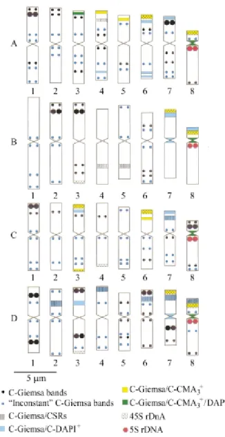
Figure 5- Idiograms and chromosomal physical mapping ofCestrum amictum (A), C. intermedium (B), C. sendtnerianum (C) and C. strigilatum(D). The ‘inconstant’ bands correspond to small C-Giemsa+
Berg and Greilhuber (1992, 1993a,b) found five dif-ferent types of heterochromatin inCestrum: (i) C-Giemsa+ bands, (ii) intercalary, proximal or distal C-Giemsa+dots, (iii) CMA3+ bands associated with NORs, (iv) CMA3+
bands not associated with NORs, and (v) DAPI+bands as-sociated with cold-sensitive regions (CSRs). Despite the different procedures employed by us our results confirm the existence of the same heterochromatic types plus two new types, i.e. C-CMA30/DAPI0 neutral bands (see also
Fregoneziet al.2004) and C-Giemsa/CSR/DAPI negative bands.
We found that the number and distribution of the CSRs varied greatly between theCestrumspecies studied,
C. strigilatumshowing a large number of CSRs while the other species showed few such sites. Variation in the size and number of CSRs has already been described in five other Cestrum species by Berg and Greilhuber (1992, 1993a, b), these authors having found a high number of CSRs in a differentC. strigilatum population and a low number in C. parqui, even though the latter species has been considered the species with the larger amount of heterochromatin. In our study qualitative and quantitative variations were evident in both the heterochromatic seg-ments and the CSRs, a fact to be expected because these regions consist of highly repetitive DNA segments. The oc-currence of CSRs in terminal positions inC. aurantiacum
(Berg and Greilhuber 1993b) and intercalary and subtermi-nal positions in the four species studied by us also indicates that CSRs can be equidistantly dispersed, except for C. amictumandC. sendtnerianumbecause CSR segments ap-pear in only one chromosome pair. In respect to AT and GC base composition, the CSRs were CMA-/DAPI+ in C. strigilatum and C. sendtnerianum but negative for both fluorochromes inC. amictum andC. intermedium. In the species studied by Berg and Greilhuber (1992, 1993a,b), CSRs were always CMA-/DAPI+. Cold-sensitive CMA3+/
DAPI0regions have been found in the genera Sambucus
andViburnum, Caprifoliaceae (Benko-Iseppon and Mora-wetz 1993). All these data suggest the existence of four CSR classes in plant genomes: (i) C-Giemsa+/DAPI+, (ii) C-Giemsa+/CMA+, (iii) C-Giemsa+/CMA0/DAPI0and (iv) C-Giemsa+ without fluorescence. However, the constitu-tion of the CSR segments does not necessarily depend on the proportion of AT or GC bases but perhaps on the type of associated proteins or the different levels of methylation among the heterochromatic segments.
Conventional staining did not allow us to visualiza-tion secondary constricvisualiza-tions in anyCestrumchromosomes, difficulty in observing satellites or secondary constrictions having previously been described in Cestrum species by Berg and Greilhuber (1992, 1993a,b) using silver nitrate (AgNO3) banding. In their study of a number ofCestrum
species Berg and Greilhuber (1992, 1993a,b) found NOR-bearing areas in the terminal region of metacentric pair 1 and subtelocentric pair 8 of all the studied species
ex-cept forCestrum parqui, the NOR region of which was not in pair 8 but in two other metacentric pairs. We found that FISH with the 45S and 5S rDNA probes was useful in local-izing rRNA genes in Cestrum. The occurrence of 45S rDNA hybridization sites in the terminal regions of Ces-trumchromosomes follows a common distribution pattern in the large majority of plant species (see Lima-de-Faria 1976). This profile has also been found in Capsicum
(Moscone et al.1995) and Nicotiana (Yoong Lim et al.
2000). In our study,C. amictumwas the only species with four 45S rDNA sites, supporting the initial proposal by Berg and Greilhuber (1992, 1993a,b). The other species showed more hybridization sites, three inC. intermedium
andC. strigilatumand four inC. sendtnerianum. Hybrid-ization also detected major and minor sites in C. strigilatum,C. sendtnerianumandC. intermedium, which could reflect intraspecific differences in the number of re-peats. Although FISH is not a completely quantitative tech-nique, the intensity of the signal can be related to the copy number of similar structures (Leicht and Heslop-Harrison 1992). We found that variation appeared not only in the number of rDNA sites but also in the organization of these segments, as seen in the occurrence of 45S rDNA segments not associated with C-CMA3+ heterochromatin. We also
found thatin situhybridization with the 5S rDNA probe re-vealed a single proximal site on the long arm of pair 8 in the four species, also associated with a proximal Giemsa+/ CMA+/DAPI+ band. In other genera such as Nicotiana
(Yoong Lim et al. 2000, Kitamura et al. 2001) and
Lycopersicon(Lapitanet al.1991, Xu and Earle 1996) 5S rDNA sites are also located preferentially in the pericen-tromeric region of the long and short arms. The available information points to the conclusion that, at least for Ces-trumspecies, the location of 45S and 5S rDNA sequences can be conserved in the chromosome field, in the terminal and subterminal region of some chromosomes (45S) and next to the centromere on the long arm of pair 8 (5S).
All theCestrumspecies so far studied exhibited a uni-form karyotype. We found that certain heterochromatic segments showed preferential localization (e.g.C-CMA3+/
NOR blocks located terminally) or appeared in the same chromosome pairs in all species (e.g.C-Giemsa+/CMA0/ DAPI0pericentromeric bands and 5S rDNA sites in pair 8). However, the majority of heterochromatic segments showed variation in size and distribution among the species studied. This suggests that differentiation amongCestrum
species occurred mainly through alterations in the organi-zation of repetitive DNA segments, with few modifications in karyotype composition as observed by conventional staining.
Acknowledgments
(ProPPG-UEL) for financial support, Edson Mendes Fran-cisco and Karina L. V. Ramalho de Sá for help with botani-cal material and Dr. Albert Leyva for help with the English.
References
Benko-Iseppon AM and Morawetz W (1993) Cold-induced chro-mosome regions and karyosystematics inSambucusand Vi-burnum. Bot Acta 106:183-192.
Berg C and Greilhuber J (1992) Cold-sensitive chromosome re-gions and their relation to constitutive heterochromatin in
Cestrum parqui(Solanaceae). Genome 35:921-930. Berg C and Greilhuber J (1993a) Cold-sensitive chromosome
re-gions and heterochromatin in Cestrum (Solanaceae): C. strigilatum,C. fasciculatum, andC.elegans. Plant Syst Evol 185:133-151.
Berg C and Greilhuber J (1993b) Cold-sensitive regions and heterochromatin in Cestrum aurantiacum (Solanaceae). Plant Syst Evol 185:259-273.
Bernardello LM and Anderson GJ (1990) Karyotypic studies in
Solanum section Basarthrum (Solanaceae). Am J Bot 77:420-431.
Bernardello LM, Heiser CB and Piazzano M (1994) Karyotypic studies inSolanumsectionLasiocarpa(Solanaceae). Am J Bot 81:95-103.
Bold HC (1949) Bristol’s solution and medium. http://www.pai. utexas.edu/research/utex/media/bristol.html. Accessed in 02/06/2003.
Cronquist A (1988) The Evolution and Classification of Flowering Plants. The New York Botanical Garden, New York, 555 pp.
Cuadrado A and Jouve N (1994) Mapping and organization of highly-repeated DNA sequences by means of simultaneous and sequential FISH and C-banding in 6x-Triticale. Chrom Res 2:231-338.
D’Arcy WG (1986) Solanaceae: Biology and Systematics. Co-lumbia University Press, New York, 603 pp.
Dyer AF (1963) Allocyclic segments of chromosomes and the structural heterozygosity that they reveal. Chromosoma 13:545-576.
Fregonezi JN, Rocha C, Torezan JMD and Vanzela ALL (2004) The occurrence of different Bs inCestrum intermediumand
C. strigilatum (Solanaceae) evidenced by chromosome banding. Cytogenet Genome Res 106:184-188.
Gerlach WL and Bedbrook JR (1979) Cloning characterization of ribosomal RNA genes from wheat and barley. Nucleic Acids Res 7:1869-1885.
Gerlach WL and Dyer TA (1980) Sequence organization of the repeting units in the nucleus of wheat which contain 5S rDNA genes. Nucleic Acids Res 8:4851-4865.
Guerra M (1986) Reviewing the chromosome nomenclature of Levanet al.Rev Bras Genet 9:741-743.
Guerra M (2000) Patterns of heterochromatin distribution in plant chromosomes. Genet Mol Biol 23:1029-1041.
Heslop-Harrison JS, Schwarzacher T, Anamthawat-Jonsson K, Leitch AR, Shi M and Leitch IJ (1991)In situhybridization
with automated chromosome denaturation. Technique 3:106-109.
Kitamura S, Inoue M, Shikazono N and Tanaka A (2001) Rela-tionship amongNicotianaspecies revealed by the 5S rDNA spacer sequence and fluorescence in situ hybridization. Theor Appl Genet 103:678-686.
Lapitan NLV, Ganal MW and Tanksley SD (1991) Organization of the 5S ribosomal RNA genes in the genome of tomato. Genome 34:509-514.
Leitch IJ and Hesolp-Harrison JS (1992) Physical mapping of the 18S-5.8S-26S rRNA genes in barley byin situ hybridiza-tion. Genome 35:1013-1018.
Lima-de Faria A (1976) The chromosome field I. Prediction of the location of ribosomal cistrons. Hereditas 83:1-22.
Masoud S, Maryam M and Mahboobeh K (1999) Karyological studies inHyoscyamusspecies of Iran. Nord J Bot 19:369-373.
Moscone EA (1993) Estudios cromosomicos en Capsicum
(Solanaceae) II. Analisis cariotipico deC. parvifoliumyC. annuumvar.annuum. Kurtziana 22:9-18.
Moscone EA, Lambrou M, Hunziker T and Ehrendorfer F (1993) Giemsa C-banded karyotypes inCapsicum (Solanaceae). Plant Syst Evol 186:213-229.
Moscone EA, Loidil J, Ehrendorfer F and Hunziker AT (1995) Analysis of active nucleolus organizing regions inCapsicum
(Solanaceae) by silver staining. Am J Bot 82:276-287. Schwarzacher TP, Ambros P and Schweizer D (1980) Application
of Giemsa banding to orchid karyotype analysis. Plant Syst Evol 134:293-297.
Schweizer D and Ehrendorfer FW (1976) Giemsa banded karyo-types, systematics, and evolution in Anacyclus
(Asteraceae-Anthemideae). Plant Syst Evol 126:107-148. Schweizer D and Loidl J (1987) A model for heterochromatin
dis-persion and the evolution of C-banded patterns. Chrom To-day 9:61-74.
Stiefkens L and Bernardello G (2002) Karyotypic studies in
LyciumsectionMesocope(Solanaceae) from South Amer-ica. Caryologia 55:199-206.
Sykorová E, Yoong Lim K, Chase MW, Knapp S, Leitch IJ, Leitch AR and Fakjus J (2003a) The absence of
Arabidopsis-type telomeres inCestrumand closely related generaVestiaandSessea(Solanaceae): First evidence from eudicots. Plant J 34:283-291.
Sykorová E, Yoong Lim K, Fakjus J and Leitch AR (2003b) The signature of theCestrumgenome suggests an evolutionary response to the loss of (TTTAGGG)ntelomeres.
Chromo-soma 112:164-172.
Xu J and Earle ED (1996) Direct FISH of 5S rDNA on tomato pachytene chromosomes places the gene at the heteromatic knob immediately adjacent to the centromere of chro-mosome 1. Genome 39:216-221.
Yoong Lim K, Matyasek R, Lichtenstein CP and Leitch AR (2000) Molecular cytogenetic analyses and phylogenetic studies in theNicotianasection Tomentosae. Chromosoma 109:245-258.