Contents lists available atScienceDirect
Vaccine
j o u r n a l h o m e p a g e : w w w . e l s e v i e r . c o m / l o c a t e / v a c c i n e
Epitope mapping and protective immunity elicited by adenovirus expressing
the
Leishmania
amastigote specific A2 antigen: Correlation with
IFN-
␥
and cytolytic activity by CD8+ T cells
Daniela M. Resende
a, Bráulia C. Caetano
h, Míriam S. Dutra
a, Marcus L.O. Penido
a,
Christiane F. Abrantes
c, Rodrigo M. Verly
d, Jarbas M. Resende
d, Dorila Piló-Veloso
d,
Simone Aparecida Rezende
e, Oscar Bruna-Romero
f,
Ana Paula Fernandes
g, Ricardo T. Gazzinelli
a,b,h,∗aDepartment of Biochemistry and Immunology, Federal University of Minas Gerais, Belo Horizonte, MG, 31270-901, Brazil
bRené Rachou Institute, Oswaldo Cruz Foundation, Belo Horizonte, MG, 30190-002, Brazil
cLaboratório Hertape Calier Saúde Animal, Juatuba, MG, 35675-000, Brazil
dDepartment of Chemistry, Federal University of Minas Gerais, Belo Horizonte, MG, 31270-901, Brazil
eSchool of Pharmacy, Federal University of Ouro Preto, Ouro Preto, MG, 354000-000, Brazil
fDepartment of Microbiology, Federal University of Minas Gerais, Belo Horizonte, MG, 31270-901, Brazil
gSchool of Pharmacy, Federal University of Minas Gerais, Belo Horizonte, MG, 31270-901, Brazil
hDepartment of Medicine, University of Massachusetts Medical School, Worcester, MA, 01605, United States
a r t i c l e
i n f o
Article history: Received 3 May 2008 Accepted 26 May 2008 Available online 25 June 2008
Keywords: Leishmaniais Recombinant vaccine Epitope mapping
a b s t r a c t
A2 was identified as an amastigote virulence factor ofLeishmania(Leishmania)donovaniand as a candidate antigen for vaccine development against visceral leishmaniasis. Here, predicted hydrophilic, class I and II MHC-binding synthetic peptides were used to define epitopes recognized by A2-specific antibodies, CD8+ T and CD4+ T cells, respectively. Immunization of BALB/c mice with adenovirus expressing A2 (AdA2) resulted in low antibody response, contrasting with high levels of IFN-␥producing CD4+ T and CD8+ T cells specific for A2. Further, A2-specific CD8+ T cells from immunized mice were capable of lysing sensitized target cellsin vivo. Finally, we demonstrated an association of A2-specific T cell responses and reduced parasitism in both liver and spleen from mice immunized with AdA2 and challenged withL.(L.)chagasi.
© 2008 Elsevier Ltd. All rights reserved.
1. Introduction
Leishmaniaparasites are distributed worldwide, and in some geographic areas more than one species can be found causing different clinical manifestations of tegumentary and visceral lesh-maniasis. Leishmaniasis is endemic in many countries in Asia and South America[1], and its control is difficult due to zoonotic fea-tures of transmission and the sylvatic nature of reservoirs and vectors[2]. In this context, an efficient vaccine would be desirable to control the disease.
A2 proteins are predominantly expressed in the amastigote stage ofLeishmaniaparasites, first described inLeishmania( Leish-mania)donovani. These proteins are composed mostly of a variable
∗Corresponding author at: Laboratory of Immunopathology, René Rachou Insti-tute, Oswaldo Cruz Foundation, Av. Augusto de Lima 1715, Centro, Belo Horizonte, MG, 30190-002, Brazil. Tel.: +55 31 3349 7774; fax: +55 31 3295 3115.
E-mail addresses:ricardo.gazzinelli@umassmed.edu,ritoga@cpqrr.fiocruz.br
(R.T. Gazzinelli).
number of 10-amino-acid repeats and their molecular weights may range from 45 to 100 kDa[3]. Thus, structurally, A2 has some resem-blance to the circumsporozoite protein (CS) fromPlasmodiumspp. [4], a main vaccine candidate for an anti-malaria vaccine[5]. A2 was identified as an important virulence factor of the parasite[6], but its role in infection process remains to be defined[7]. Inter-estingly,L.(L.)majorparasites, which express a truncated form of A2 and are associated with cutaneous leishmaniasis, after transfec-tion to express theL.(L.)donovani A2gene displayed an increased potential to survive in the resident macrophages of the spleen and liver, suggesting that expression ofA2genes may be important to visceralization of parasites in mice[8].
Antibody responses to A2 are observed in a significant num-ber of humans and dogs infected with old world and new world species ofLeishmania[9]. Moreover, in a recent study, comparing different proteins for diagnosis of leishmaniasis in dogs, A2 showed the best results to identify asymptomatic dogs, suggesting that it is associated with protective immunity[10]. In addition, A2 was identified as capable of stimulating in vitro CD4+ T and CD8+ T cells from infected C3H/HeJ mice, during a systematic screening of
L.(L.)chagasiamastigote antigens[11]. It was also demonstrated that A2, in DNA or protein formulations, can protect mice against
L.(L.)donovani[12,13],L.(L.)amazonensis[14,15]andL.(L.)chagasi
[15].
Vaccines against leishmaniasis should be capable of eliciting immune responses mediated by IFN-␥ producing CD4+ T cells [16,17]. It was also reported that CD8+ cytotoxic T lymphocytes could promote killing of infected cells, therefore contributing to disease control[18–21]. In this context, recombinant DNA plasmids and/or viruses seem attractive vehicles for generation of vaccines against this parasite, since they are efficient inducers of protective immunity mediated by both antigen-specific CD4+ Th1 cells as well as CD8+ T lymphocytes[17,22,23].
In this work, we defined and characterized the structure of the main B cell epitope in A2 and, more importantly, we mapped MHC-I, for the first time in aLeishmaniaantigen, and MHC-II binding pep-tides derived from A2 sequence. Finally, we vaccinated mice with a recombinant adenovirus encoding theA2gene fromL.(L.)donovani, and showed that homologous prime/boost protocol is effective in inducing both IFN-␥-secreting CD4+ T and CD8+ T cells as well as cytolytic CD8+ T lymphocytes, which protect mice againstL.(L.)
chagasiinfection.
2. Materials and methods
2.1. Mice
Two pairs of congenic mouse strains were used: (i) BALB/c (H2d) (parental lineage) and C.B10-H2 (H2b) (CB10); and (ii) C57BL/6 (H2b) (parental lineage) and C57BL/KsJ (BKS) (H2d); on the (BALB/c) and (C57BL/6) genetic background, respectively. Female, 6–8 weeks old mice were obtained from the animal facilities from Rene Rachou Institute, Oswaldo Cruz Foundation, Belo Horizonte, MG, Brazil and housed according to institutional standard guide-lines.
2.2. Parasites and antigen preparations
L.(L.)chagasi (MHOM/BR/1975/M2682) was kindly provided by Maria Norma Mello (Department of Parasitology, Federal Uni-versity of Minas Gerais, MG, Brazil). Parasites were grown at 23◦C in supplemented Grace’s Medium (Sigma, St. Louis, MO). Soluble Leishmania antigen (SLA) was prepared from late-log-phase promastigotes of L. (L.) chagasi after few passages in liquid culture as previously described [24]. rA2 was purified using a nickel affinity chromatography, as described elsewhere [9].
2.3. Synthetic peptides
We used the BIMAS (http://bimas.dcrt.nih.gov/molbio/hla bind/) and ProtScale (http://www.expasy.ch/cgi-bin/protscale.pl) soft-wares to predict, respectively, the MHC class I binding peptide (CD8) and B cell epitope (B-1) fromL.(L.)donovaniA2 sequence (GenBank S69693)[25]. Two MHC class II binding epitopes (CD4-1 and CD4-2), with 17 residues each, were designed by overlapping of the non-repetitive segment of the protein. Peptides B-1, CD8, CD4-1 and CD4-2 were purchased from Biosynthesis, Inc. (Lewisville, TX). Other peptides derivated from B-1 and CD4-1 (SP0511-14, SP0517-18, SP0528-29 and SP0547) were synthesized according to a standardN˛-9-ethyloxycarbonyl (Fmoc) strategy on a PSSM8 multispecific peptide synthesizer (Shimadzu, Kyoto, Japan) by solid-phase synthesis and were purified by high performance liquid chromatography and confirmed with a Micromass Q-Tof Micro (Micromass MS Technologies, Division of Waters, Milford,
MA). Peptides obtained by this method were all C-terminal amides and, for ELISA, were conjugated with bovine serum albumine (BSA), in glutaraldehyde, according to a standard protocol previously described[26].
2.4. Solution nuclear magnetic resonance (NMR)
Multidimensional solution nuclear magnetic resonance (NMR) experiments were carried out in order to determine the three-dimensional structures of SP0529 and SP0547. Total correlation spectroscopy (TOCSY), nuclear Overhauser effect spectroscopy (NOESY),1H–13C heteronuclear single quantum correlation (HSQC), and1H–15N HSQC spectra were recorded. The NMR spectra were analysed using NMRVIEW, version 5.0.3[27]. Nuclear Overhauser effect (NOE) intensities obtained at 200 ms mixing times were converted into semi-quantitative distance restrains using the calibration by Hyberts et al. [28]. The upper limits of the dis-tances restrains thus obtained were 2.8, 3.4 and 5.0 ´˚A (strong, medium, and weak NOEs, respectively). Structure calculations were performed using the Xplor-NIH software, version 2.17.0[29]. Starting with the extended structure, 200 structures were gen-erated using a simulated annealing protocol. This was followed by 18,000 steps of simulated annealing at 1000 K and a subse-quent decrease in temperature in 9000 steps in the first slow-cool annealing stage. The display, analysis, and manipulation of the three-dimensional structures were performed with the programme MOLMOL[30].
2.5. Generation of recombinant adenovirus expressing A2 (AdA2)
The gene encoding the A2 antigen was cut from construc-tion pcDNA3-A2 (pA2) (kindly provided by Dr. Greg Matlashewski, McGill University, Quebec, Canada), cloned into the expression cassete of an adenovirus shuttle vector (pAd-A2) and tested for protein expression in HEK 293 cells (CRL-1573; American Type Culture Collection [ATCC], Manassas, VA). The recombinant adenovirus was generated by intracellular homologous recom-bination between pAd-A2 and plasmid pJM17, which carries a non-replicativeE1 adenovirus type 5 genome[31], co-transfected into permissive E1-transgenic/HEK 293 cells, using calcium chlo-ride as previously described [32]. The AdA2 was purified by cesium chloride isopycnic banding, and was frozen at −70◦C, 100M Tris, pH 8.0. An adenovirus (AdCtrl) expressing the amastig-ote pramastig-otein (ASP) from Trypanosoma cruzi was used as control [33].
2.6. Western-blot and ELISA
2.7. ELISPOT
Nitrocellulose bottom 96-well plates (Millipore, Bedford, MA) were pre-incubated with an anti-IFN-␥monoclonal antibody (clone R4-6A2; BD Biosciences Pharmingen, San Diego, CA), and blocked for 2 h with Dulbecco’s modified Eagle’s medium (DMEM, Sigma). Spleen cells were prepared in complete DMEM supplemented with recombinant IL-2 (100 U/ml) and added to plates at 106cells per well, for 20 h stimulation, with A2-derived peptides (5M). A biotin conjugated monoclonal anti-IFN-␥antibody (clone XMG1.2; BD Biosciences Pharmingen) was used to detect cytokine spots in com-bination with streptavidin-peroxidase conjugate (BD Biosciences Pharmingen), and revealed with 1 mg/ml 3,3′-diaminobenzidine (DAB; Sigma)[34].
2.8. In vivo cytotoxicity assays and flow cytometric analyses
BALB/c spleen cells were divided into two populations and labeled with the fluorogenic dye carboxyfluorescein diacetate suc-cinimidyl diester (CFSE) at a concentration of 20M (CFSEhigh) or 1M (CFSElow). CFSEhighcells were pulsed for 30 min at 37◦C with A2-specific peptide, mixed with equal amounts of CFSElow, and intravenously injected at 4×10⁷ cells per mouse. Recipient mice were BALB/c, 2 weeks after the second immunization with AdA2 or AdCtrl, or immunized mice, 35 days after i.v. challenge with 107 promastigotes ofL. (L.)chagasi. Spleen cells of recipi-ent mice were collected 20 h after transfer, fixed and analysed by fluorescence-activated cell sorting (FACS), using FacScan cytometer (BD Biosciences Immunocytometry Systems, Mountain View, CA). The percentage of specific lysis was determined with the following formula: 1−[(%CFSEhigh infected−%CFSElow infected)/(%CFSEhigh naïve−%CFSElownaïve)]×100.
2.9. Depletion of CD4+ or CD8+ T cells in mice
Female BALB/c (four animals per group) were treated with three doses of 500g of anti-CD8+ (YTS169) or 200g of anti-CD4+ (GK 1.5) mAbs by intraperitoneal route, 3 days apart. Control groups received 500g of purified monoclonal antibody anti--galactosidase (GL113) or PBS. Depleted and mock-depleted mice were immunized with 109 PFU AdA2 or AdCtrl. After immuniza-tion, mice received additional doses of monoclonal antibodies once
a week. Spleens were collected for ELISPOT 2–3 weeks after immu-nization.
2.10. Immunizations and challenge
BALB/c mice received two doses of 109PFU of AdA2 or AdCtrl, 6 weeks apart, according to a protocol described elsewhere[35,36]. Serum samples were collected 11 days after booster immunization. Mice were challenged i.v., 14 days after the last vaccine adminis-tration, with 107 late-log-phaseL.(L.)chagasipromastigotes. The number of viable parasites in the liver and spleen was determined by a limiting dilution assay, as described previously[37]. The num-ber of viable parasites per milligram of tissue was determined from the highest dilution at which promastigotes had grown after 14 days of incubation. To obtain anti-rA2 sera for epitope mapping, mice were immunized s.c. in the right footpad with two doses of 50g of rA2 and 1g of alum, within a 3 weeks interval.
2.11. Statistical analysis
All data comparisons were tested for significance by non-parametric Mann–Whitney test with SigmaStat (version 2.03), and
p< 0.05 was considered significant.
3. Results
3.1. In silico analysis of T and B cell epitopes encompassed within A2 protein
Analysis of A2 sequence fromL.(L.)donovanion BIMAS and ProtScale softwares indicated the presence of, respectively, a high score class I MHC H2-Dd-binding (named CD8), located within the repetitive units, and a B cell (named B-1) epitope (Fig. 1). The epi-tope B-1 is composed of the entire non-repetitive segment plus the complete first repetitive unit, resulting in 21-amino-acid peptide. Further, two putative class II MHC-binding sequences, 17 amino-acids each, were generated by analysis of the N-terminal fragment of A2. The peptide denominated CD4-1 corresponds to a sequence running from the fifth amino-acid residue downstream the non-repetitive segment until the last amino-acid residue of the first repetition. The epitope named CD4-2 corresponds to the entire
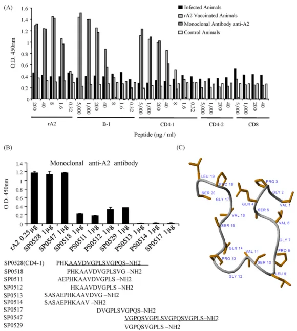
Fig. 2.B cell epitope mapping of A2. (A) The original synthetic peptides derived from A2 (B-1, CD4-1, CD4-2 and CD8) were tested in ELISA employing sera from infected and vaccinated mice (pool of three samples per group) as well as anti-A2-specific monoclonal antibody. (B) Shorter sequences based on reactive peptides CD4-1 and B-1 and peptides composed of A2 repetitions were tested in ELISA against anti-A2 monoclonal antibody in order to determine the minimal B epitope. In (A and B) results are representative of four independent experiments. (C) SP0547 was submitted to NMR, which revealed its lowest energy structure at 4.0 mM in TFE:H2O (10:90) – pH 7.0 (20 mM phosphate buffer), 20◦C as determined by NOE connectivity diagram.
non-repetitive segment of the protein plus five amino-acid residues from the first repetitive segment (Fig. 1).
3.2. Experimental analysis of B cell epitopes identified in A2 sequence
As shown inFig. 2A, the B-1 epitope reacts positively with anti-bodies present in sera from rA2-vaccinated mice and with the A2-specific monoclonal antibody. The level of reactivity observed in reactions with B-1 was similar to that observed when rA2 was used as antigen in control reactions. Sera from mice infected withL.(L.)
chagasi(Fig. 2A) did not react with B-1 or rA2. When the predicted T cell epitopes named CD8, CD4-1, and CD4-2 were tested, the only additional peptide that showed a comparable reactivity with sera from immunized mice and anti-A2 mAb (as seen with B-1 or rA2) was the CD4-1 (Fig. 2A), which except for the five last amino-acids in the N-terminal had a major aa sequence overlap with the B-1 epitope.
The peptide SP0547 was submitted to magnetic resonance. Sequence-specific chemical shift assignments have been per-formed for peptide SP0547 from the correlations observed in TOCSY and NOESY spectra using standard procedures[38]. Some inter-residual NOEs were observed in peptide SP0547 (not shown). Importantly, three correlations of the sort NN(i,i+2) observed at the central portion of the peptide sequence (Leu-9 and Val-11, Ser-10 and Gly-12, Val-12 and Pro-14) may be an indicative of a specific structural arrangement. Two long-range NOEs have been determined – H˛(i)-HN(j) Ser-5 and Gly-17, Hˇ(i)-Hˇ(j) Ser-5 and Val-16. These correlations indicate the spatial proximity between these sites and therefore characterize a sort of turn in the pep-tide structure (Fig. 2C). No inter-residual correlations have been determined in the NOESY spectrum of peptide SP0529, what is in agreement with a nonrigid conformation. Since the primary sequence of SP0547 represents the sequence of SP0529 twice, we
suggest that the activity observed for SP0547 could be attributed to its structural arrangement, once the unordered SP0529 does not show significant biological activity.
3.3. Experimental analysis of predicted T cell epitopes in A2
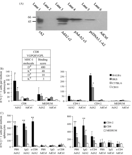
We then generated a non-replicative adenovirus encoding A2 protein (AdA2). Expression of A2 was tested in AdA2-infected cells by Western-blot with an anti-A2 monoclonal antibody. Multiple forms of rA2, with molecular masses under 53 kDa, were detected in infected cell extracts (Fig. 3A, lane 3). A similar pattern of bands was observed in rA2 purified fromE. coli(Fig. 3A, lane 1).
Based on the score indicated by software BIMAS (Fig. 3B, upper left), peptide CD8 had a high probability of binding to Ddmolecules. On the other hand, this same epitope had lower probability of show-ing affinity to molecules Db and Kb. To test this hypothesis, we
immunized with one dose of AdA2, BALB/c and C57BL/KsJ (BKS) mice, both strains showing haplotype H2d, as well as C57BL/6 and C.B10-H2 (CB10) mice, which show H2bhaplotype. In agreement with predictions, spleen cells from BALB/c and BKS mice reacted specifically (producing IFN-␥) to re-stimulation with peptide CD8 (Fig. 3B, left). We also tested if peptides CD4-1 and CD4-2 were haplotype-restricted. We demonstrated that CD4-2 peptide was presented only in association with BALB/c and BKS mice MHC-II molecules, and that peptide CD4-1 did not react with molecules Dd or Db(Fig. 3B, right).
Confirmation that peptides CD4-2 and CD8 were recognized, respectively, by CD4+ and CD8+ T cells was achieved in ELISPOT performed with spleen cells from mice submitted to selective depletion of CD4+ or CD8+ T cells, followed by immunization with one dose of AdA2 (Fig. 3C). The depletion of CD8+ T cell subpopulation abrogated only the response to the counterpart MHC-I epitope (Fig. 3C, left). In a similar extent, depletion of CD4+ T cells reduced IFN-␥production after in vitro stimulation with peptide CD4-2, since IFN-␥production detected was not sta-tistically significant when compared to control group (Fig. 3C, right).
3.4. Immune responses and protection against challenge in mice vaccinated with AdA2 in homologous prime/boost protocols
Finally, we evaluated the immune response elicited by a homol-ogous prime/boost protocol employing AdA2.Fig. 4A shows that following vaccination, high levels of A2-specific IFN-␥producing CD4+ T and CD8+ T cells from parental BALB/c lineage (H2d), but not from the congenic CB10 mice (H2b) were induced. Induction of CTLs, employing anin vivocytotoxicity assay, in which target cells were sensitized with peptide CD8 and transferred to immunized mice, was also evaluated. As seen inFig. 4B, vaccination induced activation of anti-A2-specific CTLs in BALB/c mice, but not in CB10 mice. CTLs leaded to almost 40% specific lysis of target cells. No cytotoxic activity was observed when we used the CD4-2 peptide (Fig. 4C). Even after two doses, AdA2 did not induce significant levels of anti-A2 antibodies in BALB/c or CB10 mice (not shown).
In the next step, we tested whether immunization with AdA2 is able to induce protection against challenge with a virulent strain of
L.(L.)chagasi. After i.v. challenge of BALB/c mice with 107 promastig-otes ofL.(L.)chagasi, we observed a sustained immune response, with peptide-specific cytotoxic activity (Fig. 5A, left) and IFN-␥
Fig. 5.Immune responses after challenge withL.(L.)chagasiand protection assays. (A)In vivocytotoxicity assay and ELISPOT assay performed 30 days after i.v. challenge of AdA2-immunized mice with 107L.(L.)chagasiparasites. Two animals per group were individually tested, and the experiments were repeated two times, independently. (B) Mice (six animals per group) were challenged i.v. with 107L.(L.)chagasipromastigotes 15 days after boost vaccination. Parasite burdens were determined by limiting dilution, 35 days after infection. (*) indicates statistically significant difference between experimental and control groups. These results are representative of two different experiments.
production (Fig. 5A, right). Immunization with AdA2 in a stan-dard homologous prime/boost vaccination protocol in BALB/c mice resulted in highly significant reduction of parasite burdens in liver and spleen (Fig. 5B), 35 days after infection. At this stage of infection in this model, spleen parasite burdens are low but tend to increase, while parasite numbers in liver are higher and tend to decrease[39]. Thus, demonstration of reduction of parasite burdens upon vacci-nation with A2 at both sites in this animal model is an important aspect.
4. Discussion
A2 was identified as an important amastigote virulence fac-tor ofL.(L.)donovaniparasites[6], and has recently emerged as a candidate for vaccine development against visceral leishmaniasis [12–15]. Here, we characterized the dominant B as well as CD4+ T and CD8+ T cell epitopes present in A2, and demonstrated that the adenovirus vector expressing A2 induces a strong CD4+ T and CD8+ T cell response and protective immunity against experimental infection withL.(L.)chagasi.
The B cell epitope in A2 was located within the repetitive units, since Ab recognition was dependent on maintenance of integrity of the repetitive segment. Importantly, nuclear magnetic reso-nance indicated that peptide SP0547, which has two repetitive units, folds into a beta-sheet like structure. Thus, it is possible that the activity observed for SP0547 is related to this structural fea-ture, since peptide SP0529 that consists of single repetitive unit, does not fold and is immunologically inert. Importantly, in both humans and dogs, the state of “resistance” toLeishmaniais associ-ated with development of cell-mediassoci-ated responses, whereas active disease is associated with high antibody levels and suppressed T cell
responses[40,41]. Nevertheless, a recent study has demonstrated that humoral response to A2 in dogs, although low, is associated with asymptomatic infection, and presumably, protective immu-nity againstLeishmaniaparasites[42]. Thus, the results of B cell epitope mapping of A2, obtained here, could be eventually applied in the field, to evaluate the development of protective immune responses in humans and dogs, which are the main target popula-tions for a vaccine against visceral leishmaniasis. If reactivity to the main B cell epitope in A2 is consistently traced and associated with protective immunity, for example, it would be possible to identify protected dogs vaccinated with A2 and asymptomatic dogs natu-rally infected withLeishmaniafrom naïve and susceptible animals that are infected, but did not develop protective immunity. Further, the high levels of anti-A2 antibodies in sera from dogs vaccinated with rA2 may help to discriminate these animals from naturally infected dogs, which is valuable information in geographical areas or Countries where elimination of seropositive infected dogs is the current policy.
We also successfully characterized the sequence VGPQSVGPL as a dominant CD8+ T cell epitope in A2. This peptide reacted strongly and specifically with corresponding IFN-␥producing CD8+ T cells in ELISPOT as well asin vivocytotoxic assays of mice immunized with AdA2. Indeed, as far as we know, this is the first study where a CD8+ T cell epitope is mapped in aLeishmaniaderived antigen. We also identified a CD4+ T epitope by testing overlapping epitopes of the A2 sequence. The CD4-2 peptide (SASAEPHKAAVDVGPL) was shown to be the main epitope recognized by IFN-␥producing CD4+ T cells in ELISPOT.
macrophages activated by IFN-␥[16]. The main source of IFN-␥is normally attributed to CD4+ T lymphocytes, nevertheless there are evidences that CD8+ T cells are also involved in protective immunity against leishmaniasis[43]. Thus, despite of intracellular location of parasites in the vertebrate host, the role of protective CD8+ cytolytic T cells induced by anti-leishmania vaccines is less established as compared to function of CD4+ T cells[19–21]. Importantly, peak of CTL activity coincided with regression of parasite loads in spleen and liver from mice experimentally infected withL.(L.)infantum
and were capable of killing target cells pulsed with total Leishma-niaantigen or macrophages infected withLeishmania[19]. Here, we detected for the first timein vivoCTL activity displayed by CD8+ T cells against a well-definedLeishmania target, the A2-specific VGPQSVGPL repetitive unit.
A2 has at least one epitope recognized by CD4+ T lymphocytes and, normally, has over 40 copies of the VGPQSVGPL repetitive unit that was now identified as a potent CD8+ T cell epitope. A major question here is how to induce a potent A2-specific T cell-mediated immunity in immunization protocol. Considering the well-established properties of adenovirus vector in eliciting both CD4+ T as well as CD8+ T cells in vaccine protocols, we constructed a non-replicative adenovirus vector expressing A2. AdA2 vaccina-tion induced IFN-␥production as well as CTL activity by CD8+ T cells from mice expressing the MHC haplotype H2d(BALB/c and BKS). In agreement, a homologous prime/boost protocol employing AdA2 was highly efficient in inducing A2-specific IFN-␥producing CD4+ T and CD8+ T cells as well as CD8+ T cells displaying cyto-toxic activity, leading to host protection in BALB/c mice challenged withL.(L.)chagasi. The lack of responsiveness of CD8+ T cells from CB10 or C57BL/6 mice is not surprising, since the predicted score of CD8 peptide binding to MHC class I molecule Db is only 10, as compared to 480 for binding to molecule Dd. Nevertheless,in silico analysis showed presence of other peptides in A2 that could bind to MHC class I molecule Db(not shown). Randomly designed MHC-II binding peptides were also not able to stimulate CD4+ T cells from CB10 or C57BL/6 mice. However, to thoroughly address this ques-tion, it is necessary to generate new peptides, with differences in one or two overlapping amino-acids, instead of five amino-acids, as in CD4-1 and CD4-2 sequences. In fact, vaccination of C57BL/6 mice with recombinant A2 showed to induce protection against challenge withL.(L.)donovani, which could be interpreted as indi-rect demonstration of T cell response against A2 in this mice strain [12].
In conclusion, in the present work we characterized B and T epi-topes on A2, which account for its immunogenicity in BALB/c mice. Further, we demonstrated that protective Th1-biased immune response is efficiently induced using AdA2. Further, IFN-␥ pro-duction and CTL activity mediated by CD8+ T cells, and not antibody production, are associated with induced protective immu-nity against experimental infection withL.(L.)chagasi. These results further suggest that A2 is an important candidate antigen for development of a vaccine against visceral leishmaniasis. Additional analysis of immune response to the defined B lymphocyte, CD4+ T as well as CD8+ T cell epitopes allowing comparison of symp-tomatic versus asympsymp-tomatic patients/dogs, may help evaluate the role of anti-A2 immune responses to naturalLeishmania infec-tion.
Acknowledgments
We are grateful to Greg Matlashewski for incentive and provid-ing anti-A2 mAb as well as plasmid containprovid-ingA2gene; Marcelo Porto Bemquerer for helpful discussion regarding B cell epitope structure; and Denise Golgher for advice in using monoclonal anti-bodies GK1.5 and GL113. We thank Centro Nacional de Ressonância
Magnética Nuclear (CNRMN – Brazil) for allowing us to use their 600 MHz NMR spectrometer.
The Millennium Institute for Vaccine Development and Tech-nology (CNPq - 420067/2005-1), and FAPEMIG (Rede Mineira de Biomoléculas) supported this work. Fellowships were provided by CNPq to R.T.G., A.P.F., D.M.R., M.S.D., R.M.V. and J.M.R., by FAPEMIG to M.L.O.P. and by FIOCRUZ to B.C.C.
Conflict of interest statement: We state that this work has no financial or commercial conflict of interest.
References
[1] Desjeux P. Leishmaniasis: current situation and new perspectives. Comp Immunol Microbiol Infect Dis 2004;27(5):305–18.
[2] Murray HW, Berman JD, Davies CR, Saravia NG. Advances in leishmaniasis. Lancet 2005;366(9496):1561–77.
[3] Zhang WW, Charest H, Ghedin E, Matlashewski G. Identification and overex-pression of the A2 amastigote-specific protein inLeishmania donovani. Mol Biochem Parasitol 1996;78(1–2):79–90.
[4] Nardin E, Munesinghe YD, Moreno A, Clavijo P, Calle MC, Edelman R, et al. T cell responses to repeat and non-repeat regions of the circumsporozoite protein detected in volunteers immunized withPlasmodium falciparumsporozoites. Mem Inst Oswaldo Cruz 1992;87(Suppl. 3):223–7.
[5] Gonzalez-Aseguinolaza G, Nakaya Y, Molano A, Dy E, Esteban M, Rodriguez D, et al. Induction of protective immunity against malaria by priming-boosting immunization with recombinant cold-adapted influenza and modified vaccinia Ankara viruses expressing a CD8+-T-cell epitope derived from the circumsporo-zoite protein ofPlasmodium yoelii. J Virol 2003;77(21):11859–66.
[6] Garin YJ, Meneceur P, Pratlong F, Dedet JP, Derouin F, Lorenzo F. A2 gene of Old World cutaneous Leishmania is a single highly conserved functional gene. BMC Infect Dis 2005;5(1):18.
[7] Matlashewski G. Leishmania infection and virulence. Med Microbiol Immunol (Berl) 2001;190(1–2):37–42.
[8] Zhang WW, Mendez S, Ghosh A, Myler P, Ivens A, Clos J, et al. Comparison of the A2 gene locus inLeishmania donovaniandLeishmania majorand its control over cutaneous infection. J Biol Chem 2003;278(37):35508–15.
[9] Carvalho FA, Charest H, Tavares CA, Matlashewski G, Valente EP, Rabello A, et al. Diagnosis of American visceral leishmaniasis in humans and dogs using the recombinantLeishmania donovaniA2 antigen. Diagn Microbiol Infect Dis 2002;43(4):289–95.
[10] Porrozzi R, Santos da Costa MV, Teva A, Falqueto A, Ferreira AL, dos Santos CD, et al. Comparative evaluation of enzyme-linked immunosorbent assays based on crude and recombinant leishmanial antigens for serodiagnosis of symptomatic and asymptomaticLeishmania infantumvisceral infections in dogs. Clin Vaccine Immunol 2007;14(5):544–8.
[11] Martins DR, Jeronimo SM, Donelson JE, Wilson ME.Leishmania chagasiT-cell antigens identified through a double library screen. Infect Immun 2006;74(12): 6940–8.
[12] Ghosh A, Zhang WW, Matlashewski G. Immunization with A2 protein results in a mixed Th1/Th2 and a humoral response which protects mice against Leish-mania donovaniinfections. Vaccine 2001;20(1–2):59–66.
[13] Ghosh A, Labrecque S, Matlashewski G. Protection againstLeishmania donovani infection by DNA vaccination: increased DNA vaccination efficiency through inhibiting the cellular p53 response. Vaccine 2001;19(23–24):3169–78. [14] Coelho EA, Tavares CA, Carvalho FA, Chaves KF, Teixeira KN, Rodrigues RC, et
al. Immune responses induced by theLeishmania(Leishmania)donovaniA2 antigen, but not by the LACK antigen, are protective against experimental Leish-mania(Leishmania)amazonensisinfection. Infect Immun 2003;71(7):3988–94. [15] Zanin FH, Coelho EA, Tavares CA, Marques-da-Silva EA, Silva Costa MM, Rezende SA, et al. Evaluation of immune responses and protection induced by A2 and nucleoside hydrolase (NH) DNA vaccines againstLeishmania chagasiand Leish-mania amazonensisexperimental infections. Microbes Infect 2007;9(9):1070–7. [16] Handman E. Leishmaniasis: current status of vaccine development. Clin
Micro-biol Rev 2001;14(2):229–43.
[17] Sacks D, Noben-Trauth N. The immunology of susceptibility and resistance to Leishmania majorin mice. Nat Rev Immunol 2002;2(11):845–58.
[18] Hernandez-Ruiz J, Becker I. CD8+ cytotoxic lymphocytes in cutaneous leishma-niasis. Salud Publica Mex 2006;48(5):430–9.
[19] Tzagosis P, Karagouni E, Dotsika E. CD8+ T cells with parasite-specific cyto-toxic activity and a Tc1 profile of cytokine and chemokine secretion develop in experimental visceral leishmaniasis. Parasite Immunol 2003;25:569–79. [20] Tzagosis P, Karagouni E, Dotsika E. Function of CD8+ T lymphocytes in a
self-curing mouse model of visceral leishmaniasis. Parasitol Int 2005;54: 139–46.
[21] Conceicao-Silva F, Perlaza BL, Louis JA, Romero P.Leishmania majorinfection in mice primes for specific major histocompatibility complex class I-restricted CD8+ cytotoxic T cell responses. Eur J Immunol 1994;24(11):2813–7. [22] Bruna-Romero O, Lasarte JJ, Wilkinson G, Grace K, Clarke B, Borras-Cuesta
[23] Yang Y, Li Q, Ertl HC, Wilson JM. Cellular and humoral immune responses to viral antigens create barriers to lung-directed gene therapy with recombinant adenoviruses. J Virol 1995;69(4):2004–15.
[24] Soong L, Chang CH, Sun J, Longley Jr BJ, Ruddle NH, Flavell RA, et al. Role of CD4+ T cells in pathogenesis associated withLeishmania amazonensisinfection. J Immunol 1997;158(11):5374–83.
[25] Charest H, Matlashewski G. Developmental gene expression inLeishmania donovani: differential cloning and analysis of an amastigote-stage-specific gene. Mol Cell Biol 1994;14(5):2975–84.
[26] Drijfhout JW, Hoogerhout P, Chan WC, White PD, editors. Fmoc solid phase peptide synthesis: a practical approach. New York: Oxford University Press; 2000. p. 229–42 [10 Methods of preparing peptide-carrier conjugates]. [27] Johnson BA, Blevins RA. NMR View: a computer program for the visualization
and analysis of NMR data. J Biomol NMR 1994;4(5):603–14.
[28] Hyberts SG, Goldberg MS, Havel TF, Wagner G. The solution structure of eglin c based on measurements of many NOEs and coupling constants and its com-parison with X-ray structures. Protein Sci 1992;1:736–51.
[29] Schwieters CD, Kuszewski JJ, Tjandra N, Clore GM. The Xplor-NIH NMR molec-ular structure determination package. J Magn Reson 2003;160:66–74. [30] Koradi R, Billeter M, Wuthrich K. MOLMOL: a program for display and analysis
of macromolecular structures. J Mol Graph 1996;14(1):32–51.
[31] McGrory WJ, Bautista DS, Graham FL. A simple technique for the rescue of early region I mutations into infectious human adenovirus type 5. Virology 1988;163(2):614–7.
[32] Chen CA, Okayama H. Calcium phosphate-mediated gene transfer: a highly efficient transfection system for stably transforming cells with plasmid DNA. Biotechniques 1988;6(7):632–8.
[33] Machado AV, Cardoso JE, Claser C, Rodrigues MM, Gazzinelli RT, Bruna-Romero O. Long-term protective immunity induced againstTrypanosoma cruziinfection after vaccination with recombinant adenoviruses encoding amastigote surface protein-2 and trans-sialidase. Hum Gene Ther 2006;17(9):898–908.
[34] Carvalho LH, Hafalla JC, Zavala F. ELISPOT assay to measure antigen-specific murine CD8(+) T cell responses. J Immunol Methods 2001;252(1–2): 207–18.
[35] Bruna-Romero O, Pereira BA, Caetano BC, Bouillet L, Carvalho LH, Esteban M, et al. Sufficient IL-4 production and maturation of the primary immune responses at the time of boost are key factors for efficient induction of antigen-specific T cells using recombinant adenoviruses amd poxviruses, submitted for publica-tion.
[36] Caetano BC, Bruna-Romero O, Fux B, Mendes EA, Penido ML, Gazzinelli RT. Vaccination with replication-deficient recombinant adenoviruses encoding the main surface antigens ofToxoplasma gondiiinduces immune response and pro-tection against infection in mice. Hum Gene Ther 2006;17(4):415–26. [37] Afonso LC, Scott P. Immune responses associated with susceptibility
of C57BL/10 mice to Leishmania amazonensis. Infect Immun 1993;61(7): 2952–9.
[38] Wüthrich K, editor. NMR of proteins and nucleic acids. New York: John Wiley & Sons; 1986.
[39] Carrion J, Nieto A, Iborra S, Iniesta V, Soto M, Folgueira C, et al. Immunohis-tological features of visceral leishmaniasis in BALB/c mice. Parasite Immunol 2006;28(5):173–83.
[40] Barbiéri CL. Immunology of canine leishmaniasis. Parasite Immunol 2006;28:329–37.
[41] Wilson ME, Jeronimo SM, Pearson RD. Immunopathogenesis of infection with the visceralizingLeishmaniaspecies. Microb Pathog 2005;38(4):147–60. [42] Ghedin E, Zhang WW, Charest H, Sundar S, Kenney RT, Matlashewski G.
Antibody response against aLeishmania donovaniamastigote-stage-specific protein in patients with visceral leishmaniasis. Clin Diagn Lab Immunol 1997;4(5):530–5.