RESEARCH ARTICLE
NK Cell-Mediated Regulation of Protective
Memory Responses against Intracellular
Ehrlichial Pathogens
Samar Habib2, Abdeljabar El Andaloussi2, Ahmed Hisham1, Nahed Ismail1*
1Department of Pathology, University of Pittsburgh, Pittsburgh, Pennsylvania, United States of America,
2Department of Obstetrics and Gynecology, Medical College of Georgia, Augusta University, Augusta, Georgia, United States of America
Abstract
Ehrlichiaeare gram-negative obligate intracellular bacteria that cause potentially fatal human monocytic ehrlichiosis. We previously showed that natural killer (NK) cells play a crit-ical role in host defense againstEhrlichiaduring primary infection. However, the contribution of NK cells to the memory response againstEhrlichiaremains elusive. Primary infection of C57BL/6 mice withEhrlichia murisprovides long-term protection against a second chal-lenge with the highly virulentIxodes ovatus Ehrlichia(IOE), which ordinarily causes fatal dis-ease in naïve mice. Here, we show that the depletion of NK cells inE.muris-primed mice abrogates the protective memory response against IOE. Approximately, 80% of NK cell-depletedE.muris-primed mice succumbed to lethal IOE infection on days 8–10 after IOE
infection, similar to naïve mice infected with the same dose of IOE. The lack of a recall response in NK cell-depleted mice correlated with an increased bacterial burden, extensive liver injury, decreased frequency ofEhrlichia-specific IFN-γ-producing memory CD4+and
CD8+T-cells, and a low titer ofEhrlichia-specific antibodies. Intraperitoneal infection of mice withE.murisresulted in the production of IL-15, IL-12, and IFN-γas well as an expansion of
activated NKG2D+NK cells. The adoptive transfer of purifiedE.muris-primed hepatic and splenic NK cells intoRag2-/-Il2rg-/-recipient mice provided protective immunity against chal-lenge withE.muris. Together, these data suggest thatE.muris-induced memory-like NK cells, which contribute to the protective, recall response againstEhrlichia.
Introduction
Human monocytic ehrlichiosis is an emerging, potentially fatal, tick-borne infectious disease caused byEhrlichia chaffeensis, an obligate intracellular gram-negative bacterium [1–3]. This bacterium primarily infects monocytes and macrophages and causes systemic infection of the reticuloendothelial system [3–5]. The main site of infection and pathology in ehrlichiosis is the liver. We and other investigators have shown that the development of fatal monocytic ehrlichi-osis is due to the suppression of protective immunity mediated by IFN-γand CD4+Th1 cells
a11111
OPEN ACCESS
Citation:Habib S, El Andaloussi A, Hisham A, Ismail N (2016) NK Cell-Mediated Regulation of Protective Memory Responses against Intracellular Ehrlichial Pathogens. PLoS ONE 11(4): e0153223. doi:10.1371/journal.pone.0153223
Editor:Roman R. Ganta, Kansas State University, UNITED STATES
Received:February 2, 2016
Accepted:March 25, 2016
Published:April 19, 2016
Copyright:© 2016 Habib et al. This is an open access article distributed under the terms of the
Creative Commons Attribution License, which permits unrestricted use, distribution, and reproduction in any medium, provided the original author and source are credited.
Data Availability Statement:All relevant data are within the paper and its Supporting Information files.
Funding:Funding provided by NIH/NIAID: 1R56AI097679-01, National Institute of Allergy and Infectious Diseases, PI; NI.
and excessive inflammation, which leads to immunopathology and multi-organ failure [6–11]. Currently, a broadly effective licensed vaccine is unavailable.
Natural killer (NK) cells are innate lymphocytes that provide rapid and robust host defense against infected cells, including NK cell-mediated antibody-dependent cellular cytotoxicity (ADCC) [12–14]. NK cells can also reciprocally interact with dendritic cells and B cells as well as regulate adaptive T cell-mediated and humoral immunity [15,16]. Recent data have demon-strated that NK cells share several features of the adaptive memory immune response, includ-ing their ability to promptly respond to a second challenge with the same antigen they initially encountered (16–24). Accumulating evidence shows that at least some subsets of NK cells respond to certain antigens (viruses or haptens) in a manner that has the hallmarks of adaptive immunity, namely specificity, longevity, and a recall response [13,16,17]. Notably, NK cells with memory function are more confined to the liver.
In this study, we examined the contribution of NK cells to the generation of memory T and B cell responses againstEhrlichia. In particular, we investigated whether NK cells acquire a memory-like phenotype followingE.murisinfection including their ability to provide recall response against homologousEhrlichiainfection. Our data provide the first demonstration that NK cells acquire a memory-like phenotype and mediate a protective recall response againstEhrlichiadirectly and indirectly via the regulation of memory T cell responses during
Ehrlichiare-challenge. The data obtained in this study will enhance our understanding of the different cellular mechanisms that contribute to the development of an effective and optimal memory response within peripheral organs during infection with intracellularEhrlichia, which is important for vaccine development.
Materials and Methods
Ethics Statement
This study was carried out in strict accordance with the recommendation in the guide for the Care and Use of Laboratory Animals of the National Institute of Health (NIH). The animal protocol was approved by the University of Pittsburgh Animal Care and Use Committee (Per-mit number 14125020). All efforts were made to minimize animal suffering. Mice had access to food and water ad libitum. Infected animals were observed two times/per day, in the morning and in the evening. The body weights and body temperatures were measured. There was no unexpected death. In the survival study, the humane endpoints, at which mice were euthanized, were set as excessive weight loss (>30%), decreased body temperature (drop to<32 degrees),
and development of other signs of distress (i.e. decreased movement, lethargy, and having ruf-fled hair). At that endpoint, mice were humanely euthanized by cervical dislocation. No analge-sics and anethethetics were used.
Mice and bacterial infection
Female C57BL/6J mice (8–12 weeks of age) andRag2−/−Il2rg−/−mice were obtained from
Jack-son Laboratories (Bar Harbor, ME, USA). All animals were housed under specific pathogen-free conditions at the Animal Research Facility, University of Pittsburgh. Two species of mono-cyticEhrlichiawere used in this study: the highly virulent IOE, and the mildly virulentE.
muris. IOE andE.murisstocks were propagated by passage through wild-type C57BL/6 mice. Single-cell suspensions from spleens harvested from mice 7 days post infection (DPI.) were stored in sucrose and potassium phosphate (SPK) buffer (0.5 M K2HPO4, 0.5 M KH2PO4, and
0.38 M sucrose) in liquid nitrogen and used as stocks. Mice were infected intraperitoneally (IP) with a lethal high dose of IOE (104organisms/mouse) or a high dose ofE.muris(2 X 10⁵ organisms/mouse). Mice were then monitored daily for signs of illness and survival.
In vivo
NK depletion
NK cells were depleted fromE.muris-primed mice using a non-activating polyclonal antibody (Ab) against asialo-GM1 (Wako Chemicals USA, Inc.). Infected C57BL/6 mice were IP injected with anti-asialo GM1 mAb (25 ug /mouse) or normal rabbit IgG isotype control mAb on days 22, 23, 25 and 26 after primaryE.murisinfection. Results from flow cytometry analysis indi-cated that antibody depletion resulted in a ~95% depletion of NK cells in the spleens and livers of primed mice.
Isolation of hepatic and splenic NK cells
Spleen and liver tissues were cut into small pieces with a sterile scalpel and passed through
40-μm mesh filters. Single-cell suspensions of splenocytes were prepared as previously described
[6,18]. Liver mononuclear cells (LMNCs) were enriched by density-gradient centrifugation as previously described [19–21]. Murine NK cells were isolated from splenocytes and LMNCs by negative selection using the MACS NK cell isolation kit II (Miltenyi Biotec). The purity of unla-beled NK cells was ~85%, as determined by flow cytometry. The activation status of NK cells was not affected by the negative selection process. Since the transferred cells contained ~15% cells other than NK cells, we depleted contaminating CD4+T cells in recipientRag2−/−Il2rg−/−
mice by injection of anti-CD4 mAb (200 ug/mouse) (GK1.5, Biolegend) on -1, 0, and 3 DPI.
Flow cytometry analysis
Splenocytes were harvested, counted, and resuspended in fluorescence-activated staining buffer (Dulbecco’s PBS without Mg2+or Ca2+containing 1% heat-inactivated FCS and 0.09% sodium azide) at a concentration of 106cells/well. FcRs were blocked with a mAb (clone 2.4G2) against the mouse cell surface Ags CD16 and CD32 for 15 min. The following FITC-, PE-,
PerCP-Cy5.5–, Alexa Fluor-, and allophycocyanin-conjugated Abs were purchased from BD Biosciences (San Jose, CA, USA) unless otherwise indicated: anti-CD45.2 (clone 69), anti-CD3 (clone 1452C11), anti-CD11c (clone HL3), anti-CD4 (clone RM4-4), anti-CD11b (clone M1/70), anti-NK1.1 (clone PK136), anti-IFN-γ(clone XMG102), CD62L (clone 1A8), anti-CD44 (3/23), and anti-B220 (clone 16-10A1). Isotype control mAbs, including FITC-, PE-, or allophycocyanin-conjugated hamster IgG1 (A19-3), rat IgG1 (R3-34), rat IgG2a (R35-95), mouse IgG2a (X39), mouse IgG2b (MPC-11), mouse IgG1 (X40), and rat IgG2b (A95-1), were purchased from BioLegend (San Diego, CA, USA). For intracellular cytokine staining, the sple-nocytes were incubated at 37°C for 4 h in complete medium with the addition of BD Golgi Plug (BD Biosciences, San Jose, CA, USA) according to the manufacturer’s instructions. Lymphocyte and granulocyte populations were gated based on forward and side-scatter parameters; and, 50,000–200,000 events were collected using a BD FACSCalibur flow cytometer (BD Systems, San Jose, CA, USA). Data were analyzed using FlowJo software (TreeStar, Ashland, OR, USA).
Cytokine measurement
The concentrations of IFN-γ, IL-12, IL-15 and IL-10 in the liver mononuclear cells (LMNCs) culture supernatants were determined using a Quantikine enzyme-linked immunosorbent assay (R&D Systems, Minneapolis, MN, USA) according to the manufacturer’s instructions.
Detection of nitric oxide (NO) production
NO production was determined by analyzing the level of nitrite (NO2-) in the splenocyte cul-ture supernatant from different groups of mice using a colorimetric Griess reaction. Samples were briefly mixed with an equal (1:1) volume of Griess reagent (0.1% N-(1-naphthy) ethylene
diamine hydrochloride, 1% sulfonamide, and 2.5% H3PO4) in 96-well plates, and the
absor-bance at OD540 were measured using an ELISA reader (Molecular Devices, Sunnyvale, CA, USA) with 1–50μM sodium nitrite dissolved in distilled water as the standard.
Measurement of bacterial burden by real-time PCR (RT-PCR)
The number ofEhrlichiaorganisms in frozen stocks and the bacterial burden in different organs were measured by quantitative RT-PCR using an iCycler IQ multicolor real-time detec-tion system (Bio-Rad, Hercules, CA, USA), as previously described [22]. The sequences of primer sets used that target both theE.murisand the IOEdsb(a thiol-disulfide oxidoreduc-tase) genes, the eukaryotic housekeeping gene GAPDH, and specific probes have been previ-ously described [6,22,23]. Results were normalized to the expression levels of the GAPDH gene in the same sample and were expressed as copy numbers per 104GAPDH copies. PCR analyses were considered negative forEhrlichiaDNA if the critical threshold values exceeded 40 cycles.
Histopathology staining of liver sections
Liver segments were fixed in 10% neutral buffered formalin, dehydrated in graded alcohols, and embedded in paraffin wax. Sections (3-mm thick) were collected on coated slides and stained with H&E.
Measurement of
Ehrlichia
-specific antibodies by immunofluorescence
assay (IFA)
Serum samples from infected and control mice were measured for antigen (Ag)- specific IgG Abs through an indirect immunofluorescence assay usingE.murisas anEhrlichia cross-reac-tive antigen, as previously described [3,6,23–25]. A serial two-fold dilution of serum samples was applied to fixed Ag slides. After incubation at 37°C for 30 min in a humid chamber, slides were stained with FITC-labeled anti-mouse IgG (BD eBioscience) at a dilution of 1/100. The slides were examined under a fluorescence microscope (Nikon, Tokyo, Japan). Serological titers were expressed as the reciprocals of the highest dilution at which specific fluorescence was detected.
Statistical analysis
All of the data presented are representative of two or three independent experiments that yielded similar results. Data are represented by means and standard deviations (SD). Two groups analysis was performed using an unpaired two-tailedttest. For comparison of multiple experimental groups, we used one–way analysis of variance (ANOVA) with Bonferroni’s pro-cedure. To determine whether the difference in survival between different mice groups was sig-nificant, data were analyzed by the Breslow-Wilcoxon Test. All statistical analyses were performed using GraphPad Prism (GraphPad Software Inc., La Jolla, CA, USA). Differences withPvalues of 0.05, 0.01, and 0.001 were considered slightly (), moderately (), and highly
() significant, respectively.
Results
Primary
E
.
muris
but not IOE infection induces expansion and activation
of NK cells
We previously showed that a primary non-lethal infection of wild type (WT)-B6 mice withE.
muris(EM), but not a sublethal IOE infection, provides long-term protection of primed mice
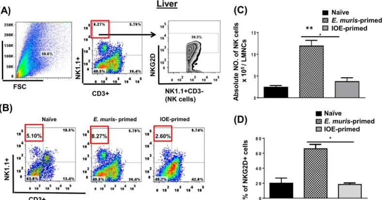
against an ordinarily lethal secondary IOE infection [23,25].E.muris, but not IOE, causes per-sistent infection, which is critical for the induction and maintenance of memory CD4+T cells. However, the adoptive transfer of memory CD4+T cells alone provides partial protection to naïve mice against lethal IOE infection [6]. Thus, we hypothesized that the protective recall response inE.muris-primed mice could be mediated by cells other than memory CD4+T cells. Because the liver, which is enriched with NK and NKT cells, serves as the main site of infection in ehrlichiosis, we examined whether cross-protection inE.muris-primed mice was due to dif-ferences in NK cell responses. To this end, we analyzed the activation status, and frequency of NK cells in the liver and spleen of mice infected withE.murisor IOE via the IP route. Our data demonstrated that NK cells expand and persist in the liverFig 1A–1CofE.muris-, but not IOE-, primed mice 21 DPI [the time point that corresponds to the contraction of effector T cells and the generation of central and effector memory T cells [23,25]. Our data further showed that approximately 70% of liver NK cells inE.muris-primed mice expressed NK cell-activating receptor (NKG2D)Fig 1D, suggesting that these cells were activatedin vivo. Similar to hepatic NK cells, the frequency of splenic NK cells was higher in the spleen ofE.muris-primed mice as compared to IOE-primed mice and uninfected controlsFig 2A–2C. Approximately 40% ofE.
muris-primed splenic NK cells expressed NKG2D, while ~ 20% of IOE-primed splenic NK cells expressed NKG2DFig 2D. Since the total number of splenic NK cells was significantly higher in
E.muris-infected mice than that detected in IOE-infected mice, these data suggest thatE.muris
also induces significant expansion of activated NKG2D+NK cells in the spleen compared to
Fig 1. NK cells expand and persist in the liver ofE.muris- but not IOE-primed mice.Liver mononuclear cells were isolated from indicated mice groups on day 21 post-infection, and the frequency and activation of NK cells were analyzed.(A)shows gating strategy on NK cells and activation marker NKG2D.
(B)The percentage and(C)absolute number of NK cells in the livers of indicated mice groups. as determined by flow cytometry.(D)The percentage of activated NK cells expressing NKG2D. The results demonstrate a higher frequency of activated NKG2D+NK cells in the livers ofE.muris-primed mice as compared to other groups (*P<0.05 and**P<0.01). The data shown are the means±SD from three mice per group and are representative of three
independent experiments.
doi:10.1371/journal.pone.0153223.g001
IOE-primed mice. These data suggest thatE.murisinfection promotes the expansion of acti-vated NK cells that persist in the primed host, in the liver and spleen.
NK cell depletion leads to an impaired protective recall response against
Ehrlichia
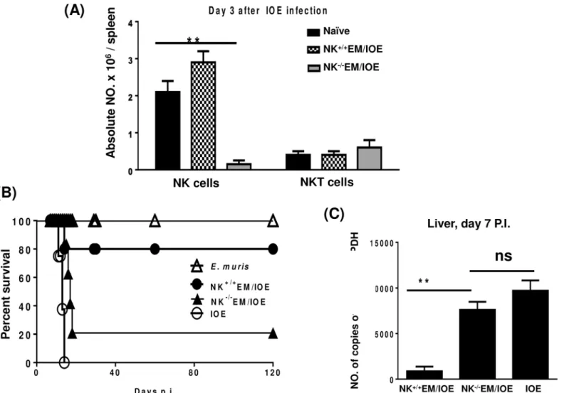
To further examine the contribution of NK cells to the memory response againstEhrlichia, we depleted NK cells fromE.muris-primed mice before we challenged these mice with IOE. The treatment ofE.muris-primed mice with anti-asialo GM1 antibodies did not influence the num-ber of T cells and NKT cells on day 3S1 Figor day 5S2 Figpost-infection, respectively. The treatment ofE.muris-primed mice with anti-asialo GM1 antibodies did not also influence the number of CD11b+macrophages (data not shown). However, anti-asialo GM1 antibodies resulted in a ~95% depletion of NK cells on day 3 after IOE infectionFig 3A.
Consistent with our previous studies [6,23], 80% of theE.muris-primed mice treated with isotype control antibody (referred to as NK+/+EM/IOE) survived re-challenge with an ordinar-ily lethal dose of IOE up to 120 days after IOE challenge. Notably, the depletion of NK cells in
E.muris-primed mice resulted in the loss of a protective recall response, such that ~80% of NK cell-depleted mice (referred to as NK-/-EM/IOE) succumbed to lethal IOE infection between days 15 and 17 post-IOE challengeFig 3B. The depletion of NK cells in NK-/-EM/IOE mice also led to an increased bacterial burden in the liver on day 7 following IOE challengeFig 3C. These data suggest that NK cells contribute to effective bacterial clearance and protective mem-ory response againstEhrlichia.
Fig 2. NK cells expand and persist in the spleen ofE.muris- but not IOE-primed mice.Splenocytes were isolated from indicated mice groups on day 21 post-infection, and the frequency and activation of NK cells were analyzed.(A)The gating strategy on NK cells and activation marker NKG2D.(B)The percentage and(C)absolute number of NK cells in the spleens of indicated mice groups as determined by flow cytometry.(D)The percentage of activated NK cells expressing NKG2D. The results demonstrate a higher frequency of activated NKG2D+NK cells in the spleens ofE.muris-primed mice as compared to other groups (*P<0.05). The data shown are the means±SD from three mice per group and are representative of three independent experiments.
doi:10.1371/journal.pone.0153223.g002
Antigen-specific CD4
+T cell responses are impaired in NK cell-depleted
EM/IOE mice
To further examine the effect of NK cell depletion on the protective memory response, we mea-sured the frequency and function of memory CD4+T cells. Splenocytes were harvested from all groups of mice 28 DPI, and stimulated withE.murisantigens (Ags). Our data show that the spleens of NK-/-EM/IOE mice had a significantly lower percentageFig 4Aand absolute number
Fig 4Bof Ag-specific CD44+CD62L-effector memory (Em) CD4+T cells 28 DPI afterE.muris
infection as compared with the number of Em CD4+T cells in sham (isotype) controlE.muris -infected mice.
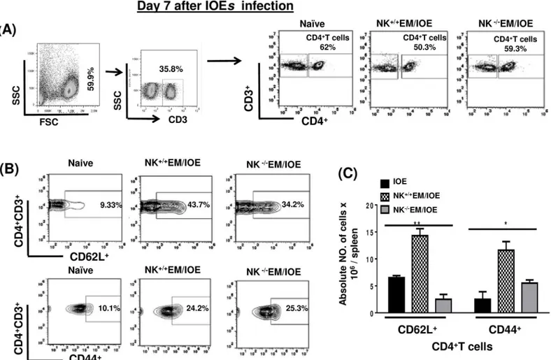
To determine the effect of NK cell depletion on the frequency of activated/effector memory CD4+T cells following secondary IOE infection, we measured the expression of CD62L and CD44 on T cells in the spleen of NK-/-EM/IOE and NK+/+EM/IOE mice 7 DPI after IOE infec-tion. Followingin vitrostimulation with IOE antigens, we detected lower a percentage and
Fig 3. Secondary high-dose challenge with IOE is fatal in NK-depleted EM/IOE-infected mice.C57BL/6 mice were infected with a high dose ofE.muris, and were either depleted of NK cells at 22, 23, 24, and 26 DPI (NK-/-EM/IOE) or treated with isotype control antibody (NK+/+EM/IOE). Both groups of mice (n = 12 mice/group) were re-challenged with a high dose of IOE on day 28 after infection. The other control group includes naïve mice infected only with IOE.
Depletion ofE.muris-primed mice with anti-asialo GM1 antibodies resulted in:A)~95% depletion of NK cells on day 3 after IOE infection;B)decreased survival of NK-/-EM/IOE mice group; andC)higher bacterial burden in the liver of NK-/-EM/IOE mice on day 7 after IOE infection as compared with NK+/+EM/
IOE-infected mice. The data represent the means±SD from four mice per group and are representative of three independent experiments.
doi:10.1371/journal.pone.0153223.g003
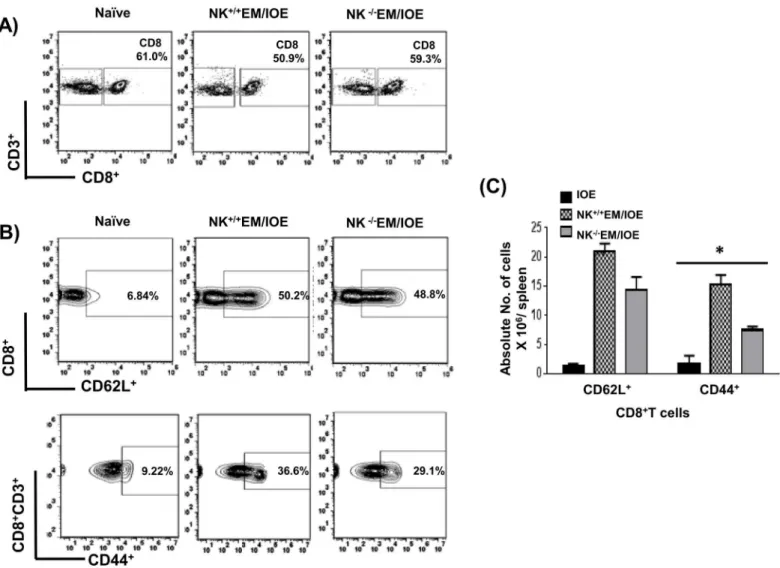
absolute number of activated CD62L+and effector/effector memory CD44+CD4+T cells in the spleen of NK-/-EM/IOE mice than that detected in the spleen of NK+/+EM/IOE miceFig 5A–
5C. NK cell depletion also significantly decreased the percentage and absolute numberFig 6A–
6Cof effector/effector memory CD44+CD8+T cells, but not CD62L+CD8+T cells, in
NK-/-EM/IOE mice when compared to NK+/+EM/IOE mice.In vitrostimulation of naïve sple-nocytes with IOE orE.muris(data not shown) did not elicit significant activation of CD4+T cells or naïve CD8+T cells as compared to primed mice. Together, these data suggest that NK cells promote expansion of effector/effector memory CD4+T cells and CD8+T cells following primary and secondaryEhrlichiainfection.
Impaired protective immunity in NK cell-depleted, EM/IOE-infected mice
is due to decreased IFN-
γ
, iNOS and NO
IFN-γis critical for effective bacterial elimination in ehrlichiosis. T cells, particularly CD4+ Th1 cells and NKT cells are major cellular subsets that produce IFN-γand thus mediate
Fig 4. NK cell depletion inE.muris-primed mice decreases expansion of memory T cells.Splenocytes from different groups of mice were stimulated withE.murisantigens and the frequency of memory cells was determined by flow cytometry. Lower percentages(A)and absolute numbers(B)of effector/ effector memory (Em) (CD44highCD62Llow), but not central memory (Cm) (CD44highCD62Lhigh), CD4+T cells in the spleens of NK-/-E.muriscompared with
those found in NK+/+E.murisor naïve mice infected with IOE on day 28 post-infection.
**indicateP<0.01. Data are presented as the means±SD of four mice per group from two independent experiments.**indicatesP<0.01.
doi:10.1371/journal.pone.0153223.g004
protective immunity againstEhrlichia. To determine the role of NK cells in host defense againstEhrlichiaduring recall response, we examined the number of antigen-specific, IFN-γ -producing T cells and NKT cells in all mice groups on day 7 after the second IOE infection. We stimulated splenocytes from naïve and infected mice withE.murisAgs, and the frequency ofE.
muris-specific, IFN-γ-producing cells was determined by flow cytometry. Impaired protective immunity in NK-/-EM/IOE mice was associated with decreases in the percentageFig 7A and 7Band absolute numberFig 7CofE.muris-specific IFN-γ-producing CD3+T cells when com-pared with those found in NK+/+EM/IOE control mice. Similarly, the percentageFig 8A and 8Band absolute numberFig 8Cof IFN-γ-producing NKT cells was decreased in NK-/-EM/IOE mice compared to NK+/+EM/IOE mice. Polyclonal stimulation of splenocytes from all mice (naïve and infected) groups with PMA/ionomycin resulted in a similar increase in the number of IFN-γ-producing T cells (data not shown), suggesting that an impaired memory CD4+Th1 response in NK-/-EM/IOE mice encompasses antigen-specific T cells. We did not find a signifi-cant difference in the frequency of antigen-specific, IL-4-producing cells or IL-10-producing T cells between NK+/+EM/IOE and NK-/-EM/IOE-infected mice (data not shown), suggesting
Fig 5. NK cell depletion in EM/IOE-infected mice impairs expansion of effector memory CD4+T cells.Splenocytes harvested on day 7 after IOE infection from NK-/-EM/IOE and NK+/+EM/IOE-mice were stimulatedin vitrowith IOE antigens and the frequency of antigen-specific activated/effector
memory T cells were determined by flow cytometry. Naïve cells were stimulatedin vitrowith IOE antigens. Gating on splenocytes and T cells was shown in
(A). The spleens of NK-/-EM/IOE-mice contained lower percentages(B)and absolute numbers(C)of CD62L+CD4+T cells compared to that detected in the
spleens of NK+/+EM/IOE-infected mice on day 7 post-IOE infection. Data are presented as the means±SD of four mice per group from two independent
experiments.**indicatesP<0.01.
doi:10.1371/journal.pone.0153223.g005
that impaired bacterial elimination is not due to altered IL-4: IFN-γor IL-10:IFN-γresponses, as suggested by other studies [18,21,26,27].
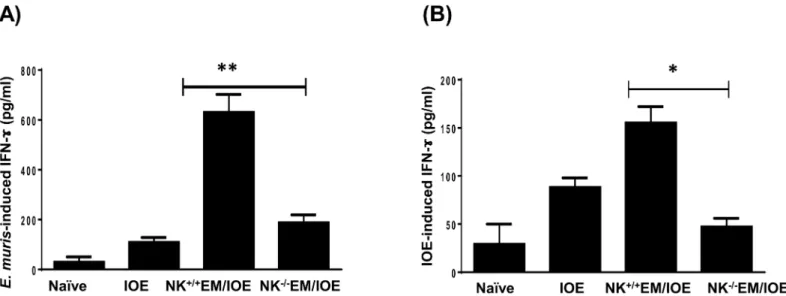
To determine the difference in total IFN-y production in the spleen of NK cell-depleted and undepleted primed mice, we stimulated splenocytes from all mice groups withE.muris
and IOE Ags, and measured IFN-γlevels in bulk culture supernatants by ELISA. Althoughin vitrostimulation of immune splenocytes with eitherE.murisor IOE Ags induced higher
IFN-γproduction as compared to that produced by naïve splenocytes, the levels of IFN-γwere lower when immune splenocytes were stimulated with IOE Ags. This is not surprising since our previous studies show that IOE is a poor inducer of IFN-γresponse. Nevertheless, deple-tion of NK cells decreased the level ofE.muris-Fig 9Aor IOE-Fig 9Binduced IFN-γ pro-duced in the spleen of NK+/+EM/IOE-infected mice compared to that produced by splenocytes from NK-/-EM/IOE mice. Ag-specific IFN-γproduction by the spleen of
NK-/-EM/IOE mice was similar to that produced in the splenocytes cultured from unprimed,
Fig 6. NK cell depletion in EM/IOE-infected mice impairs expansion of effector memory CD8+T cells.Splenocytes harvested on day 7 after IOE infection from NK-/-EM/IOE and NK+/+EM/IOE-mice were stimulatedin vitrowith IOE antigens and the frequency of antigen-specific activated/effector memory T cells were determined by flow cytometry. Naïve cells were stimulatedin vitrowith IOE antigens. Gating on splenocytes and T cells was shown in
(A). The spleens of NK-/-EM/IOE-mice contained lower percentages(B)and absolute number(C)of CD44+CD8+T cells compared to that detected in the
spleens of NK+/+EM/IOE-infected mice. No significant difference in the frequency of CD62L+CD8+T cells between NK-/-EM/IOE-mice and NK+/+
EM/IOE-mice. Data are presented as the means±SD of four mice per group from two independent experiments.**indicatesP<0.01.
doi:10.1371/journal.pone.0153223.g006
IOE-infected mice, suggesting that the absence of NK cells abrogated the protective memory response againstEhrlichia.
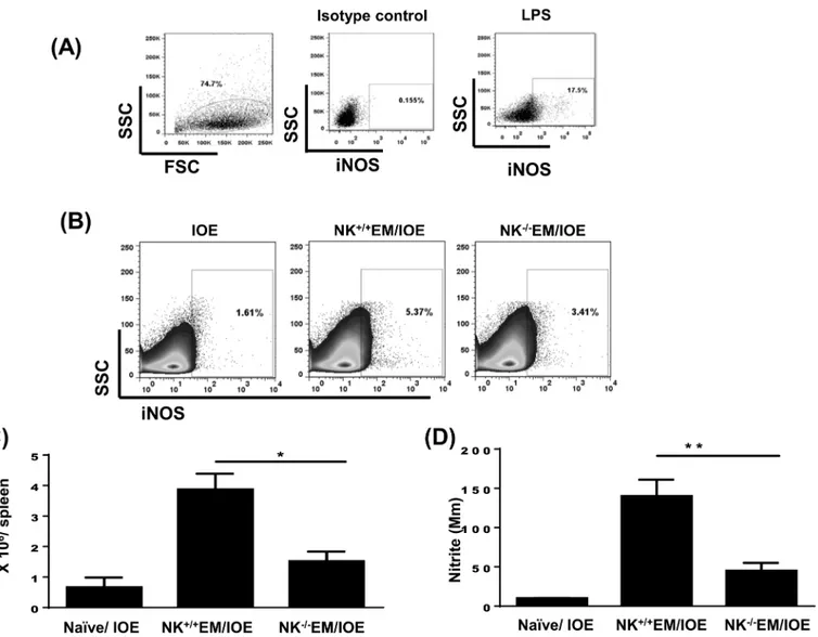
IFN-γis known to mediate activation of the bactericidal functions of macrophages, such as the induction of inducible nitric oxide synthase (iNOS) and production of nitric oxide (NO). The latter is a key antimicrobial effector molecule. Thus, we examined whether the difference in IFN-γresponse between NK depleted and NK sufficient mice influenced the production of iNOS and NO. We measured the number of CD45+leukocytes producing iNOS and the quan-tity of nitrites in the culture supernatants by flow cytometry and Griess reaction, respectively. Negligible iNOS production was detected in isotype control mAb- stained splenocytes from IOE-infected mice (negative control)Fig 10A, while a significantly high percentage of iNOS-producing cells were detected when IOE-infected splenocytes were further stimulatedin vitro Fig 7. Decreased percentage of IFN-γproducing CD3+T cells in NK-/-EM/IOE-mice.Splenocytes were harvested from the indicated mice groups on day 7 after IOE infection, and were stimulatedin vitrowithE.murisAgs or left unstimulated. Lymphocytes were gated based on the forward and side scatter, and then cells were gated on CD3. CD3+T cells were further analyzed for intracellular
IFN-γstaining as shown in the gating strategy(A). Controls shown in dot plots includes naïve splenocytes stimulatedin vitrowithE.murisAgs and IFN-γproduction by unstimulated splenocytes from IOE-infected mice.(B)Dot plots shows the percentage of gated CD3+T cells and the percentage of
IFN-γ+CD3+T cell subset in indicated mice groups followingin vitrostimulation withE.
murisAgs. (C) Data show the differences in the absolute number of IFN-γ+CD3+T cells in the spleen of indicated mice groups. Data shown are from three mice/groups with similar results in three independent experiments (n = 9 mice/group).
doi:10.1371/journal.pone.0153223.g007
Fig 8. Decreased percentage of IFN-γproducing NKT cells in NK-/-EM/IOE-mice.Splenocytes were harvested from the indicated mice groups on day 7 after IOE infection, and were stimulatedin vitrowithE.murisAgs or left unstimulated. (A) dot plots show the gating strategy where lymphocytes were first gated based on the forward and side scatter, and then cells were gated on CD3. CD3+T cells were further gated on NK1.1.+cells (NKT cells). The NKT cells
were further analyzed for intracellular IFN-γstaining. Controls shown in the dot plots includes naïve splenocytes stimulatedin vitrowithE.murisAgs and IFN-γproduction by unstimulated splenocytes from IOE-infected mice.(B)Dot plots shows the percentage of gated CD3+T cells and the percentage of IFN-γ+ producing NKT cells in indicated mice groups followingin vitrostimulation withE.murisAgs. (C) Data show the differences in the absolute number of IFN-γ+ producing NKT cells in the spleens of indicated mice groups. Data shown are from three mice/groups with similar results in three independent experiments (n = 9 mice/group).*indicatesP<0.05.
doi:10.1371/journal.pone.0153223.g008
with lipopolysaccharide (LPS) (positive control)Fig 10A. Our data showed that the spleens of NK+/+EM/IOE-infected mice contain a higher percentageFig 10Band absolute numberFig 10C
of iNOS-producing leukocytes when compared to IOE-infected mice and NK-/-EM/IOE. Con-sistent with single cell analysis, there was a higher production of nitrite in spleen bulk culture in NK+/+EM/IOE-infected mice compared to other mice groupsFig 10D. Together, these data sug-gest that NK cells promote bacterial elimination and the protective memory response against
Ehrlichiaby enhancing the induction and/or expansion of effector memory CD4+Th1 cells, IFN-γproduction, and the activation of the bactericidal function of infected phagocytic cells.
NK cells are required for the production of
Ehrlichia
-specific antibodies
Ehrlichia-specific antibodies, mainly IgG2a, are critical for the elimination of intracellular Ehr-lichiaeby enhancing opsonization of extracellularEhrlichiaeand subsequent intracellular kill-ing within phagocytes. Thus, we examined the effect of NK depletion on the production of Ag-specific antibodies in EM/IOE-infected mice following primary infection withE.murisor after re-challenge with IOE. The levels of antibodies in different mice groups were measured as the reciprocal of the dilution of serum samples. Similar to our previous findings [6,23,25], primary infection withE.murisinduced a high Ag-specific IgG titer 21 DPI (titer is 512). NK+/+EM/ IOE-infected control mice had substantially high titers ofEhrlichia-specific IgG antibodies (titer is 1024) on day 7 post IOE challenge. In contrast, NK-/-EM/IOE mice had a lower level of
Ehrlichia-specific IgG antibodies (titer is 128) on day 7 post IOE re-challenge.
Studies conducted by Winslow et al. have shown that chronicE.murisinfection elicits a pro-tective IgM response derived from extrafollicular CD11b-CD11c+B220+plasmablasts. We found that primary or secondary IOE infection in naïve orE.muris–primed mice induced similar anti-gen-dependent expansion of CD11b-CD11c+B220+plasmablasts in the spleen on day 7 after IOE infection when compared toin vitroAg-stimulated splenocytes from naïve miceFig 11A–
11C. Interestingly, depletion of NK cells in NK-/-EM/IOE-infected mice significantly increased
Fig 9. Depletion of NK cells in NK-/-EM/IOE mice decreased production of
IFN-γ.The levels of IFN-γin bulk culture of splenocytes from the indicated mice groups, harvested on day 7 after IOE infection, and stimulatedin vitrowith eitherE.muris(A) or IOE (B) Ags, were measured by ELISA. The antigen-specific IFN-γresponse was calculated by subtraction of the IFN-γconcentration produced by unstimulated cells from the Ags-stimulated cells. The data show a significantly lower production ofE.muris-(A)and IOE-(B)specific IFN-γby splenocytes from NK-/-EM/IOE mice compared with similarly-stimulated cells from NK+/+EM/IOE mice. The levels of
IFN-γin NK-/-EM/IOE-mice were similar to those detected in naïve mice infected with IOE.*and**indicate
P<0.05 andP<0.01, respectively. Data are representative of two independent experiments with four mice per group.
doi:10.1371/journal.pone.0153223.g009
the frequency of these plasmablasts compared with NK+/+EM/IOE-infected miceFig 11A–11C. On the other hand, decreased antibody productions in NK-/-EM/IOE mice were associated with decreased percentageFig 11A and 11Bas well as absolute numberFig 11Dof antigen-specific CD11b-CD11c-B220+mature B cells. These data further suggest that chronicE.murisinfection elicits NK cells that regulate antigen-specific T and B cell responses againstEhrlichia.
Depletion of NK cells in
E
.
muris
-primed mice enhanced liver injury
following lethal IOE infection
We previously showed that primary and secondary fatal ehrlichiosis is due to excessive inflam-mation, which is mediated in part by TNF-αand leads to extensive liver injury [6,10,25,28].
Fig 10. Depletion of NK cells in NK-/-EM/IOE mice decreased production of iNOS and NO.Spleen cells were harvested from the indicated mice groups on day 7 after IOE infection, and were stimulatedin vitrowithE.murisAgs or left unstimulated.(A)Dot plots shows negative control (cells stained with isotype control Ab) and positive control (splenocytes from IOE-infected mice stimulatedin vitrowith LPS). Cells were gated on leukocytes based on forward and side scatter. (B and C) Data show lower percentages and absolute number, respectively, of iNOS-producing leukocytes compared with NK+/+EM/IOE mice.(D)
The level of NO produced byE.muris-stimulated cells was measured as described in Materials and Methods. Data show a significantly lower production of NO by splenocytes from NK-/-EM/IOE mice compared with NK+/+EM/IOE mice. The levels of NO in NK-/-EM/IOE-mice were similar to those detected in naïve
mice infected with IOE.*and**indicateP<0.05 andP<0.01, respectively. Data are representative of two independent experiments with four mice per
group.
doi:10.1371/journal.pone.0153223.g010
Thus, we examined the liver pathology of undepleted and NK cell-depleted EM/IOE mice 7 DPI by H&E staining. Compared to uninfected controlsFig 12Aand unprimed/IOE-infected miceFig 12B, NK+/+EM/IOE-infected mice developed prominent lymphohistiocytic infiltrates in the liver, which were associated with minimal necrosis and apoptosisFig 12C. However, NK-/-EM/IOE-infected mice exhibited focal areas of confluent necrosis, extensive apoptosis of Kupffer cells and hepatocytes, and microvesicular steatosis and congestion 7–10 days post IOE infectionFig 12D, which was similar to that detected in unprimed, IOE-infected miceFig 12B.
Fig 11. NK cell depletion negatively affects antibody production and B cell expansion during memory response toEhrlichia.Splenocytes were harvested from uninfected mice, IOE-infected mice, NK-/-EM/IOE-mice, and NK+/+EM/IOE mice on day 7 after IOE infection. Splenocytes from all groups
were stimulatedin vitrowith IOE Ags. (A) Shows gating strategy on negative controls (naïve splenocytes that are not stimulatedin vitrowith Ags). Splenic mononuclear cells were gated based on forward and side scatter and then further analyzed for the expression of CD11c and B220. B220+CD11c+and B220+
CD11c+cells were further analyzed for the expression of CD11b to define two cellular subsets; Plasmablasts (B220+CD11c+CD11b-) and B cells
(B220+CD11c+CD11b-). The percentages(B) and absolute numbers(C)of CD11b-CD11c+B220+plasmablasts in NK-/-EM/IOE mice were significantly higher than the other groups of mice. The percentages(B) and absolute numbers(D)of CD11b-CD11c-B220+B cells in NK-/-EM/IOE mice were significantly
lower than the numbers of B cells in NK+/+EM/IOE but similar to those detected in unprimed mice infected with IOE.
*and**indicateP<0.05 andP<0.01, respectively. Data are presented as the means and SD of three mice per group and are representative of two independent experiments.
doi:10.1371/journal.pone.0153223.g011
These data suggest that NK cells prevent the development of pathology and liver injury during the recall response toEhrlichiainfection.
E
.
muris
induces an optimal cytokine environment that promotes the
induction and maintenance of memory-like NK cells
Recent studies have suggested that NK cells previously activated by cytokines or by ligation of their activating NK receptors (including FcR) display memory-like phenotypes, which cause them to respond more robustly to reactivation [29]. Because primary infection with a low dose of IOE does not confer a protective memory response, we hypothesized thatE.murisinfection may differentially activate innate immune cells and induce a different cytokine environment that promotes the differentiation of NK cells into a memory phenotype. Thus, we examined the levels of cytokines that are known to promote the survival, proliferation, IFN-γproduction and cytotoxicity of NK cells, namely IL-12, IL-15, and IL-18, in the liver of infected mice. We also measured the level of IL-10 in these mice, which is known to impair protective primary and memory immune responses againstEhrlichia[26,30]. LMNCs fromE.muris-infected mice
Fig 12. NK cells confer protection against IOE-mediated immunopathology during recall response.
Compared with naïve mice(A),and IOE-infected/unprimed mice(B),the livers of NK+/+EM/IOE mice(C)
have minimal apoptotic and necrotic cells but contain lymphohistiocytic cellular infiltration (arrowhead and inset) and minimal apoptosis. In contrast, the livers of NK-/-EM/IOE-infected mice(D)has a greater number of
apoptotic cells and foci of liver necrosis (arrows), as detected by H&E staining after challenge with IOE. (Original magnification, X40). Data are representative of sections from one mouse in each group with similar results obtained in three independent experiments with three mice per group.
doi:10.1371/journal.pone.0153223.g012
were harvested 3, 5, 7 and 14 DPI and cells were stimulated withE.murisAgs. Compared with IOE-infected mice, LMNCs fromE.muris-infected mice secreted higher levels of IL-12 and IFN-γat 7 and 14 DPI and a higher level of IL-15 at 14 DPI.Fig 13A–13C. In contrast, we detected higher production of IL-10 by LMNCs from IOE-infected mice at 7 and 14 DPI as compared withE.muris-infected miceFig 13D.
NK cells in
E
.
muris
-primed mice acquire adaptive features and a
memory phenotype
We then examined whetherE.muris-primed NK cells acquire features of memory cells, namely prolonged survival, in an antigen-free hosts and providing recall response. To this end, we adoptively transferred purified NK cells collected on day 21 from the spleens and livers ofE.
muris-primed and IOE-primed mice (~5–8 x 10⁵cells; 1:1 ratio) into recipientRag2−/−Il2rg−/−
mice (which lack T, B and NK cells). Control mice were naïveRag2−/−Il2rg−/−hosts receiving
naïve NK cells. NK cells fromE.muris-primed mice were detected in the liver ofRag2−/−Il2rg−/ −mice on day 7 after transfer, similar to the observations made in naïve NK cellsFig 14A.
Donor NK cells from IOE-primed mice were detected in the liver ofRag2−/−Il2rg−/−mice on
day 7 after transferFig 14A. However, the number of these cells was significantly lower
Fig 13.E.murisinduces an optimal cytokine environment that promotes induction and maintenance of memory-like NK cells.Cultured
supernatants of liver mononuclear cells (LMNCs) harvested 3, 5, 7 and 14 DPI fromE.muris-primed mice and stimulatedin vitrowithE.murisAg contained significantly higher levels of IL-12(A), IL-15(B)and IFN-γ(C)compared to LMNCs from naïve mice and IOE-primed mice. On the other hand, LMNC culture supernatant from IOE-primed mice contained higher levels of IL-10 as compared to naïve mice andE.muris-primed mice(D).*,**indicateP<0.05 and P<0.01, respectively. Data are presented as the means±SD of 3 mice/ group and are representative of two independent experiments.
doi:10.1371/journal.pone.0153223.g013
compared to mice receiving naïve NK cells andE.muris-primed NK cells, suggesting that these primed cells may not be able to survive in lymphopenic host. Taken all together, these data sug-gest that NK cells fromE.muris-primed mice may acquire memory-like phenotype and thus able to survive in lymphopenic hosts and in the absence of T and B cells, and in the absence of persistentE.murisinfection
To examine whether these NK cells provide a protective memory response againstEhrlichia
in lymphopenic hosts, we challenged recipient mice 24 h after transfer with a high dose ofE.
muris(104bacteria/mouse). The infection ofRag2−/−Il2rg−/−mice receivingE.muris-primed
NK cells withE.murisresulted in survival rate of a ~75% of the recipient mice transferred with
E.muris-primed NK cells as compared to ~30% survival rate ofRag2−/−Il2rg−/−mice receiving
IOE-primed NK cellsFig 14B. Protection of mice transferred withE.muris-primed NK cells
Fig 14. NK cells inE.muris-primed mice acquire adaptive features and a memory phenotype.CD45+NK1.1+CD3-NK cells were purified from the spleen and liver collected on day 21 fromE.muris-primed mice, and ~5–8 x 10⁵splenic and hepatic NK cells (1:1 ratio) were transferred into naïveRag2−/
−Il2rg−/−mice. Control mice were naïveRag2−/−Il2rg−/−hosts receiving naïve NK cells. The numbers of memory-like NK cells, as measured by flow cytometry, in recipientRag2−/−Il2rg−/−mice that received naïve,E.muris-primed or IOE-primed NK cells(A).Rag2−/−Il2rg−/−hosts receivingE.muris-primed NK cells survived longer than mice receiving naïve or IOE-primed NK cells followingE.murisinfection(B). Bacterial burden in the livers of recipientRag2−/−Il2rg−/− mice at day 7 followingE.murisinfection(C). Depletion of contaminating donor CD4+T cells in recipientRag2−/−Il2rg−/−mice did not influence mice survival followingE.murisinfection(D).**indicateP<0.01. Data are presented as the means±SD of 3 mice/ group and are representative of two independent experiments. Data are presented as means±SD from three mice per group. Data shown are representative of three independent experiments.
doi:10.1371/journal.pone.0153223.g014
infection was associated with a lower bacterial burden in the liver of these mice as compared with mice receiving naïve NK cellsFig 14C. Similar to control mice receiving naïve NK cells,
Rag2−/−Il2rg−/−mice receiving IOE-primed NK cells have a high bacterial burden, suggesting
that primed donor NK cells from these mice are either dysfunctional or ineffective in providing protective response against heterologousE.murischallenge.
Since donor NK cells were not 100% pure, we examined whether protection ofRag2−/ −Il2rg−/−mice was mediated, in part, by the contaminating CD4+lymphocytes transferred
from donorE.muris-primed WT mice. To that end, we transferred purifiedE.muris-primed NK cells, and then treated recipient mice with anti-CD4 mAb at -1, 0, and 3 DPI. As shown in
Fig 14D, depletion of contaminating CD4+lymphocytes did not influence the survival of recip-ient mice followingE.murisinfection, suggesting that protective memory response in the
Rag2−/−Il2rg−/−recipient mice is indeed mediated by memory-like NK cells. Together, these
data suggest thatE.murisinfection induces memory-like NK cells and that the survival and maintenance of these NK cells does not require persistent infection.
Discussion
Natural killer (NK) cells are innate lymphoid cells that play a role in host defense against sev-eral bacterial and viral pathogens via the production of IFN-γand cytotoxic killing of target cells [31–34]. NK cells express germline-encoded activating and inhibitory receptors and are able to respond to a diverse range of signals during their interaction with their target cells. In this study, we provide the first demonstration that NK cells generated upon primary infection with intracellularEhrlichiaacquire adaptive features that include a robust recall response, anti-gen specificity and survival. Our study demonstrates that the ability of primary infection with
E.muris, but not IOE, to provide a protective recall response is partially due to the expansion of memory-like NK cells in the liver and spleen ofE.muris-primed mice and their persistence, even after clearance of the primary infection. The activation status of NK cells depends on the expression of activating and inhibitory receptors. Hepatic NK cells inE.muris-primed mice analyzed three weeks after infection expressed the activating receptor NKG2D, suggesting that memory-like NK cells were activated.
Mechanistically, we found thatE.muris-primed, memory-like NK cells were critical for the expansion and survival of effector memory CD4+T cells during the recall response toEhrlichia. Memory CD4+T cells are the major cell subset that mediates protective cellular and humoral memory responses against several intracellular pathogens, includingEhrlichia. Other studies have suggested that cognate and non-cognate interactions of NK cells with CD4+T cells influ-ence T cell activation, differentiation and adaptive immunity via multiple mechanisms [35–
37]. Our previous studies demonstrated that primaryE.murisinfection induces the expansion and maintenance ofE.murisspecific central memory CD4+Th1 cells at 3–4 weeks after infec-tion; the time points at which NK cells were depleted in the current study. We have also previ-ously shown that central memory CD4+Th1 cells generated inE.muris-infected mice expand and differentiate into effector memory CD4+Th1 cells following a second IOE infection. Thus, it is possible that activated NK cells inE.muris-primed mice promote the induction/ mainte-nance of central memory CD4+Th1 cells, and/or expansion of effector memory CD4+Th1 cells following second IOE infection.
Although the mechanism through which NK cells promote the induction and/or mainte-nance of memory CD4+T cellsin vivois not clearly known, it is possible that the interaction of NK cells and antigen-presenting cells, such as dendritic cells (DCs), enhances the T cell-costi-mulatory function of DCs. Alternatively, NK cells could produce soluble factors, such as IL-2 and IFN-γ, that enhance the proliferation and expansion of CD4+Th1 cells. In support of the
latter conclusion, we detected a higher level of NO produced by leukocytes in the spleen of NK+/+EM/IOE-infected mice as compared with NK-/-EM/IOE-infected mice. NO is an antimi-crobial molecule produced by activated macrophages upon stimulation with IFN-γand/or TNF-α. Thus, decreased IFN-γproduction in NK-/-EM/IOE mice could result in decreased proliferation and differentiation of Th1 cells as well as ineffective activation of macrophages. Indeed, this was the case, as demonstrated by the finding that NK-/-EM/IOE mice had a lower frequency ofEhrlichia-specific, IFN-γ-producing CD4+Th1 cells than the NK+/+EM/IOE group. The lack of IFN-γand iNOS is consistent with the impaired bacterial clearance observed in NK-/-EM/IOE mice compared with the NK+/+EM/IOE controls. Notably, we did not detect significant differences in the IL-10 levels among the groups (data not shown), which further confirms that the higher bacterial burden in NK-/-EM/IOE mice is not due to increased IL-10 production, as suggested by other studies [7,18,21,27,38–40]. Together, these data suggest that NK cells are required for the induction of a strongin vivomemory CD4+Th1 response against
Ehrlichia.
Studies by our group and other investigators have demonstrated critical synergistic roles of antibodies and CD4+T cells in mediating long-term protection against fatalEhrlichiainfection. NK cells express abundant activating FcγRIII (CD16) receptors. The Fc-mediated binding of antibodies to FCγRIIIa on NK cells mediates ADCC, which lyses target cells that express the antigens recognized by these antibodies [41–44]. Our results show that the lack of NK cells in EM/IOE mice decreased the production ofEhrlichia-specific IgG antibodies following reinfec-tion, suggesting that NK cells contribute to the humoral response toEhrlichia. Notably, decreased Ag-specific antibodies in NK-/-EM/IOE mice correlated with decreased numbers of mature B cells. The exact mechanism that accounts for the decreased B cell expansion and pro-duction of Ag-specific IgG antibodies has not yet been examined. However, it is possible that the decreased antibody response is secondary to a decreased CD4+T cell response in NK-/-EM/ IOE mice because CD4+T cells are the major helper cells that induce B cell responses and iso-type switching. Alternatively,E.muris-primed NK cells can directly provide help to B cells, as suggested by other studies. Although we have not examined the contribution of plasmablasts to antibody production and/or host resistance to primary or secondary ehrlichiosis following IOE infection, our data suggest that these cells may play a role in host susceptibility to fatal ehrlichi-osis. This conclusion is merely based on the observed association between the higher expansion of splenic plasmablasts NK-/-EM/IOE mice and development of fatal disease. However, further mechanistic experiments are warranted to support this conclusion.
We and other researchers have shown that primary fatal ehrlichiosis is due to immunopa-thology [8,21,28,39,45]. In this study, we show that memory-like NK cells prevent extensive tis-sue injury during the recall response toEhrlichia, as evidenced by the presence of focal areas of confluent necrosis and apoptosis of Kupffer cells and hepatocytes in NK-/-EM/IOE mice com-pared with NK+/+EM/IOE mice. Our previous studies suggested that Foxp3+Treg cells and TGF-βplay roles in the prevention of tissue injury during secondary ehrlichiosis [25]. Studies have shown that NK cells promote the development of Tregs via the production of IL-2 as well as a cognate interaction with antigen-presenting cells (APCs). However, it remains unclear as to whether NK cells regulate the generation of Tregs during the recall response toEhrlichia.
Moreover, we investigated the potential mechanisms that could account for the survival and expansion of memory-like NK cells with protective functions duringE.murisinfection. Recent studies have shown that activation by cytokines alone leads to the generation of NK cells with memory-like properties [29,46]. Thus, we measured the levels of IL-12, IL-15, and IL-18, which are cytokines known to affect the survival, proliferation, IFN-γproduction and cytotox-icity of NK cells. Thein vitrostimulation withE.murisantigen of LMNCs fromE.muris -primed mice secreted higher levels of IL-12 and IL-15 in comparison with IOE--primed
LMNCs. In contrast, IOE infection that fails to induce memory-like NK cells or provide a pro-tective recall response toEhrlichiapromoted the production of inflammasome-dependent, pro-inflammatory cytokines, including IL-1α, IL-1β, and IL-18 (data not shown), and the immunosuppressive IL-10. These data suggest thatE.murisinfection creates the optimal cyto-kine environment for supporting the generation of NK cells with adaptive memory features, which leads to an effective memory response.
Unlike IOE infection, primaryE.murisinfection causes persistent infection in B6 mice, despite the ability of the host to mount a strong cell-mediated immune response. Our recent studies demonstrated that persistent infection plays a role in the induction and maintenance of a high frequency of effector memory CD4+T cells inE.muris-primed mice. However, the mechanism through which Ag persistence promotes memory T cell responses is not clear. The finding that hepatic NK cells expand and are activated, albeit at a lower frequency, inE.muris -primed mice at late time points after infection suggests that memory-like NK cells could be continuously activated by persistentE.murisinfection. However, persistent infection does not appear to be critical for the survival of memory-like NK cells because NK cells transferred from the spleen and liver collected on day 21 fromE.muris-primed mice into antigen–free hosts (Rag2−/−Il2rg−/−mice) survived for a similar period as naïve NK cells. This finding suggests
thatE.muris-primed NK cells can survive in lymphopenic hosts and in the absence of persis-tent infection. More importantly, transferred memory-like NK cells intoRag2−/−Il2rg−/−
recipi-ents maintained their protective functions because they were able to mediate a robust memory response against challenge with the same priming pathogen (E.muris). Our findings are consis-tent with the results obtained in other recent studies [42,47–50], which showed that infections with certain viruses, such as mouse cytomegalovirus (MCMV), induce memory-like Ly49H+ NK cells. In the present study, we did not examine specific markers expressed on memory-like NK cells induced byE.murisinfection, however, the prolonged survival ofE.murisprimed NK cells and their ability to provide recall responses againstEhrlichiasuggests that these NK cells contribute to memory response againstEhrlichia. Although we have not examined the antigen-specificity of the memory response, it is possible thatE.muris-primed NK cells may acquire the adaptive feature of memory cells, similar to conventional T cells, a possibility that will be examined further in future studies. In conclusion, our study suggests that NK cells may possess attributes of innate and adaptive immunity and contribute to recall responses against obligate intracellularEhrlichiathat target the liver.
Supporting Information
S1 Fig. Anti-asialo-GM1 antibodies did not affect the number of T cells or NKT cells inE. muris/IOE-infected mice as measured by flow cytometry analysis of splenocytes on day 3
after secondary IOE infection.
(TIF)
S2 Fig. Anti-asialo-GM1 antibodies did not affect the number of T cells or NKT cells inE. muris/IOE-infected mice as measured by flow cytometry analysis of splenocytes on day 5
after secondary IOE infection.
(TIF)
Author Contributions
Conceived and designed the experiments: SH NI. Performed the experiments: SH AH. Ana-lyzed the data: SH AH AA NI. Contributed reagents/materials/analysis tools: AA NI. Wrote the paper: SH AH AA NI.
References
1. Walker DH, Ismail N, Olano JP, McBride JW, Yu XJ, et al. (2004) Ehrlichia chaffeensis: a prevalent, life-threatening, emerging pathogen. Trans Am Clin Climatol Assoc 115: 375–382; discussion 382–374. PMID:17060980
2. Walker DH, Paddock CD, Dumler JS (2008) Emerging and re-emerging tick-transmitted rickettsial and ehrlichial infections. Med Clin North Am 92: 1345–1361, x. doi:10.1016/j.mcna.2008.06.002PMID: 19061755
3. Olano JP, Wen G, Feng HM, McBride JW, Walker DH (2004) Histologic, serologic, and molecular anal-ysis of persistent ehrlichiosis in a murine model. Am J Pathol 165: 997–1006. PMID:15331423
4. Chapman AS, Bakken JS, Folk SM, Paddock CD, Bloch KC, et al. (2006) Diagnosis and management of tickborne rickettsial diseases: Rocky Mountain spotted fever, ehrlichioses, and anaplasmosis— United States: a practical guide for physicians and other health-care and public health professionals. MMWR Recomm Rep 55: 1–27.
5. Allen MB, Pritt BS, Sloan LM, Paddock CD, Musham CK, et al. (2014) First reported case of Ehrlichia ewingii involving human bone marrow. J Clin Microbiol 52: 4102–4104. doi:10.1128/JCM.01670-14 PMID:25187638
6. Ismail N, Soong L, McBride JW, Valbuena G, Olano JP, et al. (2004) Overproduction of TNF-alpha by CD8+ type 1 cells and down-regulation of IFN-gamma production by CD4+ Th1 cells contribute to toxic shock-like syndrome in an animal model of fatal monocytotropic ehrlichiosis. J Immunol 172: 1786– 1800. PMID:14734762
7. Ismail N, Stevenson HL, Walker DH (2006) Role of tumor necrosis factor alpha (TNF-alpha) and inter-leukin-10 in the pathogenesis of severe murine monocytotropic ehrlichiosis: increased resistance of TNF receptor p55- and p75-deficient mice to fatal ehrlichial infection. Infect Immun 74: 1846–1856. PMID:16495559
8. Ismail N, Walker DH, Ghose P, Tang YW (2012) Immune mediators of protective and pathogenic immune responses in patients with mild and fatal human monocytotropic ehrlichiosis. BMC Immunol 13: 26. doi:10.1186/1471-2172-13-26PMID:22607204
9. Bitsaktsis C, Huntington J, Winslow G (2004) Production of IFN-gamma by CD4 T cells is essential for resolving ehrlichia infection. J Immunol 172: 6894–6901. PMID:15153508
10. Bitsaktsis C, Winslow G (2006) Fatal recall responses mediated by CD8 T cells during intracellular bac-terial challenge infection. J Immunol 177: 4644–4651. PMID:16982903
11. Bitsaktsis C, Nandi B, Racine R, MacNamara KC, Winslow G (2007) T-Cell-independent humoral immunity is sufficient for protection against fatal intracellular ehrlichia infection. Infect Immun 75: 4933– 4941. PMID:17664264
12. Alsharifi M, Mullbacher A, Regner M (2008) Interferon type I responses in primary and secondary infec-tions. Immunol Cell Biol 86: 239–245. doi:10.1038/sj.icb.7100159PMID:18180794
13. Carbone T, Nasorri F, Pennino D, Eyerich K, Foerster S, et al. (2010) CD56highCD16-CD62L- NK cells accumulate in allergic contact dermatitis and contribute to the expression of allergic responses. J Immu-nol 184: 1102–1110. doi:10.4049/jimmunol.0902518PMID:20008290
14. Dou Y, Fu B, Sun R, Li W, Hu W, et al. (2015) Influenza vaccine induces intracellular immune memory of human NK cells. PLoS One 10: e0121258. doi:10.1371/journal.pone.0121258PMID:25781472
15. Kalia V, Sarkar S, Gourley TS, Rouse BT, Ahmed R (2006) Differentiation of memory B and T cells. Curr Opin Immunol 18: 255–264. PMID:16632337
16. Peng H, Jiang X, Chen Y, Sojka DK, Wei H, et al. (2013) Liver-resident NK cells confer adaptive immu-nity in skin-contact inflammation. J Clin Invest 123: 1444–1456. doi:10.1172/JCI66381PMID: 23524967
17. Paust S, Gill HS, Wang BZ, Flynn MP, Moseman EA, et al. (2010) Critical role for the chemokine recep-tor CXCR6 in NK cell-mediated antigen-specific memory of haptens and viruses. Nat Immunol 11: 1127–1135. doi:10.1038/ni.1953PMID:20972432
18. Ghose P, Ali AQ, Fang R, Forbes D, Ballard B, et al. (2011) The interaction between IL-18 and IL-18 receptor limits the magnitude of protective immunity and enhances pathogenic responses following infection with intracellular bacteria. J Immunol 187: 1333–1346. doi:10.4049/jimmunol.1100092PMID: 21715688
19. Stevenson HL, Crossley EC, Thirumalapura N, Walker DH, Ismail N (2008) Regulatory roles of CD1d-restricted NKT cells in the induction of toxic shock-like syndrome in an animal model of fatal ehrlichio-sis. Infect Immun 76: 1434–1444. doi:10.1128/IAI.01242-07PMID:18212072
20. Stevenson HL, Estes MD, Thirumalapura NR, Walker DH, Ismail N (2010) Natural killer cells promote tissue injury and systemic inflammatory responses during fatal Ehrlichia-induced toxic shock-like syn-drome. Am J Pathol 177: 766–776. doi:10.2353/ajpath.2010.091110PMID:20616341
21. Yang Q, Stevenson HL, Scott MJ, Ismail N (2014) Type I Interferon Contributes to Noncanonical Inflam-masome Activation, Mediates Immunopathology, and Impairs Protective Immunity during Fatal Infec-tion with Lipopolysaccharide-Negative Ehrlichiae. Am J Pathol.
22. Stevenson HL, Jordan JM, Peerwani Z, Wang HQ, Walker DH, et al. (2006) An intradermal environment promotes a protective type-1 response against lethal systemic monocytotropic ehrlichial infection. Infect Immun 74: 4856–4864. PMID:16861674
23. Thirumalapura NR, Stevenson HL, Walker DH, Ismail N (2008) Protective heterologous immunity against fatal ehrlichiosis and lack of protection following homologous challenge. Infect Immun 76: 1920–1930. doi:10.1128/IAI.01293-07PMID:18285501
24. Zhu B, Nethery KA, Kuriakose JA, Wakeel A, Zhang X, et al. (2009) Nuclear translocated Ehrlichia chaf-feensis ankyrin protein interacts with a specific adenine-rich motif of host promoter and intronic Alu ele-ments. Infect Immun 77: 4243–4255. doi:10.1128/IAI.00376-09PMID:19651857
25. Thirumalapura NR, Crossley EC, Walker DH, Ismail N (2009) Persistent infection contributes to heterol-ogous protective immunity against fatal ehrlichiosis. Infect Immun 77: 5682–5689. doi:10.1128/IAI. 00720-09PMID:19805532
26. Yang Q, Stevenson HL, Scott MJ, Ismail N (2015) Type I Interferon Contributes to Noncanonical Inflam-masome Activation, Mediates Immunopathology, and Impairs Protective Immunity during Fatal Infec-tion with Lipopolysaccharide-Negative Ehrlichiae. Am J Pathol 185: 446–461. doi:10.1016/j.ajpath. 2014.10.005PMID:25481711
27. Chattoraj P, Yang Q, Khandai A, Al-Hendy O, Ismail N (2013) TLR2 and Nod2 mediate resistance or susceptibility to fatal intracellular Ehrlichia infection in murine models of ehrlichiosis. PLoS One 8: e58514. doi:10.1371/journal.pone.0058514PMID:23526993
28. Ismail N, Walker DH (2005) Balancing protective immunity and immunopathology: a unifying model of monocytotropic ehrlichiosis. Ann N Y Acad Sci 1063: 383–394. PMID:16481546
29. Romee R, Schneider SE, Leong JW, Chase JM, Keppel CR, et al. (2012) Cytokine activation induces human memory-like NK cells. Blood 120: 4751–4760. doi:10.1182/blood-2012-04-419283PMID: 22983442
30. Singh AK, Thirumalapura NR (2014) Early induction of interleukin-10 limits antigen-specific CD4(+) T cell expansion, function, and secondary recall responses during persistent phagosomal infection. Infect Immun 82: 4092–4103. doi:10.1128/IAI.02101-14PMID:25024370
31. Bigley AB, Lowder TW, Spielmann G, Rector JL, Pircher H, et al. (2012) NK-cells have an impaired response to acute exercise and a lower expression of the inhibitory receptors KLRG1 and CD158a in humans with latent cytomegalovirus infection. Brain Behav Immun 26: 177–186. doi:10.1016/j.bbi. 2011.09.004PMID:21933704
32. Artis D, Spits H (2015) The biology of innate lymphoid cells. Nature 517: 293–301. doi:10.1038/ nature14189PMID:25592534
33. Bellora F, Castriconi R, Dondero A, Carrega P, Mantovani A, et al. (2014) Human NK cells and NK receptors. Immunol Lett 161: 168–173. doi:10.1016/j.imlet.2013.12.009PMID:24361820
34. Achour A, Baychelier F, Besson C, Arnoux A, Marty M, et al. (2014) Expansion of CMV-mediated NKG2C+ NK cells associates with the development of specific de novo malignancies in liver-trans-planted patients. J Immunol 192: 503–511. doi:10.4049/jimmunol.1301951PMID:24307732
35. Leavy O (2013) Natural killer cells: adaptive control of NK cells. Nat Rev Immunol 13: 394.
36. Leavy O (2013) Natural killer cells: a virtual pick and mix. Nat Rev Immunol 13: 844–845.
37. Gasteiger G, Rudensky AY (2014) Interactions between innate and adaptive lymphocytes. Nat Rev Immunol 14: 631–639. doi:10.1038/nri3726PMID:25132095
38. Yang Q, Ghose P, Ismail N (2013) Neutrophils mediate immunopathology and negatively regulate pro-tective immune responses during fatal bacterial infection-induced toxic shock. Infect Immun 81: 1751– 1763. doi:10.1128/IAI.01409-12PMID:23478316
39. Ismail N, Crossley EC, Stevenson HL, Walker DH (2007) Relative importance of T-cell subsets in monocytotropic ehrlichiosis: a novel effector mechanism involved in Ehrlichia-induced immunopathol-ogy in murine ehrlichiosis. Infect Immun 75: 4608–4620. PMID:17562770
40. Lassen MG, Lukens JR, Dolina JS, Brown MG, Hahn YS (2010) Intrahepatic IL-10 maintains NKG2A +Ly49- liver NK cells in a functionally hyporesponsive state. J Immunol 184: 2693–2701. doi:10.4049/ jimmunol.0901362PMID:20124099
41. Arase N, Arase H, Hirano S, Yokosuka T, Sakurai D, et al. (2003) IgE-mediated activation of NK cells through Fc gamma RIII. J Immunol 170: 3054–3058. PMID:12626560
42. Lee J, Zhang T, Hwang I, Kim A, Nitschke L, et al. (2015) Epigenetic modification and antibody-depen-dent expansion of memory-like NK cells in human cytomegalovirus-infected individuals. Immunity 42: 431–442. doi:10.1016/j.immuni.2015.02.013PMID:25786175
43. Zhang T, Scott JM, Hwang I, Kim S (2013) Cutting edge: antibody-dependent memory-like NK cells dis-tinguished by FcRgamma deficiency. J Immunol 190: 1402–1406. doi:10.4049/jimmunol.1203034 PMID:23345329
44. Moraru M, Black LE, Muntasell A, Portero F, Lopez-Botet M, et al. (2015) NK Cell and Ig Interplay in Defense against Herpes Simplex Virus Type 1: Epistatic Interaction of CD16A and IgG1 Allotypes of Variable Affinities Modulates Antibody-Dependent Cellular Cytotoxicity and Susceptibility to Clinical Reactivation. J Immunol 195: 1676–1684. doi:10.4049/jimmunol.1500872PMID:26179905
45. Ismail N, Bloch KC, McBride JW (2010) Human ehrlichiosis and anaplasmosis. Clin Lab Med 30: 261– 292. doi:10.1016/j.cll.2009.10.004PMID:20513551
46. Strutt TM, McKinstry KK, Swain SL (2011) Control of innate immunity by memory CD4 T cells. Adv Exp Med Biol 780: 57–68. doi:10.1007/978-1-4419-5632-3_6PMID:21842365
47. Min-Oo G, Bezman NA, Madera S, Sun JC, Lanier LL (2014) Proapoptotic Bim regulates antigen-spe-cific NK cell contraction and the generation of the memory NK cell pool after cytomegalovirus infection. J Exp Med 211: 1289–1296. doi:10.1084/jem.20132459PMID:24958849
48. Hendricks DW, Min-Oo G, Lanier LL (2015) Sweet Is the Memory of Past Troubles: NK Cells Remem-ber. Curr Top Microbiol Immunol.
49. Sun JC, Lopez-Verges S, Kim CC, DeRisi JL, Lanier LL (2011) NK cells and immune "memory". J Immunol 186: 1891–1897. doi:10.4049/jimmunol.1003035PMID:21289313
50. Lanier LL (2013) Shades of grey—the blurring view of innate and adaptive immunity. Nat Rev Immunol 13: 73–74. PMID:23469373