5
Preface
The work presented in this thesis is the result of the work developed during my Ph.D. research project.
The experimental work was developed mainly at Instituto Gulbenkian de Ciência, in Oeiras, while the siRNA screen, described in Chapter 3 was performed at Imperial College London. All work was supervised by Professor Miguel C. Seabra.
Financial support was provided by Fundação para a Ciência e Tecnologia. Portugal, through the Ph.D. fellowship grant SFRH/BD/27705/2006.
This thesis is structured in 5 Chapters.
Chapter 1 compromises a general introduction to malaria, with a focus on the liver stage of the Plasmodium life cycle. A general review of well-known examples of other intracellular host-pathogen interactions is described, as well as the objectives of this thesis.
Chapter 2 characterizes the possible host-Plasmodium interactions during the liver stage of infection.
Chapter 3 shows the results obtained during the siRNA screen of host trafficking proteins and their effect on Plasmodium infection.
Chapter 4 further characterizes the role of the host-Plasmodium interactions found in Chapter 2.
Each of these results chapters includes a Summary, the relevant Materials & Methods used and a Discussion.
7
Acknowledgements
First of all, I would like to thank Miguel, my supervisor, first, for believing from the very beginning that I was able to complete a PhD. Secondly, for being the eternal optimist and motivator while understanding that, at times, EHLQJDWWKH³EHQFK´LVQWHDV\$QGDWWLPHVLWZDVQW$QGIRUEHLQJSUHVHQW HYHQZKHQKHSK\VLFDOO\ZDVQ¶W
Secondly, I would like to thank Carolina, for her positive attitude, even RQP\ZRUVWGD\VDQGIRUFDULQJHYHQLILW¶VQRWKHUH[SHULPHQW,GHILQLWHO\ would have not done half of what I did without you!
I also want to thank Elsa and Laura. Elsa for all the support during the first years, when it was just the two of us in the group, and Laura, more recently, for sharing her expertise of the Malaria world.
And to the rest of the group at the IGC and FCM, with a special thank \RX WR =p IRU DOO WKH ³KHOS´ ZLWK FRQVWUXFWV DQG WR HYHU\RQH HOVH IRU WKHLU productive comments and suggestions during lab meetings.
I also want to thank Ligia, for sharing her know-how in malaria but also for taking time to just talk about anything, especially when I needed it the most. And for the patience of teaching me how to isolate primary mouse hepatocytes.
8 whenever I was there, with a special thanks to Silene for help with the EM samples and Abul, for (very) critically reading this manuscript.
A special thanks to Dr. Sinden and Rebecca, at Imperial College, for their excellent technical help and availability to hear my story and give productive suggestions.
A big thank you to the entire Malaria group at IMM, special Maria and Cristina. Maria, for all the technical help, mosquitos and more importantly, her availability to contribute to my work. Cristina, for teaching me in the beginning, all I knew about malaria, when I knew nothing at all.
A very special thank you to all the people (past and present) in the IGC imaging facility; Nuno Moreno, Ricardo Henriques and Franscisco. All that I know about microscopes, is their fault! And for having the patience to fix SUREOHPVZKHQWKH\DURVHHYHQLIGXULQJ³LQDSSURSULDWH´KRXUV7KDQN\RX
I would also like to thank Professor Coutinho, for being my adoptive supervisor in the beginning, and allowing me to attend his labmeetings during my first months at the IGC. And to all the people (past and present) in the wider IGC community. For always being available to share their knowledge (and reagents!) and often taking time to teach me new things, when they didn´t have too.
9
Acronyms and Abbreviations
AMA1 - apical membrane antigen 1 ATG - autophagy related genes
cAMP - cyclic adenosyl monophosphate CBD - cholesterol binding domain
CelTOS - cell transversal protein for ookinetes and sporozoites COPI ± coat protein complex
CSP - circumsporozoite protein EEA1 - early endosome antigen 1 EEF - exo-erythrocytic form ER - endoplasmic reticulum FASII - type II fatty acid synthesis GAP - GTPase activating protein
GAS - genetically attenuated sporozoites GDF - GDI-displacement factor
GEF - Guanine nucleotide exchange factor GTPase - guanosine triphosphates
HGF - hepatocyte growth factor HSPG - heparin sulphate proteoglycans LAMP1 - lysosome associated membrane protein LC3 - microtubules associated protein 1 light chain 3 L-FABP - liver fatty acid synthesis
LISP1 - liver specific protein 1 LPG - lipophososphoglycan MVB ± multi vesicular bodies
PI(4)P - phosphatidylinositol 4-phosphate PL - phosoholipase
PVM - parasitophorous vacuole membrane RAS - radiation attenuated sporozoites RBC - red blood cell
10 ROP2 - rhoptry protein 2
SAP1 - sporozoite asparagines rich protein 1 SERA - serine repeat antigens
SNARE - Soluble N-ethylmaleimide-sensitive factor activating protein receptor SR-BI - scavenger receptor type B I
SPECT - sprozoite microneme protein essencial for cell transversal TRAP - thrombospin related anonymous protein
UIS - upregulated in infectious sporozoites vATPase ± vacuolar proton pump ATPase
11
List of Figures and Tables
Figure 1.1 ±Plasmodium life cycle in the human and mosquito host
Figure 1.2 ± Molecules involved in Plasmodium invasion and development in the mammalian host
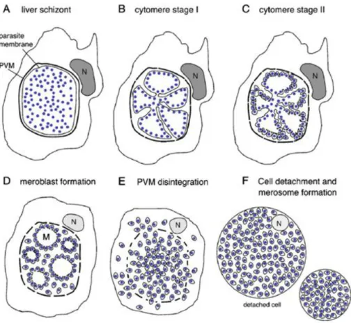
Figure 1.3 ± Model for late liver stage development
Figure 1.4 - Model for merosome dissemination and liberation into the blood Figure 1.5 ± Model for red blood cell invasion and development
Figure 1.6 - Intracellular Transport Pathways Figure 1.7 ± Model of the RabGTPase cycle
Figure 1.8 ± Map of intracellular localization of selected Rab proteins Figure 1.9 ± Stages in phagosome maturation
Figure 1.10 ± Steps in autophagosome formation and maturation Figure 1.11 ± Pathogens survival strategies to avoid lysosomal killing
Figure 1.12 ± Intracellular pathogens and membrane markers on their phagosome membrane
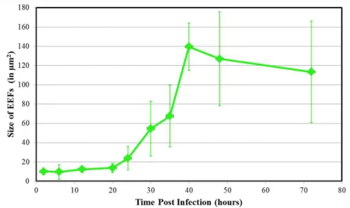
Figure 2.1 - Initial development of Plasmodium berghei parasites in Hepa1-6 cells Figure 2.2 - Late development of Plasmodium berghei parasites in Hepa1-6 cells Figure 2.3 ±P.berghei dynamic morphological modifications in Hepa1-6 cells Figure 2.4 ±P.berghei EEF size increase in Hepa1-6 cells
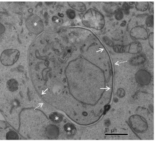
Figure 2.5 ± Transmission Electron Microscopy image of a P.berghei parasite 24 hours post infection suggests possible parasite-host vesicle interactions Figure 2.6 ±Host endoplasmic reticulum and Golgi during P.berghei liver infection Figure 2.7 - Peroxisomes during P.berghei liver infection
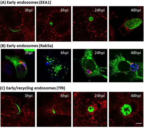
Figure 2.8 ± Early and recycling endosome do not aggregate around P.berghei parasite during liver infection
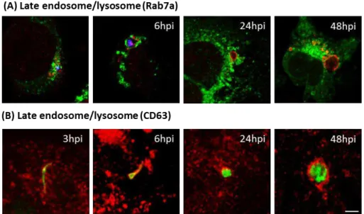
Figure 2.9 ± Late endosomes and lysosomes aggregate around P.berghei parasites Figure 2.10 ± LAMP1 vesicles aggregate around P.berghei parasite throughout the
entire liver infection.
Figure 2.11 ± Late endosomes/Lysosomes aggregate around P.berghei parasites in primary mouse hepatocytes
Figure 2.12 ± Line plot and kinetics of lysosome aggregation around P.berghei parasites throughout liver infection
Figure 2.13 ± Rab7a knock down has no effect on Plasmodium liver infection Figure 2.14 ± Knock down of proteins involved in lysosome function has no effect on
Plasmodium liver development
Figure 2.15 ± Knock down of LAMP proteins has no effect on Plasmodium liver infection
Figure 3.1 - siRNA screen strategy to identify host factors involved in Plasmodium liver infection
Figure 3.2 ± Results of siRNA screen of effect of host Rab proteins during Plasmodium liver infection
Figure 3.3± GFP-Rab localization during P.berghei liver infection
Figure 3.4 ± Rab1a downregulation affects both Plasmodium infection rate and size Figure 3.5 ± Rab1a does not affect Plasmodium migration in liver cell
Figure 3.6 ± Rab1a overexpression effects parasite size
12 Figure 3.8 ± Sar1 downregulation does not affect Plasmodium liver infection.
Figure 3.9 ± Brefeldin A treatment has no effect on Plasmodium liver infection
Figure 3.10 ± Inhibition of autophagosome formation effects Plasmodium infection and development
Figure 3.11 ± Rab1a colocalizes with autophagic vesicles which aggregate around the parasite
Figure 3.12 ±Plasmodium berghei survival kinetics in Hepa1-6 cells
Figure 4.1 ± LysoTracker®Red vesicles aggregate but do not fuse with the P.berghei PVM
Figure 4.2 ± LysoTracker®Red vesicles fuse with latex beads but not with T.gondii parasites
Figure 4.3 ±S+URGRGH[WUDQVWDLQVDFLGLFO\VRVRPes
Figure 4.4 - P.berghei PVM is surrounded by acidic vesicles but maintains a neutral pH Figure 4.5 ± Cathepsin D positive vesicles do not aggregate around P.berghei parasites Figure 4.6 ± NH4Cl treatment inhibits acidification in Hepa1-6 cells and effects
lysosome trafficking
Figure 4.7 ± Inhibition of acidification using NH4Cl effects P.berghei growth during the late stages of infection
Figure 4.8 - ± Inhibition of acidification using Concanamycin A effects P.berghei growth during the late stages of infection
Figure 4.9 - Transmission electron microscopy images suggest close interaction between P.berghei PVM and host vesicles
Figure 4.10 - Transmission electron microscopy images suggest exchange of vesicle contents between host vesicles and the P.berghei PVM
Figure 4.11 ± LAMP1 colocalizes with autophagic vesicles which aggregate around the parasite
13
Summary
Malaria is one of the world´s leading causes of death, responsible for over 700,000 deaths per year, the majority of which are African children under 5 years of age. Malaria disease is caused by the transmission of an Apicomplexa parasite, Plasmodium, through the bit of a female Anopheles mosquito, and transmitted parasites quickly reach the mammalian host liver, where the first round of replication begins.
Plasmodium sporozoites, once inside the liver, must invade and survive within hepatocytes until the first replicative stage within the mammalian host is accomplished. Upon migration through various cells, sporozoites are able to actively enter hepatocytes, forming a Parasitophorous Vacuole Membrane (PVM) around itself. Once this intracellular niche is established, parasite replication and growth is initiated. Dramatic morphological as well as gene expression modifications occur at this stage, and the parasite achieves one of the highest replication rates known within eukaryotic species (Sinnis and Sim, 1997).
Although the Plasmodium life-cycle has been extensively characterized, relatively little is known about sporozoite interaction with host organelles, vesicles and proteins. To address this issue, Plasmodium interactions with the host cell endomembranes was analyzed at various stages of liver infection using indirect immunofluorescence. Plasmodium parasites were seen closely associated with host endoplasmic reticulum (ER) and the Golgi apparatus. Surprisingly, late endosomes/lysosomes, observed with the membrane markers Rab7a, CD63 and LAMP1, aggregated around the parasite. No interaction with host peroxisomes, early and recycling endosomes was observed.
14 significantly affected liver infection rates showing a marked increase in infection. Rab1a has been implicated in ER-to-Golgi trafficking and more recently, in initial autophagosome formation in autophagy, a process by which host cytoplasmic material is degraded and recycled in response to stress. Autophagy also plays a role in antibacterial destruction and elimination, and thus, various pathogens have evolved mechanisms to avoid autophagic elimination.
Further characterization of the role of Rab1a revealed its function in autophagy, rather than ER-Golgi trafficking, affected Plasmodium liver infection. More importantly, Rab1a seems to be important during the initial stages of development by enhancing host parasite elimination, since its depletion leads to an increase in parasite infection rate. During the later stages of infection, Rab1a may be required for parasite replication, since exo-erythrocytic forms (EEFs) are smaller in Rab1a depleted cells. Colocalization with the autophagic marker LC3 revealed that LC3 vesicles aggregate very strongly around the parasite and seems to be upregulated in infected cells suggesting a novel and important role for autophagy during Plasmodium liver infection.
15 these structures could be fusing with the PVM and be an important source of nutrients during parasite growth.
16
Resumo
O agente causador da malária, o parasita Plasmodium, após ser transmitido pela picada de um mosquito, tem de invadir o mamífero hospedeiro e migrar até ao figado, onde irá infectar um hepatócito. Após migrar através de várias células no fígado, o parasita invade o hepatócito final, formando uma membrana à sua volta, a membrana vacuolar parasitária (MVP). Nesta fase occorrem alterações morfológicas, bem como modificações na expressão génica, sendo que o parasita alcança uma das maiores taxas de replicação conhecidas nas de espécies eucarióticas (Sinnis 1997).
Sabe-se pouco sobre a formação e composição da membrana vacuolar parasitária (MVP), contudo à medida que o parasita matura e replica, este requer grandes quantidades de nutrients e lípidos. Neste sentido, as proteínas e lípidos do hospedeiro podem ser uma fonte importante de nutrients para este crescimento, estando a MVP no centro desta possível troca entre a célula hospedeira e o parasita.
Muitos patogénios intracelulares, para sobreviver, conseguem subverter as vias endocíticas e fagocíticas do hospedeiro estabelecendo um nicho intracelular único no qual sobrevivem e replicam. Usando técnicas de immunoflurescência, foi possível observar que o esporozoíto de Plasmodium, em hepatócitos, encontra-se adjacente ao retículo endoplasmático (RE) e ao complexo de Golgi da célula hospedeira. Peroxissomas e endossomas primários não se agregam em redor do parasita enquanto que endossomas tardíos e/ou lisossomas rodeiam o parasita durante toda a infecção no fígado.
17 O papel da Rab1a foi caracterizado e seu efeito durante a infecção do Plasmodium no fígado foi investigada. A proteína Rab1a tem sido implicada no tráfico de RE-a-Golgi e, mais recentemente, no processo de autofagia, mais especificamente na formação de autofagosomas. Autofagia é um processo pelo qual o material citoplasmático é degradado e reciclado, tendo também uma função importante na destruição e eliminação antibacteriana, e deste modo vários patógenos desenvolveram mecanismos para evitar eliminação autofágica.
Inesperadamente, Rab1a parece estar a afectar o parasita Plasmodium através do seu papel durante a autofagia, e não pela via do tráfico retíulo endoplasmoático RE-a-Golgi. Rab1a parece ser importante para a morte de parasitas durante a fase inicial do seu desenvolvimento no hepatócito, uma vez que a sua ausência leva a um aumento na taxa de infecção parasitária. No entanto, durante os últimos estágios da infecção, Rab1a parece ser importante para a replicação do parasita, uma vez que parasitas às 40 horas após a infecção são menores nas células em que Rab1a foi silenciado. Estudos de colocalização usando LC3, como marcador autofágico, mostraram que as vesículas positivas para LC3 agregam-se em redor do parasita, sugerindo uma nova e importante função para a autofagia durante a infecção pelo Plasmodium no fígado.
18 estadios da infecção, em que o tamanho dos parasitas diminuiu significativamente. Um resultado semelhante foi obtido com concanamicina A, uma droga que actua como um inibidor da vATPase. Adicionalmente, dados obtidos usando microscopia eletrónica sugerem uma interacção próxima entre as vesiculas do hospedeiro e a MVP do parasita, propondo que aquelas estruturas podem estar a fundir com o MVP e ser uma fonte importante de nutrientes durante o crescimento do parasita.
19
Table of contents
Contents
Table of contents ... 19
1. General Introduction ... 21
1.1 History of malaria and importance in the history of humanity ... 23
1.2 Malaria endemicity and burden in the world ... 24
1.3 Overview of Plasmodium life cycle ... 26
1.4 The mammalian life cycle in more detail... 28
ϭ͘ϰ͘ϭ/ŶƐĞĐƚďŝƚŝŶŐĂŶĚƚŚĞ͞ĚĞƌŵŝƐ ƐƚĂŐĞ͟ ... 28
1.4.2 Arrest and attachment to hepatocytes ... 31
1.4.3 Liver stage migration, invasion and PVM formation ... 34
1.4.4 Development within the hepatocyte ... 35
1.4.5 Leaving the liver and into the blood ... 40
1.5 Models for liver stage infection ... 43
1.6 Eukaryotic membrane traffic ... 44
1.6.1 Rab GTPases as regulators of membrane traffic ... 46
1.7 Phagocytosis and autophagy ± the cell´s antimicrobial mechanisms ... 50
1.7.1 Phagosome formation and maturation ... 51
1.7.2 Autophagy ... 53
1.8 Intracellular pathogens and host cell subversion ... 55
1.8.1 Bacteria manipulation of the host cell ... 56
1.8.2 Apicomplexa pathogen manipulation of the host cell ... 61
1.9 Aims and objectives ... 65
2. Characterization of Plasmodium liver development and host interactions ... 67
Summary ... 69
Materials & Methods ... 71
2.1 Morphological modification and growth kinetics during Plasmodium berghei liver development ... 77
20
2.2.1 Interactions with host ER, Golgi apparatus and peroxisomes ... 84
2.2.2 Interactions with the early and recycling endosome pathway ... 88
2.2.3 Interactions with the late endosome and lysosomal pathway ... 90
2.3 Disrupting lysosome function ± a gene targeted approach ... 96
2.4 Discussion ... 102
3. siRNA Screen of host trafficking proteins and their role in P.berghei liver stage infection ... 107
Summary ... 109
Materials & Methods ... 111
3.1 siRNA screen development and results ... 114
3.2 Plasmodium colocalization with Rab proteins ... 118
3.3 siRNA screen hit ± Rab1a knock-down increases Plasmodium liver infection ... 122
3.3.1 Rab1 effects Plasmodium berghei liver infection ... 124
3.3.2 Rab1 affects Plasmodium infection via the autophagic pathway .. 128
3.4 Discussion ... 134
4. Dissecting Plasmodium-lysosome interactions ... 139
Summary ... 141
Materials & Methods ... 143
4.1 Plasmodium is surrounded by acidic vesicles but avoids acidification 145 4.2 Plasmodium is not surrounded by Cathepsin D positive vesicles ... 151
4.3 Disrupting vesicle function ± a pharmacological approach ... 153
4.4 Plasmodium parasites (may) feed on host vesicular structures ... 158
4.5 Discussion ... 162
5. General Discussion ... 165
References ... 175
21
23
1.1 History of malaria and importance in the history of humanity
The first known descriptions of malaria relate to a Chinese document from circa 2700 BC, a clay tablet from the Mesopotamia from 2000 BC, an Egyptian papyri from 1570 BC and Hindu texts from before the sixth century BC. Later Greek texts, showed that educated people were well aware of the typical symptoms associated with malaria, the fevers and the enlarged spleens, of people living around marshy areas. Interestingly the word malaria comes IURP WKH ,WDOLDQ ³PDOD DULD´ PHDQLQJ ³EDG DLU´ (Bruce-Chuvatt, 1981). Although the ethological agent was still unknown, Hippocrates, in about 400 BC, describes the occurrence of intermittent fevers which occur daily or every-other-day in malaria patients as opposed to other infectious diseases.
After the discovery of bacteria by Antoni van Leeuwenhoek in 1676 and the discovery of microorganisms as causative agents of diseases, the search for the cause of malaria intensified. The first parasite was observed in the blood of patients by Charles Louis Alphonse Laveran in 1880, who observed several different forms of erythrocytic organisms inside and outside of red blood cells. He also described clear spots that grew, acquired pigment and filled red blood cells before bursting, coinciding with the fevers associated with malaria. Importantly, he noted that quinine removed these stages from the blood. He initially named this parasitic protozoan Oscillaria malariae. Although his theories were not well accepted at first, he was later awarded the Nobel Prize for Medicine in 1907, "in recognition of his work on the role played by protozoa in causing diseases in humans" (ww.nobelprize.org).
24 Although most of the life cycle was elucidated before the 1900s, nobody knew where the parasites developed during the first 7 to 10 days after infection, as they were not detected in the blood of patients. The elusive liver stage of infection was only discovered almost 50 years later by Henry Shortt and Cyril Garnham in 1948 (Shortt and Garnham, 1948). It was only in 1982, that the dormant exo-erythrocytic stages, the hypnozoites, were discovered by Wojciech Krotoski and colleagues, which elucidated the final stage of the malaria parasite life cycle in humans (Krotoski et al., 1982).
1.2 M alaria endemicity and burden in the world
Although the widespread implementation of intervention programs has diminished the burden of malaria in most countries where it was endemic, malaria is still one of the world´s leading causes of death. According to the World Health Organization, around 3.3 billion people ± around half the world population- are at risk of malaria, and there are over 250 million new cases per year. Around 108 countries are endemic for malaria and nearly one million deaths occur every year due to this disease, the majority of which are African children under 5 years of age. (WHO Malaria Report 2009).
A recent review of malaria burden and the efficacy of intervention programs revealed that in most places, such as Southern Africa and the Horn of Africa, malaria burden has steadily and significantly decreased as intervention became more widespread and efficacy improved (O'Meara et al., 2010). Substantial increase in funding and an improvement in the procurement and distribution of effective means of prevention have been identified as the major causes for this decline. Nevertheless, in other parts of the world, such as in Central Africa, little progress has been documented (O'Meara et al., 2010).
25 sulphadoxine plus pyrimethamine or an artemisinin combination, and the scale-up of insecticide-treated bed nets and indoor residual spraying (O'Meara et al., 2010).
Alongside the human fatalities, the burden of malaria also extends to a significant economic impact in endemic countries, as it has been estimated that an average loss of 1.3% of annual economic growth is due to this disease (WHO Malaria Report 2009). This affects poor and rural areas more dramatically, where treatment is often lacking or scarce and where the population have limited access to healthcare. Pregnant women are at special risk of dying from complications of severe malaria and are also at high risk of spontaneous abortion, premature delivery and low infant birth weight. It is estimated that nearly 10 000 pregnant women and up to 200 000 infants die of malaria each year in Africa alone (WHO Malaria Report 2009).
For the past 50 years, huge efforts have been made to develop and produce a malaria vaccine although with little success. Early studies in the 1940s using irradiation attenuated parasites showed that these could indeed induce useful immunity to further infections in mice and in humans (Nussenzweig et al., 1967)(Clyde et al., 1973).
26
1.3 Overview of Plasmodium life cycle
During its life cycle, Plasmodium parasites take many forms and shapes. As the last of these stages was only discovered around 50 years ago, with the discovery of the pre-erythrocytic liver stage and the hypnozoite stage, a clear and complete image of the entire life cycle in mammals was only achieved in the second half of the last century.
In all Plasmodium species, the complete life cycle requires two different hosts, mammalian and insect, more specifically a female Anopheles mosquito. The mammalian life cycle initiates when an infected mosquito is looking for a blood meal and probes for a blood source under the dermis of a mammalian host. The sporozoites, the name given to this stage of the parasite life cycle, are injected during salivation and migrate through the dermis in order to find a blood vessel in which to travel to the host´s liver. Once in the liver, sporozoites need to transverse the liver sinousoid and reach hepatocytes, migrating through several hepatocytes before committing to infection (Mota et al., 2001)(Mota et al., 2002). Inside the final hepatocyte sporozoites invade this cell, with the formation of a Parasitophorous Vacuole Membrane (PVM) and initiate the liver stage of infection. Here the parasites, often termed exo-erythrocytic forms (EEFs), undergo distinct morphological changes and replicate for around 2 to 16 days, depending on the Plasmodium species. This stage is often called the silent stage of infection as it is asymptomatic. Once the parasites have fully replicated, producing around 10,000 to 30,000 blood stage parasites (merozoites), these are released into the vasculature (Prudêncio et al., 2006). Here, free merozoites are able to invade red blood cells (RBCs) and undergo another replication cycle, producing 16 to 32 new merozoites which rupture and infect new RBCs.
27 symptoms include rupture of the blood-brain barrier inducing coma and other QHXURORJLFDODEQRUPDOLWLHVDQGHYHQWXDOO\GHDWKDOVRFDOOHG³VHYHUHPDODULD´ as well as other milder symptoms such as fevers, headache, vomiting, metabolic acidosis which can lead to respiratory distress, severe anemia and multi-organ failure. Also during the blood stage some merozoites develop into sexual stage parasites, the male and female gametocyte, and, if taken up by another female mosquito, the life cycle can continue in the insect host (Figure 1.1).
28 Figure 1.1 ± Plasmodium life cycle in the human and mosquito host. (1) The parasite life cycle begins with the inoculation of sporozoites into the dermis of the mammalian host by an infected female Anopheles mosquito. (2) Sporozoites quickly reach the host blood stream and get to the liver, where they migrate through several hepatocytes before committing to infection, with the formation of the Parasitophorous Vacuole membrane (PVM). Here sporozoites grow and multiply, developing into large liver schizonts. (3) Liver schizonts finally develop into thousands of merozoites which are released into the blood stream, initiating the blood stage of infection and the subsequent infection of red blood cells. Some parasites will eventually develop into sexual gametocytes which can be taken up by a mosquito during a new blood meal. (4) Once in the mosquito midgut, parasites undergo a series of transformations which culminate in the development of new infectious sporozoites in the mosquito´s salivary glands, which can infect a new mammalian host. Image adapted from Prudêncio et al, 2006.
1.4 The mammalian life cycle in more detail
1.4.1 I nsect biting and th
H³GHUPLVVWDJH´
29 anticoagulants present in the saliva of the mosquito (Griffiths and Gordon, 1952) This stage of infection, recently termed tKH ³GHUPLV VWDJH´ ZDV previously thought to have no significance in the infection process although a large body of evidence has showed that the hosts immune response to sporozoites actually begins to be mounted in the skin (Sinnis and Zavala, 2008).
The number of sporozoites deposited in the dermis in one blood meal varies between different studies but between 100 to 200 sporozoites are believed to be deposited in the skin during one blood meal (Frischknecht et al., 2004)(Medica and Sinnis, 2005)(Jin et al., 2007). Although they can remain in the dermis for several hours (Yamauchi et al., 2007), most sporozoites quickly migrate through the avascular dermis in order to find a blood vessel and reach the circulatory system (Vanderberg, 1974)(Vanderberg and Frevert, 2004). Migration, a process where a parasite ruptures the cell membrane, moves through the cell cytoplasm and comes out the other end, is achieved due to a specialized and complex molecular apparatus powered by a unique subpellicular actomyosin motor that is linked to the sporozoite surface and is common to all Apicomplexa parasites (Keeley and Soldati, 2004).
Once in the dermis of the host, a parasite has one of three possible fates; close to 50% stay in the dermis. From the remaining 50%, 70% are able to invade a blood vessel whilst the remaining 30% invade a lymphatic vessel and reach a lymph node (Amino et al., 2006). Only if a parasite is able to migrate, find a blood vessel, be transported to the liver and cross the liver sinusoid, will an active infection ever occur.
30 ultimately the liver, which could aid in mounting immunity to pre-erythrocytic stages and form a basis for malaria vaccine design (Sinnis and Zavala, 2008).
Various parasite proteins essential for migration and gliding motility, both in the dermis and later in the liver, have been described (see Figure 1.2). The Thrombospondin-Related Anonymous Protein (TRAP) and the Apical Membrane Antigen 1 (AMA1), are two parasite proteins which are up-regulated during the sporozoite stage (Rogers et al., 1992b)(Rogers et al., 1992a)(Robson et al., 1997)(Silvie et al., 2004). TRAP is the primary motor-binding protein of sporozoites (Sultan et al., 1997), while AMA1 has been shown to be part of the structural component of the moving junction (Mitchell et al., 2004).
The Circumsporozoite Protein (CSP), the major sporozoite surface protein, is also essential for parasite migration not only in the mammalian but also within the mosquito host (Ménard et al., 1997)(Myung et al., 2004), but its major role in the parasite life cycle is during liver development, as will be described later.
31 Figure 1.2 ± Molecules involved in Plasmodium invasion and development in the mammalian host. The various stages of parasite development inside the mammalian host are depicted. Both parasite and host proteins known to be important for each stage of development are listed. CE, endothelial cells; LE, lymphoid endothelium; SE, fenestrated endothelia; KC, Kupffer cell; PVM, Parasitophorous vacuole membrane; PI, post infection. Image adapted from Vaughan et al, 2008.
1.4.2 Arrest and attachment to hepatocytes
Once in the host circulatory system, sporozoites may be found in hepatocytes within 2 minutes of intravenous injection into rats (Shin et al., 1982). This rapid arrest in the liver indicates that a very strong and specific interaction must occur between the parasite surface proteins and the hepatocyte surface proteins, although relatively little is known about this process.
32 CSP from circulating parasites (Sinnis et al., 1996). Although HSPGs are present in most tissues in the mammalian body, liver HSPGs are known to be more highly sulphated than in all other tissues (Lyon et al., 1994). This feature has been proposed to be responsible for the selective binding of sporozoites to the liver although more research is required to confirm this (Ying et al., 1997)(Pinzon-Ortiz et al., 2001).
More recently, another parasite protein, TRAP, has been shown to be involved in this sequestration in the liver (Pradel et al., 2002)(Pradel et al., 2004). In addition, liver stellate cells are able to synthesize eight times more sulphated HSPGs than hepatocytes and incorporate twice the amount of sulphate into heparan sulphate (Gressner and Schäfer, 1989). Thus, it has been suggested that parasite arrest in the liver may be mediated by attachment to a matrix of HSPGs that protrude from the endothelial fenestrations and are produced by a series of cells in the liver in very high amounts (Pradel et al., 2002)(Pradel et al., 2004).
In order to reach hepatocytes, sporozoites have to transverse the sinusoidal liver barrier, composed mainly of endothelial and Kupffer cells. The importance of and route by which parasite are able to transverse through either of these cells has been a topic of some controversy. Endothelial cells have fenestrations but these are too small for parasite to pass through (about one tenth of the diameter of a parasite).
33 migration through cells is an important step in establishing a successful infection (see (Frevert et al., 2006) for a good review on this topic).
The choice of hepatocytes as the preferred cell for the next stage of the Plasmodium life cycle is still a mystery. Nevertheless, compared to other cells, hepatocytes have unique metabolic properties, being particularly proficient at metabolizing compounds (e.g. lipids, purines, glucose) in large quantities (Morgan and Baker, 1986)(Pels Rijcken et al., 1993)(Klover and Mooney, 2004). The fast multiplication rate of Plasmodium parasites in the liver impose a high demand on lipids for organelle synthesis and for this reason hepatocytes represent a favorable environment within the mammalian host, especially for a parasite that is unable to synthesize sterols. Interestingly, infected cells seem to upregulate proteins involved in sterol synthesis and lipid metabolism (Albuquerque et al., 2009), suggesting that Plasmodium parasites are able to manipulate host transcription to their own advantage and survival.
Whatever the process to reach the liver hepatocytes may be, eventually, successful parasites reach the host hepatocytes where they will ultimately commit to infection. Yet, before this occurs, they migrate through a series of hepatocytes, breaching the plasma membrane and passing in direct contact with the host cytosol (Mota et al., 2001)(Mota and Rodriguez, 2004).
34
1.4.3 Liver stage migration, invasion and PVM formation
Once sporozoites have migrated through a series of hepatocytes, they are ready to invade, a process is actively controlled by the parasite, using its glideosome (motility) machinery to force its way into the host cell (Keeley and Soldati, 2004). Invasion occurs by a partial invagination process of the host cell plasma membrane (Mota et al., 2001). Specific parasite adhesins are secreted at the parasite apical end and are proteolytically processed. Processing of these proteins is believed to expose the adhesive domains which function at the sporozoite-host cell interface and help the sporozoite to adhere to the hepatocyte plasma membrane (reviewed in (Carruthers and Blackman, 2005)).
Apart from proteins involved in parasite gliding motility, only a limited number of parasite and host proteins have been shown to be involved in parasite invasion (Vaughan et al., 2008)(Ejigiri and Sinnis, 2009). P.berghei Pbs36p and Pbs36 proteins are members of a family of proteins with 6 conserved cysteine residues which are specifically produced in liver-infective sporozoites. When expression of these proteins is abolished, infectivity in the mammalian host is inhibited, although cell transversal is not affected (Ishino et al., 2005b). This result suggests that these proteins are necessary for sporozoites to recognize hepatocytes and commit to infection.
35 CD81 is a tetraspanin protein that associates with other proteins and lipids to form membrane microdomains. CD81 deficient mice were also less permissive to P.yoelii sporozoite invasion (Silvie et al., 2003) and later studies found that cholesterol was involved in the assembly of CD81 microdomains (Silvie et al., 2006). Since SR-B1 is a provider of hepatocyte cholesterol, it has been hypothesized that this protein plays a role in the formation of CD81 enriched microdomains which in turn facilitate sporozoite entry (Yalaoui et al., 2008b). SR-B1 deficient mice showed a decreased expression of CD81 on the cell surface indicating that some membrane rearrangement event had occurred, which in turn had an effect on parasite invasion. Further studies are needed to confirm all of these hypotheses.
As mentioned previously, sporozoite invasion is characterized by the formation of a PVM, yet the sequence of events leading to PVM formation are largely unknown. Three different mechanisms of PVM formation have been proposed: 1) de novo PV membrane formation solely from secreted parasite proteins, 2) PVM formation from direct invagination of the host plasma membrane or 3) a combination of both mechanisms (reviewed in (Mota et al., 2002)). Further studies are required to unravel this unique process.
1.4.4 Development within the hepatocyte
36 and ions (Bano et al., 2007) yet its composition and origin is still largely unknown.
Within the liver, sporozoites differentiate into exo-erythrocytic forms (EEFs), that grow and multiply (schizont stage) forming thousands of new erythrocyte infectious merozoites (Figure 1.3) (Sturm et al., 2009). These newly formed merozoites are subsequently surrounded by a membrane resulting in fully mature merozoites which are ready to be released into the liver blood vessels.
37 Largely due to the technical difficulties involved in studying this stage of the life cycle, to date, only a few parasite proteins have been shown to play a role in parasite liver development. Salivary gland sporozoites upregulate the expression of a unique subset of genes collectively called the UIS (upregulated in infectious sporozoites) and some of these have been shown to be essential for liver stage development (Matuschewski et al., 2002)(Mikolajczak et al., 2008). More recently it was shown that a conserved Plasmodium sporozoite low-complexity asparagine-rich protein, SAP1 (Sporozoite Asparagine-rich Protein 1) is involved in a selective post-transcriptional mechanisms to regulate the abundance of UIS transcripts (Aly et al., 2008)(Aly et al., 2011).
Up-regulated in Infective Sporozoite genes 3 and 4 (UIS3 and UIS4) are two transmembrane proteins that localize to the PVM just a few minutes after invasion. If disrupted, sporozoites are unable to establish an adequate liver infection, as knock out parasites invade hepatocytes normally but dies within the first 48 hours of infection (Mueller et al., 2005). These parasites demonstrate the importance of the PVM during the liver stage of infection. It is known that UIS3 binds directly to liver fatty acid binding protein (L-FABP) on the PVM (Mikolajczak et al., 2007)(Sharma et al., 2008) while there is still no known function for UIS4.
38 manner, as down-regulation of host L-FABP severely impairs liver development (Vaughan et al., 2009).
P36p, another parasite protein, has been shown to be important during invasion, development and PVM formation although there is conflicting evidence on its exact role (van Dijk et al., 2005)(Ishino et al., 2005a)(Labaied et al., 2007)(Ejigiri and Sinnis, 2009).
Even if many of the molecular functions of some of these proteins is not yet known, their importance in liver stage research has been immeasurable, as parasites mutated for these proteins, don´t develop fully in the liver yet illicit protective immunity to further sporozoite challenges, thus being termed genetically attenuated sporozoites (GAS). In a similar way to radiation attenuated sporozoites (RAS), this model has allowed for a deeper understanding of the basic immune mechanisms that mediate sterile immunity against reinfections (Nussenzweig et al., 1967)(Hafalla et al., 2006).
During its liver development, the parasite largely outgrows the normal host cell limits and is able to remodel and exploit the host hepatocyte (reviewed by (Mikolajczak and Kappe, 2006)). Nevertheless, very few host proteins have been implicated in this process.
Studies performed investigating the roles of the parasite UIS4 have shown that this protein colocalizes with host ApoA1 in the PVM 24 hours after parasite invasion and ApoA1 has been speculated to be important in the synthesis of large amounts of membranes essential for the PVM expansion (reviewed in (Prudêncio et al., 2006)).
UIS3 was also shown to interact directly with L-FABP using a two-hybrid system (Mikolajczak et al., 2007). L-FABP down regulation in hepatocytes severely impairs parasite growth and its overexpression promotes parasite growth. L-FABP has been speculated to be the main carrier of fatty acids from the host cell cytoplasm to the parasite yet further studies are required in order to confirm its role in parasite development.
39 rearrangements via the HGF receptor, MET, signaling pathway (Carrolo et al., 2003). It was also shown that this signaling cascade prevents infected cells from undergoing cell death by apoptosis, a mechanism that would be very detrimental for the parasite (Leirião et al., 2005). Curiously, infected hepatocytes do not display the normal signs of cell death and were actually resistant to tumor necrosis factor ((TNF)-Į'-galactosamine) stimulation (van de Sand et al., 2005), showing that the parasite is able to constantly stimulate host cell survival throughout the duration of its liver stage infection (reviewed in (Heussler et al., 2006)).
Apart from its role in parasite invasion, host SR-B1, also seems to play a role in parasite development, as parasite in SR-B1 transgenic mice had a diameter 3-fold larger than in SR-B1 knock-out mice at 48 hour post liver infection (Yalaoui et al., 2008a). Interestingly, SR-B1 was not localized around the PVM which has led to the hypothesis that SR-BI effects parasite development due to its interaction with L-FABP, although more evidence is required to confirm this hypothesis (Rodrigues et al., 2008)(Yalaoui et al., 2008b).
Taken together, these studies show that Plasmodium parasites are able to actively manipulate and hijack a wide range of host cell functions and pathways, in order to establish its own specialized intracellular niche. Host cell subversion is a common strategy used by intracellular pathogens to ensure survival and adequate growth. To achieve this pathogens utilize a wide range of techniques and secrete a multitude of proteins or effectors. Unfortunately, due to the difficulties involved in Plasmodium liver stage research, no such proteins have been identified for this intracellular pathogen.
40 vacuolar space through open channels (Sturm et al., 2009)(Bano et al., 2007). In contrast with malaria blood stages, the pores within the Parasitophorous vacuole membrane of the liver stage display a smaller size as they restrict the passage of solutes to less than 855Da. These pores are stably maintained during parasite karyokinesis until complete cellularisation. Host-derived cholesterol accumulated at the Parasitophorous vacuole membrane may modulate the channel activity (Labaied et al., 2010). These observations describe the Parasitophorous vacuole of the Plasmodium liver stage as a dynamic and highly permeable compartment that can ensure the sustained supply of host molecules to support parasite growth in the nutrient-rich environment of liver cells (Bano et al., 2007). We were thus interested in exploring this in more detail.
1.4.5 Leaving the liver and into the blood
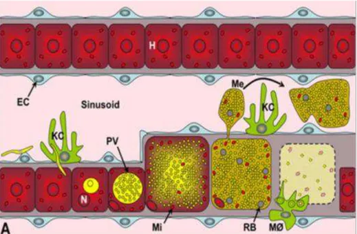
After parasites have developed in the liver, the pre-erythrocytic development comes to an end and the newly formed merozoites are ready to invade RBCs. It was initially believed that merozoites, after rupturing their PVM and coming into contact with the hepatocyte cytoplasm, were released into the blood stream by a rupture of the host cell membrane, although this did not explain how the merozoites were able to cross the extracellular matrix and pass through the endothelium sinusoid layer. Early studies found merozoite filled membrane bound vesicles within the extracellular matrix (Meis and Verhave, 1988). This membrane was later found to be of host origin, which explains why these vesicles, termed merosomes, are not attacked by phagocytic cells in the liver, and are able to pass unharmed through the sinusoidal space and into the blood vessels (Sturm et al., 2006) (Figure 1.4).
41 liberates the fully formed merozoites into the lung circulation (Baer et al., 2007). This phase of the life-cycle is believed to enhance the dissemination of merozoites into the blood circulation where they will encounter red blood cells and initiate the next round of infection in the mammalian host.
Figure 1.4 - Model for merosome dissemination and liberation into the blood. Model for merosome dissemination and liberation, surrounded by hepatocyte plasma membrane, avoiding recognition by Kupffer cells in the sinusoidal space; M, meroblast; H, hepatocyte; EC, endothelial cell; N, nucleus; KC, Kupffer cells; PV, Parasitophorous vacuole; Mi, host cell mitochondria; Me, merosomes; RB, remenant bodies; MØ, mononuclear phagocytes. Image adapted from Baer et al, 2007.
42 Exit from infected host cells also appears to be mediated by a class of papain-like cysteine proteases called Serine Repeat Antigens (SERAs). Members of this family, such as PbSERA1 and PbSERA2, are abundantly expressed in the final stages of merozoite formation and are targeted to the PVM. Single loss-of-function mutant parasites for PbSERA1 or PbSERA2 progressed normally throughout the parasite life cycle although the expression of other members of this family, namely PbSERA3 was upregulated, suggesting that members of this family can compensate for each other (Putrianti et al., 2010).
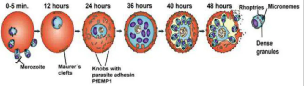
The next phase of the Plasmodium life cycle starts with the invasion of uninfected red blood cell (RBC) in the blood. The erythrocytic development initiates with parasites attaching and repositioning themselves on the RBC membrane. Once inside, parasites develop into the ring stage (0-24h), followed by the trophozoite (24-36h) and finally the schizont stage (40-48h) (Maier et al., 2009). After the first round of RBC development, parasites are released into the blood stream and initiate the new cycle of RBC invasion, giving rise to all the malaria associated symptoms (Figure 1.5).
43
1.5 M odels for liver stage infection
One of the most important breakthroughs in malaria research was the development of in vitro blood culture systems, pioneered by William Trager and J.B. Jensen in 1976 (Trager and Jensen, 2005). This opened the path to vaccine and drug development research. Yet, models for liver stage development were more difficult to develop. The liver stage is still the most elusive stage of the entire life cycle of the Plasmodium parasites. Although it was identified more than 50 years ago (Shortt and Garnham, 1948), technical constrains has made it very difficult to study the molecular mechanisms involved in this stage of infection. Human liver stage research is not feasible in vivo, although some studies have been able to mimic the complete liver stage development in human primary hepatocytes (Mazier et al., 1984)(Mazier et al., 1985)(Mazier et al., 1987), yet these experiments rely on the often scarce and unpredictable availability of human material, making this type of experiments very technically challenging. Complete Plasmodium falciparum and P vivax liver stage development has been achieved in human hepatocyte cell lines, HUH7 and HepG2-A16 respectively (Uni et al., 1985)(Calvo-Calle et al., 1994). More recently, the HC-04 cell line was able to support the complete liver stage development of both P.falciparum and P.vivax (Sattabongkot et al., 2006) although in all these experiments, mosquito breeding and infection with human parasites is required, all of which involve very specific and controlled safety protocols, thus making it very difficult and expensive to perform.
44 (Mota and Rodriguez, 2000). P.berghei is known to be more promiscuous than P.yoelii as it is also able to infect human hepatoma cell lines such as HepG2 and HUH7. More recently, a humanized mouse model was developed to study P.falciparum liver infection, providing a new tool to study this part of the life stage using this human pathogen (Morosan et al., 2006).
Both rodent models have different infection efficiencies, with P.berghei having the highest efficiency in vitro and P.yoelii having the highest efficiency in vivo (Khan and Vanderberg, 1991)(Briones et al., 1996). Most of the work in this project involved in vitro work and P.berghei parasites were used throughout.
Plasmodium berghei in vitro infection rates vary between 1-5% while Plasmodium yeolli infection rate are often even lower. This makes using cell biology and molecular biology techniques very difficult, which ultimately slowed down research in this area. Liver stage research also involves the growth and maintenance of live and infected Anopheles mosquitoes, a technique that is not easy to master. All these factors together, has made malaria liver stage research slow and difficult, even during the past 20 years where the number of publications in this area has skyrocketed.
1.6 Eukaryotic membrane traffic
45 the correct destination. It is the precise regulation between these different processes that ensures efficient intracellular transport while preserving correct organelle identity.
In order to transport cargo between organelles, vesicle budding or formation needs to occXUIURPD³GRQRU´FRPSDUWPHQWE\DSURFHVVWKDWDOORZV for the sorting of the received cargo. The vesicles are later targeted and fuse ZLWK D VSHFLILF ³DFFHSWRU´ FRPSDUWPHQW YHVLFOH WDUJHWLQJ DQG IXVLRQTo ensure correct organelle homeostasis, these transport machinery components are recycled back from the acceptor compartment in a pathways termed ³UHWURJUDGH WUDQVSRUW´Figure 1.6 depicts the various intracellular transport pathways.
Figure 1.6 - I ntracellular Transport Pathways. The three most important intracellular transport pathways are depicted in this scheme; the secretory or exocytic pathway, where cargo is delivered to the plasma membrane via secretory granules; the endocytic pathway, where cargo is internalized from the extracellular space; and the lysosomal/vacuolar pathway where cargo that has been internalized is processed to the cell interior. Adapted from Bonifacino and Glick, 2004.
46 vesicle machinery and can also actively remodel membranes (Scales et al., 2000).
Once formed, vesicle targeting and fusion is largely mediated by proteins termed the SNAREs (Soluble N-ethylmaleimide-sensitive factor activating protein receptor) (Gerst, 1999)(Duman and Forte, 2003). SNAREs have been extensively studied and mediate vesicle fusion by bridging two membranes (Ungar and Hughson, 2003). Individual SNARE family members tend to be compartment-specific and are thus thought to contribute to the specificity in docking and fusion events between organelles (Pelham, 2001) although it has been shown that some SNAREs can compensate for the loss of another or even act in multiple distinct complexes (reviewed in (Ungar and Hughson, 2003)).
Another family of proteins, the Rab GTPases, is also involved in multiple regulatory processes along these transport pathways as well as mediating the interaction of membranes with cytoskeletal motors. This family of proteins and their role in membrane traffic will be discussed further.
1.6.1 Rab GTPases as regulators of membrane traffic
A hallmark of eukaryotic cells is that they are highly compartmentalized and have evolved mechanisms to regulate intracellular transport between compartments. A number of intracellular pathogens are able to manipulate components of these mechanisms to aid their survival. Rab proteins, the largest branch of the Ras superfamily of small guanosine triphosphates (GTPases), are master regulators of organelle identity and play a key role in intracellular trafficking by controlling vesicle fusion and motility.
47 to specific cell types, reflects the great complexity and organization required in higher eukaryotic organisms (Pereira-Leal and Seabra, 2001).
Rab proteins function as molecular switches, cycling between their active GTP-bound state and their GDP-bound inactive state. As Rabs do not have high intrinsic guanine nucleotide exchange or hydrolysis activities, they require the recruitment of guanine exchange factors (GEFs) and GTPase-activating proteins (GAPs) respectively to switch between their active and inactive forms (Pfeffer and Aivazian, 2004). Once in the active GTP-bound form Rab proteins can recruit a diverse array of effector molecules such as tethering factors, molecular motors and enzymes to perform their downstream biological activity. It is the coordinated spatial and temporal control between each Rab, its effector and the recruitment of specific GEFs and GAPs that gives Rabs the ability to tightly and specifically regulate intracellular membrane traffic (Barr and Lambright, 2010) (see Figure 1.7).
48 Figure 1.7 ± Model of the RabGTPase cycle. After synthesis Rab proteins are recognised by REP which presents the Rab to RabGGTase (Rab geranyl geranyl transferase), the enzyme that prenylates the Rab protein (1). REP then escorts the Rab protein to the appropriate donor membrane where it is recognised by a GDF (GDI-displacement factor) and a putative specific Rab receptor . The GDF destabilises the REP-Rab complex allowing membrane association of the Rab protein (2). The Rab protein is then activated by a GEF (Guanine nucleotide exchange factor) which catalyses the removal of GDP and allows binding of GTP. Once in its active form the Rab protein can interact with multiple effectors (3) to mediate vesicle formation and budding (4), vesicle motility usually through the action of a molecular motor like myosin or kinesin (5) and vesicle docking (5). Rab proteins and their effectors may also interact with SNARE complexes and influence membrane fusion. During or following fusion Rab proteins are inactivated by a GAP (GTPase activating protein) which catalyses the Rab proteins own GTPase activity. Once in its inactive, GDP-bound form the Rab protein can be extracted from the acceptor membrane by GDI (guanine nucleotide dissociation inhibitor) (6). GDI then recycles the Rab protein to its donor membrane where GDF catalyses the dissociation of the Rab-GDI complex and allows membrane association of the Rab protein (7) and the commencement of a new cycle.
49 The mechanism by which Rab proteins achieve their specific intracellular localizations is largely uncharacterized although putative membrane-bound targeting factors, Rab-GEFs, effector binding and lipid composition of membranes have all been implicated. Rabs function by coordinating a continuum of membrane trafficking events in a mechanism termed Rab conversion. This involves controlling the tethering/docking of vesicles to their target membranes, which ultimately leads to their fusion (Markgraf et al., 2007). Rabs also control the vesicle budding and motility, by regulating binding and movement along both the microtubule an actin cytoskeleton. This often involves direct as well as indirect binding with motor proteins such as myosins and dyneins (Pfeffer, 2003)(Pfeffer and Aivazian, 2004).
50 Figure 1.8 ± Map of intracellular localization of selected Rab proteins. Summary of the intracellular location of selected Rab proteins in mammalian cells. CCV, clathrin-coated vesicle; CCP, clathrin-clathrin-coated pit; EC, epithelial cells; IC, ER±Golgi intermediate compartment; M, melanosomes; MTOC, microtubule-organizing centre; SG, secretory granules; SV, synaptic vesicles; T, T-cell granules; TGN, trans-Golgi nextwork. Image adapted from Zerial & McBride, 2001.
1.7 Phagocytosis and autophagy
±
the cell´s antimicrobial
mechanisms
51 subverting host immune mechanisms to their advantage. At the level of the cell, ingested particles are normally processed by the cell through the endosomal-lysosomal degradation pathway, where pathogens are degraded and digested, and antigens are presented on the surface of the cell to trigger an immune response (Aderem and Underhill, 1999)(Houde et al., 2003). More recently, autophagy has also been described as a pathway used by cells for pathogen degradation and elimination (Levine and Kroemer, 2008)(Subauste, 2009).
1.7.1 Phagosome formation and maturation
Phagocytosis is an essential process that triggers the activation of multiple transmembrane signaling pathways leading to the reorganization of the actin cytoskeleton and the formation of a unique intracellular compartment, termed the phagosome. It is believed that phagocytosis was already present in early eukaryotic ancestors (Dacks and Field, 2007) thus it is not surprising that it is a very tightly regulated endomembrane system.
It is now widely accepted that phagosome maturation occurs through a strictly choreographed sequence of fusion and fission events involving defined compartments of the endocytic pathway. Effective phagocytosis therefore requires two distinct phases; particle internalization and phagosomal maturation. Internalization depends on the type of particle being internalized as well as the target cell, but can be generalized to two general mechanisms; direct, through recognition of specific receptors at the surface of cells by pathogen associated molecules, or indirectly, by opsonins, which trigger specific signaling cascades depending on the nature of the molecules involved. (See (Flannagan et al., 2009) for an excellent review on the subject).
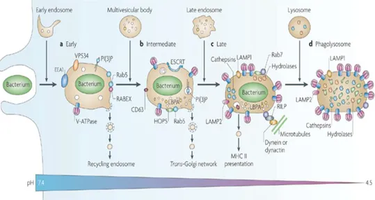
52 Rab7 (Pitt et al., 1992)(Desjardins et al., 1994)(Desjardins, 1995). The late endosome-phagosome then becomes acidified due to the acquisition of the vacuolar proton pump ATPase (vATPase). The phagosome lumen is acidified due to the pumping of hydrogen protons by this protein before fusion with lysosomes, becoming a hydrolase-rich phagolysosome. Here, ingested particles are degraded and processed pathogen antigens are then ready to be presented on the cell surface via recycling endosomes (see Figure 1.9).
Figure 1.9 ± Stages in phagosome maturation. Shortly after pathogen uptake, the phagosome undergoes a series of sequential fusion events with subcompartments of the endocytic pathway. Various stages may be identified, based on a series of protein markers, including an early (a), intermediate (b) and late (c) stage of maturation. The final stage involves fusion with lysosomes, forming a phagolysosome (d) that acquires various hydrolases and becomes acidic, culminating with the degradion of the internalized pathogen. EEA1, early endosome antigen 1; ESCRT, endosomal-sorting complex required for transport; HOPS, homotypic protein sorting; LAMP, lysosomal-associated membrane protein; LBPA, lysobisphosphatidic acid; PI(3)P, phosphatidylinositol-3-phosphate; MHCII, major histocompatibility complex II; RILP, Rab-interacting lysosomal protein. Image adapted from Flannagan et al, 2009.
53 by phagolysosomes by manipulating these series of sequential fusion events since proper phagosome maturation would ultimately lead to pathogen degradation and elimination (see (Hackstadt, 2000)(Tjelle et al., 2000)(Kahn et al., 2002)for good reviews on the subject).
1.7.2 Autophagy
Macroautophagy (referred to as autophagy hereafter) is an evolutionary conserved catabolic process whereby cytoplasmic materials, including organelles, reach lysosomes for degradation (Mizushima et al., 2008)(Levine and Kroemer, 2008). The metabolic roles of autophagy can be classified in two types: basal autophagy, which occurs at low levels constitutively and is believed to be important in the internal quality control of intracellular components, and induced autophagy, where cells, under nutrient deprivation, self-digest cytoplasmic components to maintain the amino acid pool during stress conditions (Kuma and Mizushima, 2010).
54 Figure 1.10 ± Steps in autophagosome formation and maturation. Autophagy is initiated by the nucleation of a phagophore, which elongates and closes on itself to form and autophagosome. This vesicle then fuses with the endocytic pathway (early and late endosomes and MVBs), in a process termed maturation. The resulting amphisomes is more acidic than the autophagosome and begins to acquire hydrolytic enzymes. This vesicle finally fuses with lysosomes and degradation of the internalized material occurs. Shown in the scheme are some of the molecular players involved at the different stages of this process. Image adapted from Mehrpour et al, 2010.
55 Just like proper phagosome maturation, correct autophagic maturation is an essential pathway for intracellular pathogen elimination, making them two ideal pathways to be manipulated by intracellular pathogens.
1.8 I ntracellular pathogens and host cell subversion
Several pathogens have evolved to enter, survive and replicate within mammalian cells. Most of these intracellular pathogens use existing cellular pathways not only to enter host cells, exploiting existing receptors at the surface to attach and enter, but also to acquire nutrients and survive once inside the cell. Because most of these pathways end in the formation of phagolysosomes, which are capable of degrading microorganisms, pathogens have evolved remarkable ways to interact with the host cell phagosome maturation machinery to ensure survival.
Although it seems that each organism seems to have developed its own unique mechanism to evade lysosomal destruction, these strategies may be summarized in five categories: 1) lysis of the phagosomal membrane and escape to the host cytosol, 2) avoidance of the host autophagy pathway, 3) delay of the phagosomal maturation process, 4) subversion of the phagocytic pathway and 5) survival within the harsh phagolysosomal environment (Luzio et al., 2007) (Figure 1.11 summarizes these alternative strategies used by pathogens to evade host killing). Examples of bacterial and parasite organisms that employ each of these strategies will be discussed as well as the molecular players involved, where known.
56 Figure 1.11 ± Pathogens survival strategies to avoid lysosomal killing. Pathogens have evolved different strategies to prevent lysosome degradation and killing. These include (1) escape into the cytosol, (2) avoidance of autophagy, (3) delay in phagosomal maturation (4) subversion from the normal phagocytic pathway and (5) ability to survive in the harsh phagolysosomal environment. Image adapted from Luzio et al, 2007.
1.8.1 Bacteria manipulation of the host cell
57 Figure 1.12 shows a summary table of an extensive list of intracellular bacterial pathogens and specific proteins markers that have been described to be on their respective phagosome membrane. Specific examples will be discussed further below.
In next page:
Figure 1.12 ± I ntracellular pathogens and membrane markers on their phagosome membrane. Summary table of various intracellular bacterial and parasites pathogens and various protein markers described to be on their phagosome membrane. Red boxes represent markers NOT found on the membrane, green boxes represent marker found on the membrane and white boxes represent missing data. Yellow boxes represent acidic phagosomes while blue boxes represent neutral phagosomes. Pale green boxes in the last column show pathogens that, upon invasion with the formation of a vacuole membrane around them, later escape and survive in the host cytosol. The results obtained in the context of this project, related to membrane markers found surrounding Plasmodium berghei PVM are also included. References from this image are listed in
59 As mentioned previously, one mechanism used by bacteria to survive in host cells is to enter the host cell with the formation of a vacuole around it, but quickly escape to the host cytosol. This is the case with Listeria, Burkholderi, Francisella, Shigella and Rickettsia species. Listeria monocytogenes initially inhabits a membrane bound vacuole that acquires markers of early endosomes such as Rab5, but later secretes a pore-forming toxin called Listeriolysin O which causes a delay in phagosomal maturation by causing alterations in both vacuolar pH and Ca2+ concentration, giving the bacteria time to escape into the host cytosol (Henry et al., 2006). Once in the cytosol, Listeria is able to escape autophagic death by secreting ActA protein, which recruits the Arp2/3 complex and Ena/VASP to the bacteria surface, disguising it from autophagic recognition (Yoshikawa et al., 2009). Remarkably, Ricketssia conorii also secretes an ActA related protein, RickA, a protein that recruits the host Arp2/3 complex, inducing actin nucleation and generation of actin filaments, but possibly, also protecting it from autophagic killing (Gouin et al., 2004)(Jeng et al., 2004).
Shigella flexneri is also able to evade autophagic delivery to the lysosome after escaping to the cytosol by a secreting IcsB, which avoids bacterium autophagic death, although the exact mechanism by which IcsB achieves this is still largely unknown (Ogawa et al., 2005)(Kayath et al., 2010). Curiously, Burkholderi pseudomallei, another intracellular bacteria, secretes BopA, which is a IcsB homologue, which also contains the essential cholesterol binding domain (CBD) required for autophagic escape (Kayath et al., 2010).