Smooth ER in Mouse Hepatocytes and Is Important in the
Regulation of Glucose and Lipid Metabolism
Karin Berger1.*, Rebecka Lindh1., Nils Wierup2
, Emilia Zmuda-Trzebiatowska1,3, Andreas Lindqvist2,
Vincent C. Manganiello3, Eva Degerman1
1Department of Experimental Medical Sciences, Lund University, BMC C11, Lund, Sweden,2Department of Experimental Medical Sciences, Lund University, BMC B11, Lund, Sweden,3Translational Medicine Branch, National Heart, Lung, and Blood Institute (NHLBI), National Institutes of Health (NIH), Bethesda, Maryland, United States of America
Abstract
Cyclic nucleotide phosphodiesterases (PDEs) are important regulators of signal transduction processes mediated by cAMP and cGMP. One PDE family member, PDE3B, plays an important role in the regulation of a variety of metabolic processes such as lipolysis and insulin secretion. In this study, the cellular localization and the role of PDE3B in the regulation of triglyceride, cholesterol and glucose metabolism in hepatocytes were investigated. PDE3B was identified in caveolae, specific regions in the plasma membrane, and smooth endoplasmic reticulum. In caveolin-1 knock out mice, which lack caveolae, the amount of PDE3B protein and activity were reduced indicating a role of caveolin-1/caveolae in the stabilization of enzyme protein. Hepatocytes from PDE3B knock out mice displayed increased glucose, triglyceride and cholesterol levels, which was associated with increased expression of gluconeogenic and lipogenic genes/enzymes including, phosphoenolpyruvate carboxykinase, peroxisome proliferator-activated receptorc, sterol regulatory element-binding protein 1c and hydroxyl-3-methylglutaryl coenzyme A reductase. In conclusion, hepatocyte PDE3B is localized in caveolae and smooth endoplasmic reticulum and plays important roles in the regulation of glucose, triglyceride and cholesterol metabolism. Dysregulation of PDE3B could have a role in the development of fatty liver, a condition highly relevant in the context of type 2 diabetes.
Citation:Berger K, Lindh R, Wierup N, Zmuda-Trzebiatowska E, Lindqvist A, et al. (2009) Phosphodiesterase 3B Is Localized in Caveolae and Smooth ER in Mouse Hepatocytes and Is Important in the Regulation of Glucose and Lipid Metabolism. PLoS ONE 4(3): e4671. doi:10.1371/journal.pone.0004671
Editor:Silvana Gaetani, Sapienza University of Rome, Italy
ReceivedNovember 14, 2008;AcceptedJanuary 28, 2009;PublishedMarch 5, 2009
Copyright:ß2009 Berger et al. This is an open-access article distributed under the terms of the Creative Commons Attribution License, which permits unrestricted use, distribution, and reproduction in any medium, provided the original author and source are credited.
Funding:This work was supported by the Swedish Research Council Project 3362 to E.D; K.B. was supported by Lund University Diabetes Center (LUDC); Grants were obtained from the following foundations: Swedish Diabetes Association; Novo Nordisk, Denmark; The Swedish Society of Medicine, Dr. P. Hakansson, Albert Pahlsson, Fredrik och Ingrid Thuring, Wiberg, Lars Hierta, AhlA˜ßn, Tore Nilsson and Magn. Bergvall. The funding agencies did not take any part in the design and conduct of the study, in the collection, analysis, and interpretation of the data, or in the preparation, review, or approval of the manuscript.
Competing Interests:The authors have declared that no competing interests exist.
* E-mail: karin.berger@med.lu.se
.These authors contributed equally to this work.
Introduction
Cyclic nucleotide phosphodiesterases (PDEs) are important regulators of signal transduction processes mediated by cAMP and cGMP. The PDE family contains eleven structurally related and functionally distinct subfamilies (PDE1-11) that differ in their primary structures, affinities for cAMP and cGMP, responses to specific effectors and inhibitors, as well as mechanisms through which they are regulated [1]. PDE3 isoforms are encoded by two similarly organized genes, PDE3A and PDE3B. These enzymes hydrolyze cAMP and cGMP with high affinity in a mutually competitive manner and are inhibited by compounds such as cilostamide, cilastazol and milrinone [2,3]. The structural organization of PDE3A and PDE3B proteins is identical with the catalytic domain found in all PDEs located in the C-terminal portions of the molecules [2,3]. The catalytic domains of PDE3A and B are highly conserved, except for an insertion of 44 unique amino acids that is not found in the catalytic domains of other PDE families and that also differs in, and thus distinguishes, PDE3A and B isoforms [2,3]. Their N-terminal regulatory
PDE3A and PDE3B exhibit cell-specific differences in expres-sion. PDE3A is highly expressed primarily in the cardiovascular system, for example in platelets, smooth muscle cells and cardiac myocytes [2,3]. PDE3B on the other hand is relatively highly expressed in cells important in energy metabolism, such as white and brown adipocytes, pancreaticb-cells and liver [2,3] indicating a role for this enzyme in the regulation of metabolism.
Recent results from PDE3B transgenic mouse models do indicate that PDE3B plays an important role in overall regulation of energy metabolism [10,11]. For example, mice that specifically overexpress PDE3B in pancreaticb-cells demonstrate glucose intolerance and impaired insulin response to glucose and glucagon-like peptide-1 (GLP-1) [10]. The phenotype of PDE3B knock out (KO) mice is complex. Hence, on one hand PDE3B KO mice are lean and have improved insulin secretion but they also exhibit glucose intolerance, insulin resistance and increased lipolysis [11]. The role of hepatocyte PDE3B in the regulation of lipid and glucose metabolism remains unknown. However, clamp studies in PDE3B KO mice show increased glucose production and reduced ability of insulin to suppress glucose production indicating multiple roles for this enzyme in hepatocytes [11].
In this study we demonstrate that, in hepatocytes, PDE3B is localized to caveolae and smooth ER and that the enzyme has an important role in the regulation of triglyceride, cholesterol and glucose metabolism in these cells.
Materials and Methods
Ethics statement
All animals were handled in strict accordance with good animal practice as defined by the national and local animal welfare bodies, and all animal work was approved by the Ethics Committee at Lund University, Lund, Sweden.
Materials
C57BL/6 male mice were purchased from Taconic (Skensved, Denmark). Caveolin-1 KO mice on the C57BL/6 background, obtained from the Jackson Laboratory (Bar Harbor, Maine, USA), were further backcrossed on the same background and genotyped as described by Razani et al [12]. PDE3B deficient mice were generated and characterized as previously described in [11] (a fragment containing exon 1 of the mousePde3bgene was cloned into
the pBluescript and used to construct the targeting vector). Antibodies against caveolin-1 (rabbit polyclonal) and adenylyl cyclase (AC) V were obtained from Santa Cruz Biotechnology (Santa Cruz, CA, USA); flotillin-1, Na+
K+
-ATPase, nucleoporin p62 and BiP from BD Transduction Laboratories. Antibodies against phosphoenolpyruvate carboxylase [PEPCK (PCK1)] were obtained from Abgent (San Diego, CA, USA). Anti-PDE3B antibodies used for immunoblot analysis were raised in rabbits against the peptide CGYYGSGKMFRRPSLP and affinity purified. For electron microscopy affinity purified antibodies against the C-terminal (CT) part of PDE3B were generated in rabbit using the peptide NASLPQADEIQVIEEA. As secondary antibody, HRP-goat anti-rabbit antibody (BioSource, Invitrogen, Carlsbad, CA, USA) was used. If not otherwise stated, reagents and chemicals were obtained from Sigma-Aldrich (Stockholm, Sweden).
Subcellular fractionation
Hepatocyte subcellular fractions were prepared according to Fleisher and Kervina [13] with a few modifications based on a study by Tuma et al [14]. Isolated and washed hepatocytes (35– 1006106cells) from C57BL/6 mice or washed whole livers from
10–12 weeks old caveolin-1 KO and wild type (WT) mice were
homogenized with a Dounce homogenizer (10–15 strokes) in 10 ml buffer A (10 mM HEPES pH 7.5, 0.25 M sucrose, 1 mM EDTA, 1 mM NaF, 0.2 mM sodium orthovanadate, 1mg/ml pepstatin A, 10mg/ml leupeptin and 10mg/ml antipain). The homogenate was centrifuged (2806g, 5 min). The supernatant was saved and the pellet rehomogenized in 2.5 ml buffer A and centrifuged as above (2806g, 5 min). The first and second supernatants were combined and further centrifuged (1 5006g for 10 min). The resulting supernatant (sup 1) was further centrifuged as described below and the pellet was homogenized in 2 ml buffer B (10 mM Tris pH 7.4, 0.25 M sucrose, 0.5 mM MgCl2, 3 mM NaF, 0.6 mM sodium orthovanadate, 3mg/ml pepstatin A, 30mg/ml leupeptin and 30mg/ml antipain). The homogenate was supplemented with 6.75 ml of a sucrose rich buffer (2 M sucrose, 10 mM Tris (pH 7–8) and 0.5 mM MgCl2in order to yield a final concentration of 1.6 M sucrose. The suspension was transferred into a rotor tube, overlayed with 2 ml buffer B and centrifuged (SW41, 70 9006g, 70 min). The interphase with plasma membranes was collected and the pellet containing nuclei was rehomogenized in buffer A supplemented with sucrose to a final concentration of 2 M, and centrifuged (70 9006g, 60 min). The pellet, referred to as the nuclei fraction,
was suspended in 1 ml buffer C (50 mM TES pH 7.4, 50 mM sucrose, 1 mM EDTA, 0.1 mM EGTA, 1mg/ml pepstatin A, 10mg/ml leupeptin and 10mg/ml antipain). The interphase containing plasma membranes was resuspended in 6 ml buffer A and centrifuged (1 7006g, 10 min). The pellet was used for caveolae enrichment as described in the next section, or further purified with regard to the plasma membrane on a 1.45 M sucrose gradient according to Fleisher and Kervina [13]. The purified plasma membrane fraction was finally suspended in 1 ml buffer C. The supernatant from the first centrifugation step (sup1) was centrifuged twice (8 0006g, 15 min) to isolate mitochondria. The combined pellets were resuspended in buffer A and centrifuged (25 0006g, 10 min), the lower brown part of the pellet was
resuspended in 1 ml buffer C and is referred to as the mitochondria fraction. The supernatant from the 8 0006g centrifugation was further centrifuged (124 0006g, 60 min) and
the pellet referred to as internal membranes were suspended in 1 ml buffer C. Each step was performed at 4uC.
Caveolae enrichment
Plasma membranes (originating from 50–1156106 isolated
hepatocytes) prepared as described above were resuspended in 1 ml 0.5 M Na2CO3pH 11 with 1mg/ml pepstatin A, 10mg/ml leupeptin and 10mg/ml antipain and sonicated with a probe-type sonifier (soniprep 150) 3620 sec according to previously well
described procedures [15–17]. The sonicated plasma membranes were thereafter placed in the bottom of a tube and mixed with 1 ml 90% sucrose in 25 mM MES and 0.15 M NaCl to yield a final concentration of 250 mM Na2CO3and 45% sucrose. On top of this solution, 4 ml 35% sucrose in 25 mM MES, 0.15 M NaCl and 250 mM Na2CO3pH 11 was added. Finally, 4 ml of 5% sucrose in 25 mM MES, 0.15 M NaCl and 250 mM Na2CO3 pH 11 was layered on top. The gradient was centrifuged using a SW41 Beckman rotor at 39 0006g for 18–19 hrs at 4uC. From the top of
the tube, 1 ml fractions were collected. Before measuring PDE3 activity, pH was neutralized (pH 7–8) using approximately 30ml of 5.0 M HCl per ml sample. Each step was performed at 4uC.
Preparation of detergent resistant membranes and Superose-6 chromatography
Isolated mouse hepatocytes (20–1006106) were homogenized in
was further centrifuged (175 0006g, 40 min) to obtain a pellet containing total membranes. The pellet was homogenized in 1.5 ml of detergent containing buffer, buffer D (25 mM HEPES pH 7.4, 150 mM NaCl, 1 mM EDTA, 10 mM Na4O7P2, 1% NP-40, 5 mM NaF, 1 mM PMSF, 1 mM Na3VO4, 1mg/ml pepstatin A, 5mg/ml leupeptin and 5mg/ml antipain). Half of the homogenized pellet was immediately subjected to centrifugation (10 0006g for 10 min, 4uC). The supernatant was filtered (0.2mm pore size) and thereafter subjected to gel filtration chromatography on a Superose-6 (10/300) column with a separation range of 5– 4 000 kDa (Amersham Pharmacia Biotech AB, Uppsala, Sweden) in a fast protein liquid chromatography system. The remaining part of the homogenized pellet from the 175 0006g centrifugation
was incubated for 1 h at 4uC and then re-centrifuged (175 0006g for 50 min, 4uC). The subsequent supernatant contained solubi-lized membranes and the pellet detergent resistant membranes (DRM). The pellet was homogenized in 0.75 ml buffer D and thereafter subjected to centrifugation (10 0006g for 10 min, 4uC). The supernatants containing either solubilized membranes or DRM were filtered (0.2mm pore size) and thereafter subjected to gel filtration chromatography on a Superose-6 (10/300) column. The column was equilibrated and eluted with buffer D and the flow rate was set to 0.5 ml/min and 0.4 ml fractions were collected. Absorbance at 280 nm was monitored on-line and gel filtration standards (Bio-Rad, Hercules, CA, USA) ranging from 1–670 kDa was used.
Immuno-electron microscopy
Livers from C57BL/6 mice were perfused in situ with 50 ml 37uC phosphate buffered saline (PBS) followed by 100 ml 1.5% paraformaldehyde, 0.5% glutaraldehyde in 0.1 M phosphate buffer, pH 7.2. Liver pieces were dissected and left in fixative for 1 h. The tissue pieces were washed in 0.1 M phosphate buffer pH 7.2, dehydrated in graded ethanol concentrations to 100% ethanol and embedded in Lowicryl HM120 (TAAB, Reading, UK) as previously described [18,19]. Ultrathin sections were cut and placed on gold grids. Sections were blocked with PBS (pH 7.2) containing 0.5% bovine serum albumin (BSA) and incubated over night at 4uC with primary antiserum (PDE3B-CT diluted 1:50 in PBS containing 0.25% BSA and 0.25% Triton X-100) or PBS containing 0.25% BSA and 0.25% Triton X-100 (as negative control). The sections were washed thoroughly in PBS and thereafter incubated for 1 h with gold-conjugated (10 nm diameter gold particles) goat-anti-rabbit IgG (diluted 1:20, Amersham Pharmacia Biotech AB, Uppsala, Sweden) and washed again in PBS. All sections were contrasted with 0.5% lead citrate and 4% uranyl acetate before examination in a Philips CM10 transmission electron microscope.
PDE-assay
PDE activity was measured in duplicate in the presence or absence of 3mM of the specific PDE3 inhibitor OPC3911 [20] (Otsuka Pharmaceuticals Co., Tokyo, Japan), as described previously [10]. The assay was performed at 30uC in a total volume of 300ml containing 50 mM TES pH 7.4, 250 mM sucrose, 1 mM EDTA, 0.1 mM EGTA, 8.3 mM MgCl2, 0.5mM cAMP, 1mCi/ml3H-cAMP and 0.6mg/ml ovalbumin.
Immunoblot analysis
Different hepatocyte fractions were mixed with Laemmli sample buffer and subjected to SDS-polyacrylamide gel electrophoresis (PAGE) (9–12% acrylamide) before electrotransfer of the proteins to hybond-C extra (Amersham Bioscienses, Buckinghamshire, UK) (fractions from subcellular fractionation) or PVDF
mem-branes. Membranes were blocked for 1 h with 5–8% fat free milk in 20 mM Tris pH 7.6, 137 mM NaCl, and 0.1% Tween-20 before incubation with antibodies as indicated at 4uC over night. The analyses were performed using Super Signal reagents (Pierce, Rockford, USA) and CCD camera (LAS 1 000 Plus, Fuji, Tokyo, Japan) or exposure to Kodak autoradiographic film.
Preparation of primary hepatocytes
PDE3B KO and WT mice (3–5 months old) were anesthetized with an intraperitoneal injection of midazolam (Dormicum, Hoffman- La Roche, Basel, Switzerland) and a mixture of fluanison/fentanyl (Hypnorm, Janssen, Beerse, Belgium). The livers were subjected to non-recirculating two-step collagenase perfusion. The perfusion was done through the portal vein according to a protocol modified from Carlsson et al [21]. In the first step, Hank’s Balanced Salt Solution (HBSS) without calcium and magnesium (Invitrogen Corporation, Carlsbad, CA, USA) supplemented with 2 mM EGTA, 20 mM HEPES and 10 mM NaHCO3, pH 7.4, was infused with a pump for 6 minutes with a flow rate of 12–15 ml/min. In the second step, the liver was perfused with William’s E medium with Glutamax (Invitrogen Corporation, Carlsbad, CA, USA) supplemented with 50 U/ml, penicillin, 50mg/ml streptomycin, 0.28 mM sodium ascorbate, 0.1mM Na2SeO3and 0.45 mg/ml collagenase type IV (Sigma) for 6 min at the same flow rate as above. The perfusion solutions were kept in a 37uC water bath and continuously infused with 95% O2 and 5% CO2. After the perfusion, the livers were excised and the hepatocytes were flushed out from the liver with ice cold William’s E medium supplemented as described (without collagenase) and filtered through a 250mm pore size mesh nylon filter (Sintab Produkt AB, Oxie, Sweden) followed by a 100mm pore size nylon filter (Falcon, BD Bioscience, Stockholm, Sweden). The cells were washed with William’s E medium supplemented as previously described but with the addition of 3 nM insulin (Actrapid, Novo Nordisk, Denmark) and 1 nM dexamethasone. The cells were centrifuged at 506g for 2 minutes between the washes. The viability of the cells was more than 70% as determined by trypan blue exclusion. Hepatocytes used for triglyceride and RNA extraction were collected at this step and not cultured. For other experiments, hepatocytes were seeded at a density of 106106
cells/10 cm or 26106cells/well in 6-well PrimariaTMplates (BD Bioscience, Stockholm, Sweden). The cells were allowed to attach for 4–5 hours, thereafter the medium and the non-attached cells were aspirated and fresh medium added for culture over night.
Activation of PDE3B in primary hepatocytes
Hepatocytes were cultured overnight on 10 cm PrimariaTM dishes and thereafter washed twice with PBS. The cells were treated without or with 10 nM insulin or 10 nM glucagon for 15 minutes in 5 ml DMEM medium without glucose (Invitrogen Corporation, Carlsbad, CA, USA) supplemented with 5 mM glucose, washed in PBS and thereafter scraped and homogenized in homogenization buffer (50 mM TES, 250 mM sucrose, 1 mM EDTA, 0.1 mM EGTA, 1mg/ml pepstatin A, 10mg/ml leupep-tin and 10mg/ml antipain, pH 7.4) with a glass/teflon homog-enizer. The homogenate was centrifuged (2806g, 10 min) and the supernatant was analyzed for PDE3 activity as previously described. The PDE3 activity was normalized to protein content using Bradford protein assay.
RNA isolation and quantitative real-time RT-PCR
Non-cultured hepatocytes (106106cells/ml) (nine month old
frozen in280uC until use or total RNA was immediately isolated using RNeasy kit (Qiagen, Solna, Sweden). Primers were designed on the basis of the sequences available at the NCBI gene bank and produced by DNA technologies (Aarhus, Denmark). Primer sequences and accession numbers are presented in Table 1. The real-time RT-PCR analyses were performed on a Miniopticon Thermal Cycler (BioRad, Hercules, CA, USA) and the software MJ Opticon Monitor (BioRad) was used for analysis. SYBR Green I was used as the source of fluorescence and cyclophilin A (CypA) was used as reference gene. The concentration of primers was 400 nM and 250 ng of template was used under the following conditions: 25uC for 5 min, 50uC for 10 min, 95uC for 5 min and then 55 cycles of 94uC for 10 sec, 53uC for 10 sec and 72uC for 1 min. The specificities of the PCR products were confirmed by single dissociation curves. The mRNA expression was calculated using the 22DDC(t)
-formula and expressed as arbitrary units in relation to cyclophilinA mRNA expression.
Glucose production
Hepatocytes were cultured overnight in 6-well plates, washed twice with PBS, and thereafter incubated in Krebs-Ringer bicarbonate buffer (120 mM NaCl, 5 mM NaHCO3, 5 mM KCl, 1.2 mM KH2PO4, 2.5 mM CaCl2, 1.2 mM MgSO4, 0.2% BSA, 10 mM HEPES, pH 7.2–7.4) supplemented with 5 mM Na-pyruvate and 5 mM Na-lactate as gluconeogenic substrate. The cells were incubated for 4 h after which the culture medium was collected and centrifuged and glucose concentration was deter-mined with a reagent based on glucose oxidase/hydrogen peroxidase enzymatic reaction (DiaSys Diagnostic Systems, Holz-heim, Germany). The glucose concentration was normalized to cell density determined as protein content using Bradford protein assay.
Hepatocyte triglyceride content
Non-cultured hepatocytes (about 26106cells) were homogenized in 200ml William’s E medium supplemented with Complete protease inhibitor cocktail (Roche, Bromma, Sweden), 5 mM NaF and 1% Triton X-100. The homogenate (25 or 50ml) was transferred to a glass tube containing 3 ml chloroform/methanol (2:1) solution and stored in a N2environment at 4uC overnight. Water (1.5 ml) was added and the tubes were centrifuged at 20006g for 10 min. After removal of the upper layer the wash step was repeated once. The samples were dried under N2and resuspended in chloroform. Aliquots were transferred into Eppendorf tubes and air-dried. Thesit (Bio Chemika, Sigma-Aldrich, Stockholm, Swe-den; 20%, v/w, in chloroform) was added and thereafter the sample was solubilized in water and incubated at 37uC for 10 min. A standard curve was prepared with use of different concentrations of triolein treated in parallel with the samples. Triglyceride reagent (InfinityTM, Thermo Electron, Melbourne, Australia) was added according to the manufacturer and absorbance was measured at 510 nm. The triglyceride content was normalized to protein content using Bradford protein assay.
Cholesterol and protein measurement
Livers previously fixed in formaldehyde (150 mg of dried tissue/ sample) were chopped with a scalpel into cubes of around 1 mm3. Lipids were extracted with methanol:chloroform (2:1) as described in the section ‘‘Hepatocyte triglyceride content’’. Cholesterol levels were determined by measuring the amount of H2O2produced by cholesterol oxidase using InfinityTM Cholesterol Liquid Stable Reagent (Thermo Electron, Melbourne, Australia). This assay was optimized for measuring cholesterol in tissue samples. Protein was measured using a bicinchoninic acid (BCA) protein reagent kit from Pierce (Rockford, USA).
Statistics
Data are presented as mean6standard error of the mean (SEM) from the indicated number of experiments. Data was analyzed using a two-tailed paired student’s t-test (hepatocyte stimulation and glucose output) or a two-tailed non-parametric Mann-Whitney test (triglyceride and cholesterol content and mRNA expression). Differences with a p,0.05 were considered signifi-cant. *p,0.05, **,0.01 and ***,0.001.
Results
Hepatocyte PDE3B is distributed in plasma membrane and internal membrane fractions
To examine the intracellular localization of PDE3B in mouse hepatocytes, we performed subcellular fractionation of isolated mouse hepatocytes, as described in Materials and Methods. Subcellular fractions were first analyzed with regard to subcellular markers; Na+K+-ATPase for plasma membrane, BiP (an ER chaperone) for
ER and Nucleoporin p62 (nucl) for the nucleus (Figure 1b). As seen in Figures 1a and 1b, PDE3B was found in the plasma membranes and internal membranes, the latter consisting, to a large extent, of ER. According to Figure 1a, the proportion of PDE3B in the plasma membrane and internal membrane fraction is 1:5. When PDE3B activity in subcellular fractions was expressed per mg protein the following results were obtained for plasma membranes, internal membranes, mitochondria and nucleus, respectively; 5.561.7, 10.263.2, 4.461.2 and 2.960.83 pmol PDE3B/mg protein (n = 3, mean6SEM). The subcellular fractions were also analyzed for the caveolae marker caveolin-1 and the lipid-raft marker flotillin-1. Both these proteins were present selectively in the plasma membrane (Figure 1b). PDE4, another major hepatocyte PDE, was mainly found in the cytosolic fraction (data not shown).
Plasma membrane PDE3B is associated with caveolae in hepatocytes
In order to investigate the possibility that PDE3B is localized to caveolae in hepatocytes, we isolated caveolae from the plasma membrane of isolated mouse hepatocytes. The plasma membranes were sonicated in high concentration of Na2CO3 and put on a
Table 1.Primer sequences used for quantitative real-time RT-PCR.
Gene Primer fwd Primer rev Accession nr
Phosphoenolpyruvate carboxy kinase (PEPCK) tatctggaggaccaggtcaa tagatctcagcgcatgctgt NM_011044
Peroxisome proliferator activated receptor gamma (PPARc) gaactgcagctcaagctgaa tgaaggctcatgtctgtctc NM_011146
Hydroxy-3-methylglutaryl coenzyme A reductase (HMGCR) gcagtcagtgggaactattgca cggcttcacaaaccacagtct NM_008255
Sterol regulatory element binding transcription factor 1 (SREBF-1) ggagccatggattgcacatt ggaagtcactgtcttggttgttga NM_011480
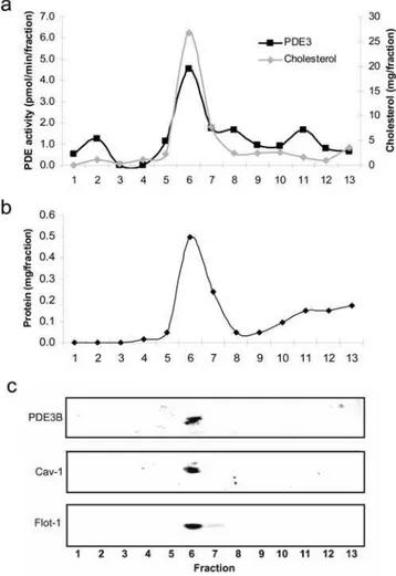
sucrose gradient to separate caveolae and lipid rafts from other components of the plasma membrane fraction. After sucrose gradient fractionation of the sonicated plasma membrane a milky band was detected at the interface of 5 and 35% sucrose. All of the PDE3B activity appeared together with flotillin-1, caveolin-1 and cholesterol in the caveolae/raft enriched fraction (Figures 2a and 2c). This fraction (fraction 6) contained 33% of total proteins whereas factions 7–8 contained 20% and fractions 9–13 contained 42% of total proteins (Figure 2b). When caveolae were enriched from detergent resistant plasma membrane of hepatocytes the same results were obtained (data not shown).
The importance of caveolin-1 for PDE3B expression was studied using caveolin-1 KO mice. The lack of caveolin-1 results in the absence of caveolae [12]. Thus, we isolated plasma membranes from livers from caveolin-1 KO mice and measured the level of PDE3B protein and activity. As seen in Figure 3, PDE3 protein and activity were significantly lower in plasma membranes from the caveolin-1 KO mice compared to WT mice. In total
homogenates from caveolin-1 KO mice PDE3 activity was slightly lower (3.8860.22) compared to WT mice (4.4060.65) (n = 3, not significant). These results show that caveolin-1 and caveolae are important for determining the amount of PDE3B protein in the plasma membrane of hepatocytes.
To further study the possible association of PDE3B with caveolae, we took advantage of the fact that caveolae are detergent resistant. Total membranes, solubilized membranes and detergent resistant membranes (DRM) were prepared from mouse hepato-cytes. The solubilized membranes and DRM fractions were subjected to superose-6 gel filtration chromatography. PDE3 activity in the total membrane fraction (detergent-treated, 10 0006g supernatant) eluted in two peaks, one with a Mw slightly above 670 kDa (elution volume 10–12 ml) and one corresponding to the void volume (.4 000 kDa, elution volume 7–8 ml) (Figure 4). PDE3 from the solubilized membranes (detergent-treated, 100 0006g supernatant) eluted with the ‘‘10–
12 ml peak’’ whereas PDE3 in the DRM (detergent-treated, 100 0006g pellet) eluted primarily with the void volume which may represent PDE3 associated with caveolae and/or lipid rafts. Figure 1. Localization of PDE3B in isolated hepatocytes.
Hepatocytes (35–1006106) were isolated from C57BL/6 mice and subjected to subcellular fractionation. Four compartments were isolated; the plasma membranes (PM), internal membranes (IM), mitochondria (Mit) and Nuclei (Nucl). The fractions were subjected to PDE3 activity measurements (a) and immunoblot analysis (b) with antibodies against PDE3B, caveolin-1 (Cav-1, a caveolae marker), flotillin-1 (Flot-1, a lipid raft marker), Na+
K+
-ATPase (NaKA, a PM marker), BiP (ER marker) and nucleoporin p62 (Nucl, a nucleus marker). PDE3 activities are shown as mean6SEM and immunoblots as one representative experiment (n = 4). Amount of proteins loaded on the gel: PM, 79mg; IM, 82mg; Mitochondria, 31mg; Nuclei, 31mg. doi:10.1371/journal.pone.0004671.g001
Figure 2. PDE3B is present in caveolae-enriched fractions of hepatocyte plasma membranes.Plasma membranes isolated from mouse hepatocytes (50–1206106) were sonicated in NaCO3 and
subjected to sucrose gradient fractionation as described in Materials and Methods. Fractions of 0.8 ml were collected starting from the top. The fractions were analyzed for PDE3 activity and cholesterol (a), proteins (b) and subjected to immunoblot analysis (90ml of each
The proportion of total PDE3B activity in the void volume in relation to the ‘‘10–12 ml peak’’ was estimated to be 1:2.
PDE3B is associated with smooth endoplasmic reticulum
In order to study PDE3B localization in internal membranes at the ultrastructural level, transmission electron microscopy (TEM) with immunogold labeling for PDE3B was employed. Thorough examination of several liver sections from different mouse livers revealed that the majority of the labeling for PDE3B was localized to smooth ER (Figure 5). In addition, weak PDE3B labeling was detected in rough ER as well as in mitochondria (Figure 5). Plasma membranes displayed weak labeling only (data not shown). Control sections displayed randomly scattered labeling only. Smooth ER has previously been shown to be in close association with glycogen particles [22–24]. As seen in Figure 5, PDE3B is located in close proximity to the glycogen particles which appear as pale areas among the smooth ER.
Basal glucose production is elevated in PDE3B KO mice
Hepatocytes isolated from C57BL/6 mice were cultured over night and thereafter stimulated without or with insulin or glucagon
for 15 minutes. Cell homogenates were analyzed for PDE3 activity. As seen in Figure 6a, stimulation of hepatocytes with either insulin or glucagon induced activation of PDE3 by 35 and 45%, respectively, in agreement with previous results from rat hepatocytes [25–26]. Activation of PDE3B by insulin and glucagon has previously been suggested to play a role in hormone-mediated regulation of rat hepatocyte glucose produc-tion [27]. Indeed, as shown in Figure 6b, PDE3B KO mouse hepatocytes showed increased glucose production as compared to WT hepatocytes which is in agreement with previously performed clamp studies in these mice [11]. Furthermore, PDE3B KO hepatocytes showed increased mRNA (Figure 6c) and protein levels (Figure 6d) of the key gluconeogenic enzyme phosphoenol-pyruvate carboxykinase (PEPCK).
Triglyceride- and cholesterol contents are increased in hepatocytes isolated from PDE3B KO mice
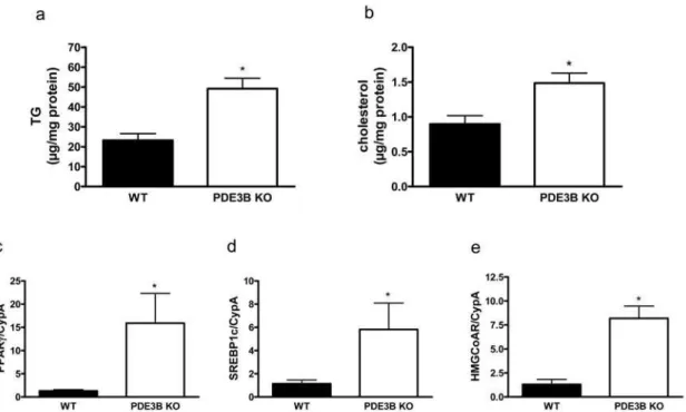
Triglycerides (Figure 7a) and cholesterol (Figure 7b) were analyzed in isolated hepatocytes. Both triglycerides and cholesterol were increased in PDE3B KO hepatocytes. The increments were associated with elevated levels of peroxisome proliferator-activated receptorc(PPARc) and sterol regulatory element-binding protein 1c (SREBP1c) mRNAs, both key lipogenic transcriptional factors (Figures 7c and 7d), as well as mRNA for hydroxyl-3-methylglu-taryl coenzyme A (HMG CoA) reductase, a rate limiting enzyme in cholesterol synthesis (Figure 7e).
Discussion
In this study we demonstrate that PDE3B is localized to caveolae/lipid raft regions in the plasma membrane as well as in smooth ER. Further, we show that PDE3B plays important roles in the regulation of glucose, triglyceride and cholesterol metab-olism in hepatocytes.
The localization of PDE3B to caveolae is highly interesting, since caveolae have been suggested to be important in the organization of cellular signalling as well as in lipid synthesis and cholesterol homeostasis [7]. The presence of caveolae in hepatocytes has previously been demonstrated using different approaches. Plasma membranes of isolated rat hepatocytes has been shown to contain sphingolipid-enriched microdomains with high amounts of cholesterol and caveolin-1 [28], and the presence of caveolae in the hepatocyte plasma membrane has been shown directly using rapid-freeze, deep-etching electron microscopy [29]. In agreement with the presence of caveolae in hepatocyte plasma membranes, in this study we detect caveolin-1 and flotillin-1, markers for caveolae and lipid rafts, respectively, specifically in the plasma membrane of isolated mouse hepatocytes and fractionation of sonicated plasma membranes in a sucrose gradient resulted in co-migration of caveolin-1, flotillin-1 and cholesterol. Further-more, results from caveolin-1 KO mice suggest an important role of caveolin-1 in hepatocyte lipid droplet formation and liver regeneration [30–32]. The finding that caveolin-1 KO livers show reduced levels of both PDE3B activity and protein expression indicates a role of caveolin-1/caveolae in the stabilization of the PDE3B protein which has also been demonstrated for the adipocyte PDE3B [5]. Our results demonstrating that PDE3B in hepatocytes is localized to distinct cellular locations, plasma membrane/caveolae as well as smooth ER, is in agreement with recent results in adipocytes [5,6]. Exactly which is the role for PDE3B at these cellular locations needs to be further investigated. In this study, we demonstrate increased glucose production and increased level of PEPCK mRNA and protein in hepatocytes from PDE3B KO mice, which is in agreement with previous studies in Figure 3. PDE3B protein is reduced in plasma membranes
isolated from livers of caveolin-1 KO mice.Plasma membranes isolated from WT and caveolin-1 KO livers were analyzed for PDE3 activity (a) and subjected to immunoblot analysis with antibodies against PDE3B, flotillin-1 (flot-1) and caveolin-1 (cav-1) (b). PDE3 activities (mean6SEM) were significantly different in the two groups (p,0.046). One immunoblot (34mg protein/lane) with two represen-tative experiments are shown (n = 5).
Figure 4. PDE3B is in large size complexes after gelfiltration.Total membranes were prepared from isolated mouse hepatocytes (20– 1006106) and treated with detergent. A portion was used to isolate non-solubilized detergent resistant membranes (DRM) and solubilized membranes. The different membranes were subjected to Superose-6 chromatography. Fractions were collected and analyzed for PDE3 activity. One representative experiment is shown (n = 4).
doi:10.1371/journal.pone.0004671.g004
Figure 5. Transmission electron micrographs of mouse hepatocytes; PDE3B is localized to the smooth ER. Sections of lowicryl embedded mouse livers with immunogold labeling for PDE3B (A and C). B and D: negative control with omission of the primary antibody. C and D are higher magnifications of the indicated areas in A and B, respectively. Arrows show examples of gold labeling. Representative micrographs are shown (n = 4). G; Glycogen storage M; Mitochondria, Nucl; nucleus, rER; rough endoplasmic reticulum, sER; smooth ER.
these mice (clamp studiesin vivoand PEPCK expression in intact
liver) and with studies using PDE3 inhibitors [11,27]. The increase in glucose production observed in isolated hepatocytes is not extensive and whether this increase can fully explain the increased production of glucose seenin vivoin PDE3B KO mice is difficult to
know. Although we culture the hepatocytes on PrimariaTMdishes to keep as much as possible of the original properties of the cells [33], the hepatocytes most likely have reduced biological responsiveness compared to the in vivo situation in PDE3B KO
mice. Furthermore, a number of defects related to the storage of triglycerides and cholesterol were identified in hepatocytes from PDE3B KO mice. Thus, we found up-regulation of triglyceride levels in hepatocytes as was the case in PDE3B KO livers [11] and of SREBP1c, one of two transcriptional factors encoded by SREBF1 [34]. The expression of SREBP1c predominates in the liver and has previously been shown to be regulated by insulin and increased intracellular cAMP [34]. In this study we demonstrate a specific role of PDE3B in cAMP mediated regulation of SREBP1c. Nuclear SREBPs is known to interact with cAMP response element binding protein (CREBP)-binding protein (CBP) and PPARc–regulated coactivator-1b (PGC-1b) which leads to upregulation of fatty acid synthase and suppression of PEPCK gene expression in liver in response to insulin [34]. Cyclic AMP has also been shown to contribute to the regulation of HMG CoA reductase [35], however, little is known regarding involvement of specific PDEs, such as PDE3B, in this process and in the
regulation of cholesterol homeostasis. In this study we show that indeed PDE3B seems to be important in this context but the exact signalling pathways in the regulation of HMG CoA reductase and cholesterol synthesis needs to be further evaluated. Thus, in hepatocytes from PDE3B KO mice, increased triglyceride and cholesterol biosynthesis occurred in parallel with elevated gluconeogenesis. At this point we can not completely exclude that decreased breakdown and/or release of triglycerides and choles-terol contribute to the increased levels of the lipids in the PDE3B KO hepatocytes. However, the increased expression of PPARc, SREBP1c and HMG CoA reductase together with previous results [11] showing increased expression of fatty acid synthase (FAS) in PDE3B KO liver, indicate that increased biosynthesis contribute to the fatty liver phenotype. Indeed increased accumulation of fat in the liver is highly relevant in the context of type 2 diabetes (T2D) and the metabolic syndrome [36,37]. It has been estimated that 70–80% of T2D patients have non alcoholic fatty liver disease. This disease covers a spectrum of liver diseases from steatosis to nonalcoholic steatohepatitis and cirrhosis. Indeed, increased fat accumulation in the liver appears to be a marker of hepatic insulin resistance and a close correlate of all components of the metabolic syndrome independent of obesity. The role of defect/dysregulated PDE3B in fatty liver development needs to be further investigated.
In summary, we demonstrate that, in mouse hepatocytes, PDE3B is localized in caveolae/raft regions in the plasma Figure 6. Hormonal regulation and glucose production in mouse hepatocytes.Hepatocytes were isolated from C57BL/6 mice and cultured on Primaria plates. After 18 hours the hepatocytes were incubated for 10 min with 10 nM insulin, 10 nM glucagon or without stimuli (Ctrl). PDE3 activity was measured in total homogenates (a). Values represent mean6SEM (p,0.032 for Insulin, p,0.024 for Glucagon, n = 5). Hepatocytes were isolated from PDE3B KO and WT mice. For glucose production (b) and Western blot analysis of PEPCK (d), the hepatocytes were cultured on Primaria plates over night before the experiment. RNA for PEPCK mRNA expression analysis (c) was isolated from non-cultured hepatocytes. Values are means6SEM. Glucose production: p,0.006, n = 7. PEPCK mRNA: p,0.02, analyzed in duplicate in two independent experiments from two mice of each genotype. PEPCK western blot: one representative experiment is shown, n = 4.
membrane as well as in smooth ER and that the enzyme has an important role in the regulation of triglyceride, cholesterol and glucose metabolism in these cells.
Acknowledgments
We thank Dr. Karl Swa¨rd for supplying the PDE3B KO livers. We thank Ann-Ki Pa˚lbrink for her excellent technical assistance with the isolation of primary hepatocytes and RNA preparation and Eva Ohlson for excellent
technical assistance. The authors also thank Professor A˚ ke Nilsson for valuable input regarding hepatocytes.
Author Contributions
Conceived and designed the experiments: KB RL NW EZT AL VM ED. Performed the experiments: KB RL NW EZT AL VM ED. Analyzed the data: KB RL NW EZT AL VM ED. Contributed reagents/materials/ analysis tools: KB RL NW EZT AL VM ED. Wrote the paper: KB RL NW EZT AL VM ED.
References
1. Conti M, Beavo J (2007) Biochemistry and physiology of cyclic nucleotide phosphodiesterases: essential components in cyclic nucleotide signaling. Annu Rev Biochem 76: 481–511.
2. Degerman E, Manganiello V (2007) Phosphodiesterase 3B; an important regulator of energy homeostasis. In Cyclic Phosphodiesterases in health and disease. Beavo, Houslay, Frances, eds. Broca Raton, FL USA: CRC Press. pp 79–99.
3. Thompson PE, Manganiello V, Degerman E (2007) Re-discovering PDE3 inhibitors–new opportunities for a long neglected target. Curr Top Med Chem 7(4): 421–36.
4. Choi YH, Ekholm D, Krall J, Ahmad F, Degerman E, et al. (2001) Identification of a novel isoform of the cyclic-nucleotide phosphodiesterase PDE3A expressed in vascular smooth-muscle myocytes. Biochem J Jan 1;353(Pt 1): 41–50. 5. Nilsson R, Ahmad F, Swa¨rd K, Andersson U, Weston M, et al. (2006) Plasma
membrane cyclic nucleotide phosphodiesterase 3B (PDE3B) is associated with caveolae in primary adipocytes. Cell Signal 18(10): 1713–21.
6. Ahmad F, Lindh R, Tang Y, Weston M, Degerman E, et al. (2007) Insulin-induced formation of macromolecular complexes involved in activation of cyclic nucleotide phosphodiesterase 3B (PDE3B) and its interaction with PKB. Biochem J 404(2): 257–68.
7. Cohen AW, Combs TP, Scherer PE, Lisanti MP (2003) Role of caveolin and caveolae in insulin signaling and diabetes. Am J Physiol Endocrinol Metab 285(6): E1151–60.
8. Lindh R, Ahmad F, Resjo¨ S, James P, Yang JS, et al. (2007) Multisite phosphorylation of adipocyte and hepatocyte phosphodiesterase 3B. Biochim Biophys Acta Apr;1773(4): 584–92.
9. Pozuelo Rubio M, Campbell DG, Morrice NA, Mackintosh C (2005) Phosphodiesterase 3A binds to 14-3-3 proteins in response to PMA-induced phosphorylation of Ser428. Biochem J Nov 15;392(Pt 1): 163–72.
10. Ha¨rndahl L, Wierup N, Enerba¨ck S, Mulder H, Manganiello VC, et al. (2004) Beta-cell-targeted overexpression of phosphodiesterase 3B in mice causes impaired insulin secretion, glucose intolerance, and deranged islet morphology. J Biol Chem 279(15): 15214–22.
11. Choi YH, Park S, Hockman S, Zmuda-Trzebiatowska E, Svennelid F, et al. (2006) Alterations in regulation of energy homeostasis in cyclic nucleotide phosphodiesterase 3B-null mice. J Clin Invest 116(12): 3240–51.
12. Razani B, Combs TP, Wang XB, Frank PG, Park DS, et al. (2002) Caveolin-1-deficient mice are lean, resistant to diet-induced obesity, and show hypertri-glyceridemia with adipocyte abnormalities. J Biol Chem 277(10): 8635–47. 13. Fleischer S, Kervina M (1974) Subcellular fractionation of rat liver. Methods
Enzymol 31(Pt A): 6–41.
14. Tuma PL, Hubbard AL (2001) Isolation of rat hepatocyte plasma membrane sheets and plasma membrane domains. Curr Protoc Cell Biol. May; Chapter 3: Unit 3.2. 15. Song KS, LiShengwen, Okamoto T, Quilliam LA, Sargiacomo M, et al. (1996) Co-purification and direct interaction of Ras with caveolin, an integral membrane protein of caveolae microdomains. Detergent-free purification of caveolae microdomains. J Biol Chem Apr 19;271(16): 9690–7.
16. Smart EJ, Ying YS, Mineo C, Anderson RG (1995) A detergent-free method for purifying caveolae membrane from tissue culture cells. Proc Natl Acad Sci U S A. Oct 24;92(22): 10104–8.
17. Ortegren U, Karlsson M, Blazic N, Blomqvist M, Nystrom FH, et al. (2004) Lipids and glycosphingolipids in caveolae and surrounding plasma membrane of primary rat adipocytes. Eur J Biochem May;271(10): 2028–36.
18. Wierup N, Sundler F (2005) Ultrastructure of islet ghrelin cells in the human fetus. Cell Tissue Res 319(3): 423–8.
19. Bo¨ttcher G, Alumets J, Ha˚kanson R, Sundler F (1986) Co-existence of glicentin and peptide YY in colorectal L-cells in cat and man. An electron microscopic study. Regul Pept 13(3–4): 283–91.
Figure 7. Triglyceride and cholesterol contents are increased in hepatocytes isolated from PDE3B KO mice.Hepatocytes (a, c–e) and liver (b) from PDE3B KO and WT mice were analyzed with regard to triglyceride (a) and cholesterol (b) content. Furthermore, PPARcmRNA (c), SREBP1c mRNA (d) and HMG CoA reductase mRNA (e) were analyzed in hepatocytes. Values are means6SEM. Triglyceride content was analyzed as duplicate lipid extractions from four animals of each genotype, p,0.03. Cholesterol content was measured in duplicate in six mice of each genotype, p,0.03. mRNA expressions were analyzed in duplicate in two independent experiments from two mice of each genotype.
20. Komas N, Movsesian M, Kedev S, Degerman E, Belfrage P, et al. (1996) cGMP-inhibited phosphodiesterases (PDE3). In: Phosphodiesterase Inhibitors, Schudt, Dent, Rabe, eds. Academic Press Chapter 6. pp 89–109.
21. Carlsson L, Nilsson I, Oscarsson J (1998) Hormonal regulation of liver fatty acid-binding protein in vivo and in vitro: effects of growth hormone and insulin. Endocrinology 139(6): 2699–709.
22. Cardell R Jr, Michaels JE, Hung JT, Cardell EL (1985) SERGE, the subcellular site of initial hepatic glycogen deposition in the rat: a radioautographic and cytochemical study. The journal of Cell Biology 101: 201–206.
23. Garfield SA, Cardell RR Jr (1979) Hepatic glucose-6-phosphatase activities and correlated ultrastructural alterations in hepatocytes of diabetic rats. Diabetes 28(7): 664–79.
24. Hammad ES, Striffler JS, Cardell RR Jr (1982) Morphological and biochemical observations on hepatic glycogen metabolism in mice on a controlled feeding schedule. II. Streptozotocin-diabetic mice. Dig Dis Sci 27(8): 692–700. 25. Zhao AZ, Shinohara MM, Huang D, Shimizu M, Eldar-Finkelman H, et al.
(2000) Leptin induces insulin-like signaling that antagonizes cAMP elevation by glucagon in hepatocytes. J Biol Chem 275(15): 11348–54.
26. Pyne NJ, Cooper ME, Houslay MD (1987) The insulin- and glucagon-stimulated ‘dense-vesicle’ high-affinity cyclic AMP phosphodiesterase from rat liver. Purification, characterization and inhibitor sensitivity. Biochem J 242(1): 33–42. 27. Abdollahi M, Chan TS, Subrahmanyam V, O’Brien PJ (2003) Effects of phosphodiesterase 3,4,5 inhibitors on hepatocyte cAMP levels, glycogenolysis, gluconeogenesis and susceptibility to a mitochondrial toxin. Mol Cell Biochem 252(1–2): 205–11.
28. Tietz P, Jefferson J, Pagano R, Larusso NF (2005) Membrane microdomains in hepatocytes: potential target areas for proteins involved in canalicular bile secretion. J Lipid Res 46(7): 1426–32.
29. Calvo M, Tebar F, Lopez-Iglesias C, Enrich C (2001) Morphologic and functional characterization of caveolae in rat liver hepatocytes. Hepatology 33(5): 1259–69.
30. Mayoral R, Ferna´ndez-Martı´nez A, Roy R, Bosca´ L, Martı´n-Sanz P (2007) Dispensability and dynamics of caveolin-1 during liver regeneration and in isolated hepatic cells. Hepatology 46(3): 813–22.
31. Frank PG, Lisanti MP (2007) Caveolin-1 and liver regeneration: role in proliferation and lipogenesis. Cell Cycle 6(2): 115–6.
32. Ferna´ndez MA, Albor C, Ingelmo-Torres M, Nixon SJ, Ferguson C, et al. (2006) Caveolin-1 is essential for liver regeneration. Science 313(5793): 1628–32. 33. Dohda T, Nakamura Y, Kamihira M, Iijima S (2004) Functional role of RhoA
in growth regulation of primary hepatocytes. J Biochem 135: 631–637. 34. Raghow R, Yellaturu C, Deng X, Park EA, Elam MB (2008) SREBPs: the
crossroads of physiological and pathological lipid homeostasis. Trends Endocrinol Metab 19(2): 65–73.
35. Bifulco M, Perillo B, Saji M, Laezza C, Tedesco I, et al. (1995) Regulation of 3-hydroxy-3-methylglutaryl coenzyme A reductase gene expression in FRTL-5 cells. Identification and characterization of a cyclic AMP-responsive element in the rat reductase promoter. J Biol Chem 270(25): 15231–6.
36. Kotronen A, Yki-Ja¨rvinen H (2008) Fatty liver: a novel component of the metabolic syndrome. Arterioscler Thromb Vasc Biol 28(1): 27–38.