A
rt
igo
Received 06/18/06. Accepted 01/25/07
Brazilian Journal of Pharmacognosy 17(1): 17-22, Jan./Mar. 2007
Antimicrobial activity of
Trembleya lani
fl
ora
,
Xyris platystachia
and
Xyris pterygoblephara
Claiton Pires Ventura, Alaíde Braga de Oliveira, Fernão Castro Braga*
Faculdade de Farmácia, Universidade Federal de Minas Gerais, Av. Antônio Carlos 6627, Campus Pampulha, 31270-901, Belo Horizonte, MG, Brazil
RESUMO: “Atividade antimicrobina de Trembleya lanifl ora, Xyris platystachia e Xyris pterygoblephara”. As espécies Trembleya lanifl ora (Melastomataceae), Xyris platystachia
(Xyridaceae) e Xyris pterygoblephara foram coletadas na Serra do Cipó, região considerada hotspot
para conservação de biodiversidade. A atividade antimicrobiana dessas espécies foi avaliada em ensaiosin vitro de difusão em ágar frente a linhagens padronizadas de Staphylococcus aureus e
Micrococcus luteus. Todos os extratos, avaliados na concentração de 2000 g/disco, foram ativos contraM. luteus, enquanto a inibição de crescimento de S. aureus somente foi observada para os extratos de T. lanifl ora (folhas) e X. platystachia (partes aéreas). A partição dos extratos brutos entre solventes imiscíveis resultou na obtenção de frações ativas, oriundas de extratos originalmente inativos frente a S. aureus, observando-se atividade principalmente para as frações de baixa e média polaridade. O extrato de folhas de T. lanifl ora foi adicionalmente fracionado por cromatografi a em coluna de sílica gel e as frações resultantes apresentaram atividade antimicrobiana e perfi s por CLAE distintos daquelas obtidas pela partição entre solventes imiscíveis.
Unitermos: Trembleya lanifl ora, Xyris platystachia; Xyris pterygoblephara, atividade antimicrobiana.
ABSTRACT: Trembleya lanifl ora (D. Don) Cogn. (Melastomataceae), Xyris platystachia
Alb. Nilss. (Xyridaceae) and Xyris pterygoblephara Kunth., Brazilian species collected from a biodiversity hotspot for conservation priority, had their antimicrobial activity evaluated against standardized strains of Staphylococcus aureus andMicrococcus luteus, by the agar diffusion assay. All extracts, assayed in the concentration of 2000 g/disc, were active against M. luteus, whereas S. aureus growth was inhibited only by T. lanifl ora leaves and X. platystachia aerial parts. Fractionation of the extracts by partition between immiscible solvents resulted in active fractions from extracts originally inactive against S. aureus. Activity was mainly found in low and medium polar fractions. The extract of T. lanifl ora leaves was also fractionated by silica gel column chromatography and both the HPLC fi ngerprint and antimicrobial activity of the obtained fractions were distinct of those originated from the partition process.
Keywords:Trembleya lanifl ora,Xyris platystachia,Xyris pterygoblephara, antimicrobial activity.
INTRODUCTION
Brazil is recognized as one of the megadivesity countries, concentrating around 10 to 20% of all plant species in the world (Mittermeier et al., 1997). A total of 10,000 plant species are estimated to occur in Minas Gerais (Mendonça; Lins, 2000). Serra do Cipó is a national park located in this state, in a region classifi ed as a biodiversity hotspot for conservation priority (Myers et al., 2000). The area presents exceptional concentrations of endemic species and is experiencing loss of habitats, contributing to drive many species into extinction. We have previously investigated the antifungal and antibacterial activity of 20 plant species from Serra do Cipó (Cota et al., 2002). Among the active species, three were selected for study in the present work.
Trembleya lanifl ora (Melastomataceae) is a
shrub popularly named “fl or-de-lã” (wool fl ower), used as ornamental species (Pio Corrêa, 1969). T. lanifl ora grows mainly in rocky soils from campos rupestres, an altitudinal ecosystem covered by open vegetation, being the genus endemic in Brazil. A chemotaxonomic study carried out for the leaves of Melastomataceae species, belonging to the closely related genera Lavoisiera, Microlicia and Trembleya, resulted in the identifi cation of 116 fl avonoids, comprising 69 fl avonol and 47 fl avone glycosides, including kaempferol 3-O-glycosides and quercetin 3-O-glycosides in T. lanifl ora (Bomfi m-Patrício et al., 2001). A literature search indicated no ethnomedical use or biological activity other than antimicrobial described for T. lanifl ora (Cota et al., 2002).
uses, to treat eczemas and dermatitis (Pio Corrêa, 1969). The identifi cation of Xyris species based solely on morphological characters is rather limited (Varanda et al., 2002). Around 90% of the Xyris found in Brazil are endemic (Sajo et al., 1997) and over harvesting has put several species in risk of extinction.
The chemistry and biological activity of Xyris have been poorly investigated. Metabolites obtained from this genus include isocoumarins from X. indica (Ruangrungsi et al., 1995), anthraquinones from X. semifuscata (Fournier et al., 1975) and fl avonoids from X. itatiayensis, X. longiscapa and X. obtusiuscula (Varanda et al., 2002). Recently, we reported the isolation of a new anthraquinone from X. pilosa, active against Fusarium oxysporum (Cota et al., 2004). Except the antimicrobial effect (Cota et al., 2002; Cota et al., 2004), no other biological activity or any chemical data has been described for X. platystachia and X. pterygoblephara, species selected for the study.
The diversity of compounds found in plant species make these organisms promising sources of new antimicrobial agents, with general or specifi c effects (Amaral et al., 2006; Leitão et al., 2006). The interest in plant secondary metabolites with antimicrobial properties has revived as a consequence of microbial resistance development against the antibiotics in clinical use (Rocha et al., 2004; Lima et al., 2006; Oliveira et al., 2006), especially in the case of opportunistic infections affecting immunocompromised patients (Klausmeyer et al., 2004). Considering that the species selected for the study occur in an ecosystem with a high degree of endemism, it is feasible to infer that they are a potential source of bioactive compounds. Hence, the main goal of this work was to assay the antimicrobial activity of extracts and fractions from T. lanifl ora,X. platystachia andX. pterygoblephara.
MATERIAL AND METHODS
Plant materials
The species Trembleya lanifl ora (D. Don) Cogn., Xyris pterygoblephara Kunth. and Xyris platystachia Alb. Nilss. were collected in Minas Gerais state, Brazil, at Serra do Cipó National Park and APA Morro da Pedreira. The plants were identifi ed by botanists from the Fundação Zoo-Botânica, Belo Horizonte, Brazil, where voucher specimens are deposited, under numbers BHZB 2340, BHZB 2496 and BHZB 2492, respectively.
Plant material extraction and preliminary fractionation
The plants were dried separately, at 40 ºC, for 72 h. The extracts of T. lanifl ora (leaves, stems), X. pterygoblephara (aerial parts) and X. platystachia (aerial parts) were prepared by exhaustive percolation
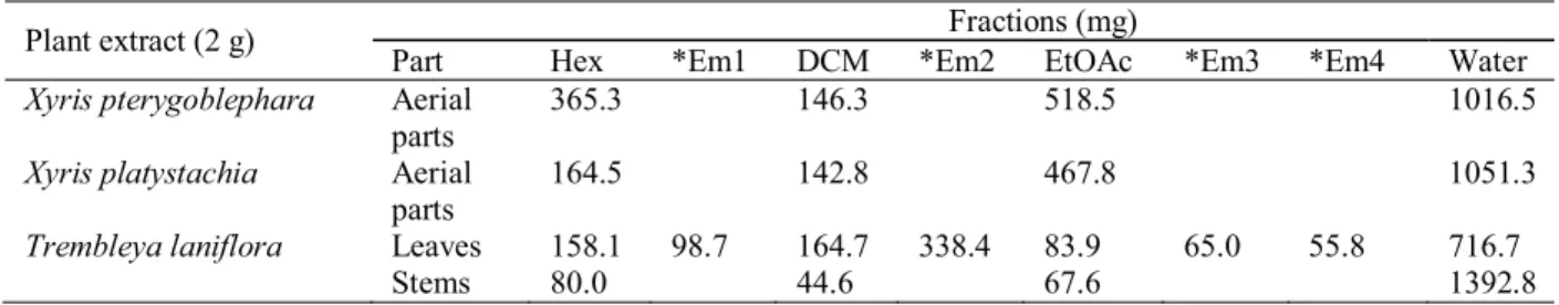
with ethanol. The extracts were concentrated to residue by removing the solvents in a rotavapor, at 50 ºC. Data obtained for the dry extracts are shown in Table 1. Portions (2 g) of the dry extracts were suspended in MeOH/H2O (1:1; 120 mL) and sequentially partitioned with equal volumes (120 mL) of n-hexane, CH2Cl2 and EtOAc. MeOH was removed in a rotavapor, before partitioning the extract suspension with CH2Cl2 and EtOAc. Solvents were removed in a rotatory evaporator, at maximum temperature of 50 °C, and the obtained residues are displayed in Table 2. Emulsions were generated during the partition of the extract from T. lanifl ora leaves between n-hexane (Emulsion 1), dichloromethane (Emulsion 2) and ethyl acetate (Emulsions 3 and 4). HPLC analysis carried out for the emulsions indicated distinct profi les from the obtained fractions and for this reason they were concentrated separately and had their residues evaluated in the antimicrobial assays.
Chromatographic fractionation of the extract from T.
laniforaleaves
The crude extract of T. lanifl ora leaves (22.8 g) was chromatographed on a silica gel column (70-230 mesh, Merck), employing a gradient elution of n-hexane (79.6 mg, TL1), n-hexane:CH2Cl2(1:1) (361.7 mg, TL2), CH2Cl2(689.4 mg, TL3), CH2Cl2:EtOAc (1:1) (1639.3 mg, TL4; 1239.7 mg, TL5), EtOAc (2205.2 mg, TL6), MeOH (80.9 mg, TL7; 11589.8 mg, TL8) and MeOH: H2O (1:1) (1201.6 mg, TL9).
Bacterial cultures and growth conditions
Staphylococcus aureus ATCC 25923 and Micrococcus luteus ATCC 9341 were employed as test organisms. The cultures were grown in agar medium, in tubes kept in a slating position, at 36 ºC, for 24 h. Cultures were maintained in plates, at 4 °C, in nº 1 antibiotic agar.
Antimicrobial assay
Plant name Part Dry vegetal
material (g) Dry extract (g) Extractive (%)
Xyris pterygoblephara Aerial parts 43.3 4.30 9.93
Xyris platystachia Aerial parts 100.0 13.31 13.31
Trembleya laniflora Leaves Stems
200.0 100.0
28.50 15.34
14.25 15.34
Plant extract Part Extract / fractions
Microbial inhibition (mm diameter zone ± rsd)
M. luteus S. aureus
Trembleya laniflora stems acrude extract 9.2 ± 0.3 b
−
n-hexane 12.8 ± 1.0 9.0 ± 0.0
DCM 11.5 ± 0.5 9.3 ± 0.3
EtOAc 13.5 ± 0.9 10.2 ± 0.6
water − −
leaves crude extract 12.8 ± 0.6 9.5 ± 0.5
n-hexane 21.0 ± 0.9 14.3 ± 0.8
Emulsion 1 13.0 ± 0.9 10.2 ± 0.3
DCM 16.2 ± 0.8 14.2 ± 0.8
Emulsion 2 12.7 ± 0.8 8.3 ± 0.8
EtOAc 13.2 ± 0.6 12.7 ± 0.3
Emulsion 3 − 8.2 ± 0.3
Emulsion 4 7.3 ± 0.3 −
water 9.2 ± 0.6 −
Xyris platystachia aerial parts crude extract 10.3 ± 0.8 7.7 ± 0.6
n-hexane − −
DCM 12.2 ±0.8 11.2 ± 0.7
EtOAc 9.7 ± 0.3 −
water - −
Xyris pterygoblephara aerial parts crude extract 10.0 ± 0.5 −
n-hexane − −
DCM 9.2 ± 0.6 8.3 ± 0.3
EtOAc 11.0 ± 0.5 7.7 ± 0.6
water − −
Chloramphenicol 21.0 ± 0.9 12.6 ± 0.7
Table 1. Ethanol extractives obtained from the plants in study.
Plant extract (2 g) Fractions (mg)
Part Hex *Em1 DCM *Em2 EtOAc *Em3 *Em4 Water
Xyris pterygoblephara Aerial parts
365.3 146.3 518.5 1016.5
Xyris platystachia Aerial parts
164.5 142.8 467.8 1051.3
Trembleya laniflora Leaves Stems
158.1 80.0
98.7 164.7 44.6
338.4 83.9 67.6
65.0 55.8 716.7 1392.8 Table 2. Fractions resulting from partition of plant extracts between immiscible solvents.
*Emulsions formed during the partition process. See experimental for details.
Table 3. Antimicrobial activity of plant ethanol extracts and fractions obtained by partition of crude extract between immiscible solvents, assayed by the agar diffusion method.
aPaper discs were impregnated with 2000 g of the extracts or 1000 g of the fractions. b(
Fractions
Microbial inhibition (mm diameter zone ± rsd)
M. luteus S. aureus
TL1 (n-hexane)a b
− −
TL2 (DCM:n-hexane, 1:1) − −
TL3 (DCM) − −
TL4 (DCM:EtOAc, 1:1) 14.0 ± 0.9 −
TL5 (DCM:EtOAc, 1:1) 20.0 ± 0.5 9.2 ± 0.8
TL6 (EtOAc) 12.7 ± 1.0 8.5 ± 0.0
TL7 (MeOH) 8.6 ± 1.0 7.8 ± 0.6
TL8 (MeOH) − −
TL9 (MeOH:water, 1:1) − −
Chloramphenicol 21.4 ± 0.9 10.5 ± 0.7
Table 4. Antimicrobial activity of chromatographic fractions from the extract of Trembleya lanifl ora leaves, assayed by the agar diffusion method
aPaper discs were impregnated with 1000 g of the fractions. b(
−) no detected activity at the assayed concentrations. MeOH (control) did not show any inhibitory activity. plus standard deviation) were recorded by measuring
the zones of growth inhibition surrounding the discs. Chloramphenicol (3 g/disc) was included in the assays as positive control, whereas control disks contained solvent only (MeOH) as negative control.
HPLC characterization of fractions
Analysis were carried out on a Merck-Hitachi apparatus (Germany) composed of pump L-6200A, automatic injector AS-2000A, UV-VIS detector L-4250 and integrator D-2500. An ODS column (150 × 4.0 mm I.D., 5 M) was employed (Merck, Germany) at a temperature of 40 °C and fl ow rate of 1.0 mL/min. Analysis were performed at 220 nm. A linear gradient of H2O (A) and CH3CN (B) was employed: 0 min 90% A, 10% B; 60 min 10% A, 90% B, followed by 10 min of isocratic elution. Solvents used were of HPLC grade (Merck, Germany) and were degassed by sonication before use. Fractions were dissolved in MeOH to a concentration of 5 mg/mL. After centrifugation at 10,000 r.p.m, the sample solutions (30 L) were automatically injected.
Phytochemical analysis
The presence of saponins, alkaloids, coumarins, anthraquinones, flavonoids, triterpenes and tannins was evaluated in the ethanol extracts, by TLC analysis, according to Wagner et al. (1984).
RESULTS AND DISCUSSION
The ethanol extracts of Trembleya lanifl ora,
Xyris platystachia and Xyris pterygoblephara have been previously evaluated in antimicrobial assays, against different strains of bacteria and fungi (Cota et al., 2002; Cota et al., 2004). The three species were particularly active against S. aureus and M. luteus, and for this reason both were selected as test organism in the present work. Aliquots from the crude ethanol extracts were initially fractionated by partition between immiscible solvents and the obtained results are displayed in Table 2.
All assayed extracts were active against M. luteus, whereas S. aureus growth was inhibited only by the extracts from T. lanifl ora leaves and X. platystachia aerial parts. In the previous work (Cota et al., 2002), the extracts from T. lanifl ora stems and X. pterygoblephara aerial parts were active against S. aureus, while X. platystachia did not show inhibitory effect against this microorganism. Such contradictory results may be explained by differences in extract compositions, since the plant materials were collected in distinct locations.
Phytochemical analysis (Wagner et al., 1984) of the ethanol extracts gave positive results for saponins, triterpenes and tannins. Flavonoids were also detected in the three species, except in the stems of T. lanifl ora, whereas coumarins were solely present in the aerial parts of X. platystachia and X. pteryglobephara. Several flavonoids (Harborne; Williams, 2002), coumarins (Borges et al., 2005), tannins (Chung et al., 1998), saponins (Wallace, 2004) and triterpnes (Katerere et al., 2003) have been reported to posses antimicrobial activity. Therefore, the presence of metabolites from these classes in the assayed species might explain their antimicrobial activity here reported.
(a)
(c)
(b)
Figure 1. HPLC profi les of fractions from Trembleya lanifl ora leaves. (a) Dichloromethane fraction obtained by partition between immiscible solvents; (b) dichloromethane (TL3) and (c) dichloromethane / ethyl acetate (1:1) (TL4) fractions from silica gel column chromatography. HPLC conditions: see experimental.
activity evaluated. Active fractions were obtained from extracts originally inactive against S. aureus (Table 3). This result demonstrates the importance of a preliminary fractionation when assaying the antimicrobial activity of plant extracts, once the low concentration of the active compounds may impair their detection in crude extracts.
Partition between immiscible solvents is an adequate approach for the preliminary separation of
solubility in the fi rst and adsorption in the second. Hence, applying these methods to the same matrix might result in fractions with distinct compositions and activities.
In order to confi rm this supposition, fi ngerprint profi les were registered by HPLC for fractions of the extract from T. lanifl ora leaves obtained by partition between immiscible solvents and by fractionation on a silica gel column. Besides, the antimicrobial activity of the chromatographic fractions was also assayed.
Dichloromethane fractions originated from both approaches showed distinct HPLC profi les (Figure 1) and antimicrobial effects: while the fraction obtained by partition was signifi cantly active against M. luteus and S. aureus (Table 3), the chromatographic one (TL3) showed no activity against both microorganisms (Table 4). On the other hand, HPLC analysis of the dichloromethane / ethyl acetate (1:1) fraction (TL4), originated from the chromatographic fractionation, showed a more related profi le to that of the dichloromethane fraction obtained by partition between immiscible solvents (Figure 1). However, TL4 was active solely against M. luteus. These results clearly confi rm our hypothesis that fraction constitution, and therefore biological activity, depends on the procedure adopted for fractionation.
In conclusion, the results here reported corroborate the popular use of the species to treat microbial diseases and also demonstrate the relevance of a preliminary fractionation for detecting active fractions, when assaying the antimicrobial effect of plant extracts.
ACKNOWLEDGEMENTS
This work was fi nanced by funds from Fundo Fundep, UFMG, Brazil. CNPq is also acknowledged for a research fellowship (F.C.B.).
REFERENCES
Amaral FMM, Ribeiro MNS, Barbosa-Filho JM, Reis AS, Nascimento FRF, Macedo RO 2006. Plants and chemical constituents with giardicidal activity. Rev Bras Farmacogn 16(Supl.): 696-720.
Bomfi m-Patrício MC, Salatino A, Martins AB, Wurdack JJ, Salatino MLF 2001. Flavonoids of Lavoisiera,
Microlicia and Trembleya (Melastomataceae) and their taxonomic meaning. Biochem Syst Ecol 29: 711-726.
Borges F, Roleira F, Milhazes N, Santana L, Uriarte E 2005. Simple coumarins and analogues in medicinal chemistry: occurrence, synthesis and biological activityCurr Med Chem 12: 887-916.
Chung KT, Wong TY, Wei CI, Huang YW, Lin Y 1998. Tannins and human health: a review. Crit Rev Food Sci Nutr 38: 421-464.
Cota BB, Oliveira AB, Ventura CP, Mendonça MP, Braga FC 2002. Screening for antimicrobial activity of plant species from a Brazilian hotspot for conservation priority. Pharm Biol 40: 542-547.
Cota BB, Oliveira AB, Guimarães KG, Mendonça MP, Souza
Filho JD, Braga FC 2004. Chemistry and antifungal activity of Xyris species (Xyridaceae) a new antraquinone from Xyris pilosa. Biochem Syst Ecol 32: 391-397.
Fournier G, Bercht CAL, Paris RR, Paris MR 1975. 3-Methoxychrysazin, a new antraquinone from Xyris semifuscata.Phytochemistry 14: 2099.
Harborne JB, Williams CA 2000. Advances in fl avonoids research since 1992. Phytochemistry 55: 481-504. Katerere DR, Gray AI, Nash RJ, Waigh RD 2003. Antimicrobial
activity of pentacyclic triterpenes isolated from African Combretaceae. Phytochemistry 63:81-88. Klausmeyer P, Chmurny GN, McCloud TG, Tucker KD,
Shoemaker RH 2004. A novel antimicrobial indolizinium alkaloid from Aniba panurensis. J Nat Prod 67: 1732-1735.
Leitão SG, Castro O, Fonseca EM, Julião LS, Tavares ES, Leo RRT, Vieira RC, Oliveira DR, Leitão GG, Martino V, Sulsen V, Barbosa YAG, Pinheiro DPG, Silva PEA, Teixeira DF, Lourenço MCS 2006. Screening of Central and South American plant extracts for antimycobacterial activity by the Alamar Blue test.
Rev Bras Farmacogn 16: 6-11.
Lima MRF, Ximenes CPA, Luna JS, Sant’Ana AEG 2006. The antibiotic activity of some Brazilian medicinal plants.
Rev Bras Farmacogn 16: 300-306.
Mendonça MP, Lins LV 2000. Lista vermelha das espécies ameaçadas de extinção da fl ora de Minas Gerais. Belo Horizonte: Fundação Biodiversitas, Fundação Zoo-Botânica de Belo Horizonte.
Mittermeier RA, Gil PR, Mittermeier CG 1997. Megadiversity: Earth’s Biologically Wealthiest Nations. México City: CEMEX, Agrupacíon Sierra Madre.
Myers N, Mittermeier RA, Mittermeir CG, Fonseca GAB, Kent J 2000. Biodiversity hotspots for conservation priorities.Nature 403: 853-858.
Oliveira RAG, Lima EO, Vieira WL, Freire KRL, Trajano VN, Lima, IO, Souza EL, Toledo MS, Silva-Filho RN 2006. Estudo da interferência de óleos essenciais sobre a atividade de alguns antibióticos usados na clínica. Rev Bras Farmacogn 16: 77-82.
Pio Corrêa M 1969. Dicionário de plantas úteis do Brasil e das exóticas cultivadas. Rio de Janeiro: Di Giorgio. Rocha AD, Oliveira AB, Souza Filho JD, Lombardi JA, Braga
FC 2004. Antifungal constituents of Clytostoma ramentaceum and Mansoa hirsuta.Phytother Res 18: 463-467.
Ruangrungsi N, Toshikazu S, Phadungcharoent T, Suriayagan S, Murakoshi I 1995. Isocoumarins from Xyris indica.
Phytochemistry 38: 481-483.
Sajo MG, Wanderley MGL, Menezes NL 1997. Observações anatômicas sobre a vascularização fl oral em Xyris L. (Xyridaceae).Bol Bot USP 16: 15-19.
Varanda EM, Rondinoni C, dos Santos DYAC 2002. Flavonoids fromXyris species (Xyridaceae). Biochem Syst Ecol 30: 997-998.
Wagner H, Bladt S, Zgainski EM 1984. Plant drug analysis. Berlin: Springer-Verlag.