online | memorias.ioc.fiocruz.br
Deciphering the contribution of lipid droplets in leprosy: multifunctional
organelles with roles in
Mycobacterium leprae
pathogenesis
Katherine Antunes de Mattos1, Euzenir Nunes Sarno2, Maria Cristina Vidal Pessolani1, Patricia T Bozza3/+
1Laboratório de Microbiologia Celular 2Laboratório de Hanseníase
3Laboratório de Imunofarmacologia, Instituto Oswaldo Cruz-Fiocruz, Rio de Janeiro, RJ, Brasil
Leprosy is an infectious disease caused by Mycobacterium leprae that affects the skin and nerves, presenting a singular clinical picture. Across the leprosy spectrum, lepromatous leprosy (LL) exhibits a classical hallmark: the presence of a collection of M. leprae-infected foamy macrophages/Schwann cells characterised by their high lipid content. The significance of this foamy aspect in mycobacterial infections has garnered renewed attention in leprosy due to the recent observation that the foamy aspect represents cells enriched in lipid droplets (LD) (also known as lipid bodies). Here, we discuss the contemporary view of LD as highly regulated organelles with key func-tions in M. leprae persistence in the LL end of the spectrum. The modern methods of studying this ancient disease have contributed to recent findings that describe M. leprae-triggered LD biogenesis and recruitment as effective mycobacterial intracellular strategies for acquiring lipids, sheltering and/or dampening the immune response and favouring bacterial survival, likely representing a fundamental aspect of M. leprae pathogenesis. The multifaceted functions attributed to the LD in leprosy may contribute to the development of new strategies for adjunctive anti-leprosy therapies.
Key words: leprosy - lipid droplet - eicosanoids - bacterial survival - pathogenicity - nutritional source - inflammation
Leprosy, or Hansen’s disease, is an ancient chronic infectious disease caused by Mycobacterium leprae, a slow-growing obligate intracellular bacterial patho-gen. Despite the “elimination of leprosy” slogan that has dominated the priorities of leprosy work and the success of multidrug therapy (MDT), which has suc-ceeded in reducing the leprosy burden over the years, this disease remains a public health problem in several countries (WHO 2010). The fact that the elimination of leprosy has not yet been achieved suggests that the in-fection has not been adequately controlled by antibiot-ics alone. Thus, it is critical to invest in broad-based re-search studies that examine the interaction of M. leprae
with host cells to identify new approaches to improve our understanding of the pathophysiology of leprosy and to identify new therapeutic targets.
Leprosy manifests as a spectrum of clinical forms dictated by the type and magnitude of the immunologi-cal response mounted by the host against the infection. The tuberculoid (TT) and lepromatous (LL) polar clinical forms are at the two extremes of the spectrum, whereas the intermediate clinical forms present with hybrid char-acteristics (Ridley 1974). TT patients develop a specific, strong T-helper (Th)1-type response, which restricts M. leprae multiplication and dissemination and limits the
Financial support: IOC/FIOCRUZ, CNPq, FAPERJ, CAPES PTB is a Guggenheim fellow.
+ Corresponding author: [email protected] Received 25 May 2012
Accepted 30 August 2012
Contemporary evidence supports a key role for the regulation of lipid metabolism and LD in the host re-sponse to intracellular pathogens; also termed lipid bod-ies or adiposomes, LDs are dynamic lipid-enriched or-ganelles found in different cell types. Pathogen-induced increases in LD formation have been described in
bac-terial, viral, fungal and parasitic infections and a role for this organelle in intracellular survival and pathogen replication has been proposed for all of these infections (reviewed in D’Avila et al. 2008a, Bozza et al. 2009a, van der Meer-Janssen et al.2010). Therefore, the aim of this review article is to discuss the roles of LD in M.
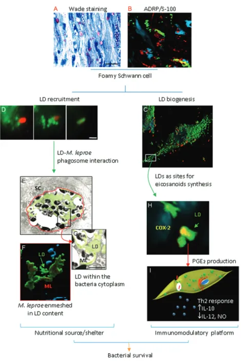
Fig. 1: overview of lipid droplet (LD) function in leprosy disease. Foamy phenotype from a lepromatous leprosy nerve visualized by Wade stain-ing (A) and immunohistochemistry confirmstain-ing the LD accumulation in Schwann cells (SC) usstain-ing anti-adipose differentiation-related protein (ADRP) and anti-S-100 antibodies (B). In vitro model of Mycobacterium leprae (ML)-trigger biogenesis (C) and recruitment (D). Lipid recruit-ment culminates in LD-M. leprae phagosome interaction (E) with consequent enmeshment of bacilli in LD (F) and possible incorporation of this lipid content into the bacterial cytoplasm (G). This sequence of events suggests the role of LD as a nutritional source and/or shelter in the host cell. In addition, LD organelles compartmentalize the cyclooxygenase (COX)-2 enzyme (H), reinforcing its role as an immunoinflamma-tory platform to prostaglandin E2 (PGE2) production and upregulation of T-helper (Th)2/Th1 cytokines (I). Together, these data suggest LDs are
leprae infection, focusing on the mechanisms of lipid droplet formation and lipid droplet composition and function within the context of leprosy. Indeed, recent findings have demonstrated that LDs are highly induced in a regulated manner during M. leprae infection. The LD formed in leprosy are emerging as organelles with major functions in disease pathogenesis, including roles in pathogen evasion and as a rich source of nutrients that allow the persistence of M. leprae.
Lipid droplet biogenesis - Lipid droplet biogenesis is a biological phenomenon that has gained attention over the last few years, mainly due to its intimate relationships with inflammatory and infection processes. Although different hypotheses regarding lipid droplet biogenesis have been described and the subject is still highly con-troversial, there is a consensus that both pathogen and host factors govern the molecular mechanisms of this process. Renewed attention is being given to the for-mation of LD within host cells infected by intracellular pathogens, a phenomenon that has been observed for all of the four main groups of pathogens (i.e., viruses, bac-teria, fungi and parasites) (Bozza et al. 2009a, b, van der Meer Janssen et al. 2010, Saka & Valdivia 2012). In most cases, it appears that the pathogen takes advantage of the LD formed to support its own life cycle. This intriguing concept points to new, previously unrecognised aspects of lipid droplet biology. In this section, we focus on the current knowledge of the mechanisms that regulate lipid droplet formation during the host response to M. leprae.
The pathogen-triggered dysregulation of host cell lipid metabolism is an emerging feature in the pathogen-esis of mycobacterial infections. Accumulating evidence
suggests that mycobacterial pathogenesis is directly re-lated to the modulation of host lipid metabolism. During the infectious process, LDs are directly triggered to form by pathogenic mycobacterial species, including Myco-bacterium bovis (D’Avila et al. 2006, 2008b, Almeida et al. 2009), Mycobacterium tuberculosis (Peyron et al. 2008, Cáceres et al. 2009) and M. leprae (Tanigawa et al. 2008, Mattos et al. 2010, 2011a, b); in contrast, such non-pathogenic species as Mycobacterium smegmatis
are not able to induce these organelles (D’Avila et al. 2006, Peyron et al. 2008, Almeida et al. 2009, Mattos et al. 2010). However, as summarised in Table, differential mechanisms govern this phenomenon. This differential profile of lipid droplet induction among mycobacteria may be related to the obligate intracellular lifestyle and the levels of pathogenicity of these species, reinforcing the idea that lipid accumulation is intimately related to bacterial survival and persistence in the host cell.
Regarding leprosy, in particular, our own studies, which have focused on the lipid accumulation in mac-rophages, as associated with the cutaneous lesions of LL, have revealed that these foamy-appearing, lipid-laden cells originate, at least in part, from the lipid droplet for-mation triggered by an M. leprae infection. The mecha-nisms that govern the lipid droplet formation process in
M. leprae-infected macrophages have been investigated using in vitro models (human and murine macrophag-es). These mechanisms exhibit similarities to M. bovis
BCG infection during which lipid droplet biogenesis is induced (i) in a time and dose-dependent manner, (ii) by both live and dead mycobacteria, (iii) by mycobacterial cell wall components and (iv) in bacterium-associated
TABLE
Differential regulatory mechanism of mycobacteria-trigger host lipid droplet (LD) biogenesis and their functions during infection
Mycobacterium sp.
Mycobacterium smegmatis
Mycobacterium bovis BCG
Mycobacterium tuberculosis
Mycobacterium leprae
Avirulent Virulence-attenuated Virulent Virulent
LD biogenesis/recruitment
Cell type Mφ SC Mφ SC Mφ SC Mφ SC
Host LD induction Noa,b Nob Yesa Noc Yesd ND Yesb Yesc
Bacterial viability dependency - - Noa - Nod - Nob Yesc
TLR signalling - - TLR2a - ND - TLR2/6b TLR6c
Paracrine signalling - - Yesa - ND - Yesb Noc
Host LD-mycobacterial
phagosome interaction - - Yes
a - Yesd - Yesf Yesg
LD functions
Immunomodulators - - PGE2/ LTB4a - PGE
2/LTA4
e - PGE
2
b PGE
2
c
Shelter/nutrition - - Yesa - Yesd,e - Yesg Yesc,g
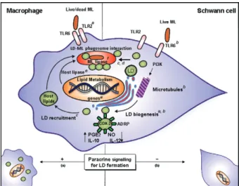
cells and in cells with no internalised bacteria, indicat-ing that soluble factors secreted by neighbourindicat-ing in-fected cells are able to induce the bystander formation of these organelles (Fig. 2). However, differential lipid droplet-formation signalling induced by these mycobac-terial species has also been noted. Whereas lipid droplet formation induced by M. bovis BCG involves a Toll-like receptor (TLR)2-dependent signalling process, this phe-nomenon is induced in a TLR2/TLR6-dependent manner during M. leprae infection (D’Avila et al. 2006, Almeida et al. 2009, Mattos et al. 2010).
Interestingly, we have shown that the mechanisms of lipid droplet induction by M. leprae in Schwann cells share similarities with those observed in macrophages, but also exhibit some differences (Table) (Mattos et al. 2010, 2011b). Lipid droplet biogenesis is induced in a time and dose-dependent manner and requires bacterial phagocytosis. Moreover, using selective drugs, we have
shown that lipid droplet biogenesis in M. leprae-infected Schwann cells follows a classical route. LDs are known to originate in the endoplasmic reticulum (ER)-Golgi com-plex and the capacity of LD to form and grow in size is dependent on motor proteins and microtubules (Pacheco et al. 2007, Zehmer et al. 2009). The induction of LD in Schwann cells by M. leprae is dependent on the ER and Golgi apparatus because it is inhibited by the drugs brefeldin and monensin, which are known to disrupt the ER-Golgi complex (Fig. 2). In addition, the disturbance of microtubule and actin organisation by specific drugs reveals that M. leprae-induced lipid droplet biogenesis also signals through a pathway that is dependent on cy-toskeleton dynamics. Lastly, the capacity of M. leprae to induce LD in Schwann cells can be abrogated with in-hibitors of phosphoinositide 3-kinase (PI3K), an enzyme that participates in several signalling pathways, including cytoskeletal organisation (Fig. 2) (Mattos et al. 2011a).
However, the induction of LD in Schwann cells ex-hibits interesting and unique features when compared to induction in macrophages. First, in contrast to mac-rophages, live M. leprae, but not dead bacteria or iso-lated mycobacterial components, are able to induce LD in in vitro-cultured Schwann cells, suggesting that the bacteria may play an active role in lipid droplet forma-tion in this cell type (Mattos et al. 2011b). Second, in M. leprae-infected cultures, M. leprae-induced lipid droplet formation is observed in cells associated with bacteria, but not in cells without internalised bacteria (Mattos et al. 2011b). In deep contrast to the observations in mac-rophages, a culture medium conditioned by M. leprae -stimulated Schwann cells is unable to mimic the ability of the bacteria to induce LD, confirming the absence of any soluble factors secreted by infected Schwann cells that are able to promote lipid droplet biogenesis in unin-fected neighbouring cells (Mattos et al. 2011a, b) (see be-low for further discussion). A third important difference between the M. leprae-induced lipid droplet biogenesis in macrophages and Schwann cells is related to the dif-ferential requirement of pattern recognition receptors (Mattos et al. 2011a, b), as discussed below.
The role of innate immune receptors in lipid droplet formation - The first level of mycobacterial interaction and recognition by host cells consists of pattern recog-nition receptors, such as the TLRs. Different members of the TLR family, including TLR1, TLR2, TLR4 and TLR6, have been implicated in mycobacterial recogni-tion (Means et al. 1999, Takeuchi et al. 2002, Krutzik et al. 2003, Oliveira et al. 2003). The stimulation of TLR signalling by bacteria and bacterial molecules has been shown to promote the accumulation of lipids in mac-rophages in the form of lipid droplet organelles. Experi-ments performed using macrophages from TLR-deficient mice have revealed that lipid droplet formation in response to bacteria is dependent on TLR signalling, particularly via TLR2 (Pacheco et al. 2002, D’Avila et al. 2006, Cao et al. 2007, Almeida et al. 2009, Mattos et al. 2010, 2011b). Moreover, it has been shown that killed bacteria and pu-rified molecules derived from bacteria are also capable of promoting lipid accumulation in treated macrophages (Pacheco et al. 2002, Kazemi et al. 2005, D’Avila et al. Fig. 2: a:Mattos et al. (2010); b:Mattos et al. (2011b); c:Mattos et
al. (2011a); d: Tanigawa et al. (2008); e: Cruz et al. (2008); f: Cole et al. (2001). Molecular mechanisms regulating the Mycobacterium lep-rae (ML) trigger of lipid droplet (LD) biogenesis/recruitment in host cells. In leprosy, the mechanisms of LD formation by ML in Schwann cells share similarities, but also exhibit some differences compared with those observed in macrophages. Macrophages induced LD in a Toll-like receptor (TLR)2/TLR6 mechanism, independent of bacterial viability and dependent of phagocytosis and paracrine signalling (a). Schwann cells induced LD in a TLR6-dependent and TLR2-indepen-dent mechanism, depenTLR2-indepen-dent of bacterial viability and phagocytosis and independent of paracrine signalling (b). LDs are promptly recruit-ed to bacterial-containing phagosomes, where they accumulate (c, d). ML-induced LD biogenesis is an endoplasmic reticulum-Golgi and actin or microtubule-dependent event (b). LD recruitment may consti-tute an intracellular strategy for ML persistence within host cells by subversion of the host lipid metabolism (c). The proposed mechanism of lipid acquisition from LDs is reinforced by the presence of bacterial and host lipases (d, e). In addition, new LDs constitute active catalytic sites of prostaglandin E2 (PGE2) synthesis and contribute to the
2006, Chen et al. 2008, Almeida et al. 2009, Mattos et al. 2010). These findings suggest that the mechanisms responsible for bacteria-induced lipid droplet formation involve the recognition of bacterial pathogen-associated molecular patterns (PAMPs) by TLRs. These data are re-inforced by observations that non-virulent bacterial spe-cies only induce foamy macrophages after transforma-tion with a gene encoding an enzyme that catalyses the oxygenation of mycolic acids (Russell et al. 2009).
Specifically in leprosy, the role of TLRs in M. lep-rae-induced lipid droplet biogenesis is unlike the tion described for BCG and there is differential regula-tion depending on the cell type and bacterial viability (Table). BCG and the purified cell component lipoarabi-nomannan are potent inducers of lipid droplet formation in macrophages from wild-type mice, but fail to induce lipid droplet biogenesis in TLR2-deficient mice, though not TLR4 or TLR6-deficient mice, suggesting a role for TLR2 in this mechanism (D’Avila et al. 2006, Almeida et al. 2009, Mattos et al. 2010). Interestingly, the stimu-lation of macrophages with M. smegmatis does not in-duce lipid droplet formation under the same conditions in which BCGinfection is highly effective (D’Avila et al. 2006, Almeida et al. 2009, Mattos et al. 2010). In par-ticular, we have demonstrated that the formation of LD in response to M. leprae is dependent on TLR2/TLR6 in infected macrophages (Mattos et al. 2010). In addi-tion, although Schwann cells express TLR2 (Oliveira et al. 2003), signalling through this receptor is not es-sential for lipid droplet biogenesis. Corroborating these data, BCG is unable to trigger lipid droplet formation signalling in Schwann cells, despite the fact that BCG contains the signature molecules recognised by TLR2, reinforcing the exclusion of TLR2-dependent signalling in this cell type. Moreover, although TLR6 is essential for sensing M. leprae in both cell types, a potential ad-ditional role for TLR6 as a phagocytic receptor has been observed in Schwann cells. Interestingly, the signalling that excludes the TLR2 pathway and involves a TLR6 heterodimer during inflammation in response to micro-bial infection has only just been explored for the first time, but it has also recently been described in scenarios of sterile inflammation, such as in atherosclerosis and Alzheimer’s disease (Stewart et al. 2010). The question of whether other coreceptors in conjunction with TLR6 participate in the recognition and signalling of live M. leprae in Schwann cells deserves further investigation. Additionally, the specific M. leprae chemical signatures recognised by TLR6 should be characterised.
Given the importance of TLR-mediated signalling for host resistance to microbial pathogens, much of the research exploring the influence of the genetic back-ground on leprosy has focused on polymorphisms in various TLR genes (Alcais et al. 2005, Cardoso et al. 2011) or other PAMP receptors (de Messias et al. 2009). Genetic factors almost certainly also play a key role in determining whether an infection leads to the TT form of the disease, the LL form of the disease or an interme-diate manifestation. In view of the discussed findings, it would not be surprising to discover that the genetic
variations that control the tendency to form LD in mac-rophages/Schwann cells might also be quite important in the foamy phenotype.
Paracrine signalling - The recent observation that the secreted factors from macrophages that stimulate through TLRs also contribute to foamy cell formation suggests that a paracrine or autocrine signal may be in-volved in this phenomenon, which is of particular inter-est in the study of foam cell biogenesis (Pacheco et al. 2002, 2007, D’Avila et al. 2006, Montoya et al. 2009). Specifically, it was shown that LD form both in the mac-rophages that take up M. leprae in a TLR2-dependent manner and in neighbouring macrophages that do not take up the bacteria (Mattos et al. 2010). Moreover, the supernatant from challenged macrophages promotes lipid droplet formation in unchallenged macrophages in a TLR2-independent manner, indicating that lipopeptide carryover cannot account for this finding (Mattos et al. 2010). A secondary autocrine mechanism of lipid droplet formation may help to explain a number of paradoxes in current lipid droplet research. In contrast to macrophag-es, media conditioned by M. leprae-stimulated Schwann cells is unable to replicate the lipid droplet induction ob-served with bacteria, confirming the absence of soluble factors secreted by infected Schwann cells that are able to promote lipid droplet biogenesis in uninfected neigh-bouring cells (Table) (Mattos et al. 2011b).
the direct interaction between the pathogen and host cell and also on indirect mechanisms in which neighbouring cells sense an infection through a bystander amplifica-tion-induced system triggered by bacterial components and/or host generated cytokines and chemokines.
M. leprae takes advantage of lipid droplet induc-tion for its persistence - The modulation of the host lipid pathway during host-pathogen interactions and viru-lence will be discussed in this section. Overall, a model can be proposed in which the lipid storage phenomenon observed in M. leprae-infected cells plays a key role in leprosy pathogenesis by facilitating bacterial persis-tence in the host via at least two mechanisms (Fig. 1). First, during M. leprae attachment and internalisation, the LDs are promptly recruited to and accumulate in bacteria-containing phagosomes (Mattos et al. 2011a). Lipid droplet recruitment likely constitutes an effective
M. leprae intracellular strategy for acquiring host cell lipids as a nutritional source and/or shelter to promote bacterial survival. Second, the accumulation of host-derived lipids in infected cells favours the generation of an innate immune response that may contribute to a per-missive environment for M. leprae proliferation within lepromatous skin and nerves.
Host LD as shelter and/or nutrition for M. leprae - We propose that the foamy phenotype (Fig. 1A) observed in LL is a consequence of lipid droplet biogenesis (Fig. 1B, C) and that lipid droplet recruitment (Fig. 1D) may constitute an effective M. leprae intracellular strategy for acquiring host lipids and promoting bacterial sur-vival. Blocking lipid droplet formation enables infected cells to kill the invading bacteria, supporting a model in which induced lipid droplet biogenesis is part of the bacterial strategy for persistent infection (Fig. 1C) (Mat-tos et al. 2011b). Indeed, we have shown that the LD in cells infected with M. leprae are found in close proxim-ity to, attached to and even internalised into the parasite-containing vacuole (Fig. 1D). Schwann cells also exhibit a relocalisation of adipose differentiation related protein (ADRP)-positive LD to bacteria-containing phagosomes in cultured cells and in nerve biopsies from infected pa-tients (Fig. 1E, F) (Mattos et al. 2011a). This lipid droplet relocalisation depends on PI3K signalling and requires cytoskeletal rearrangements (Mattos et al. 2011a) and blocking lipid droplet movement by interfering with cytoskeleton function decreases bacterial survival in in-fected cells (Mattos et al. 2011a). These data suggest that
M. leprae intercepts exocytic vesicles from the secre-tory pathway and reinforces the lipid droplet organelle as a possible source of host cell-derived lipid nutrients. Electron microscopy suggests that lipid droplet content is found within the bacterial cytoplasm (Fig. 1G). M. bovis BCG and M. tuberculosis-infected cells also dis-play a close association of the mycobacterial phagosome with LD (D’Avila et al. 2006, Peyron et al. 2008) and this close association of LD with the pathogen-containing vacuole is thought to constitute an important mechanism for lipid acquisition in vivo (D’Avila et al. 2006, Peyron et al. 2008, Russell et al. 2009, Daniel et al. 2011). In ad-dition, macrophage LD have been shown to accumulate
mycobactin (the lipophilic siderophore of mycobacteria)-iron complexes and ferric mycobactin-enriched LDs are found in close contact with phagosomes, suggesting that this organellar association enables the delivery of iron to the mycobacteria (Luo et al. 2005). To reinforce these data, which describe the hijacking of LD by pathogens for their own growth, we utilised experimental TB mod-els of BCG infection to demonstrate that LDs are also frequently found in close proximity to bacteria-contain-ing phagosomes and that the inhibition of lipid droplet formation greatly reduces BCG viability (D’Avila et al. 2006, Almeida et al. 2009), suggesting a potential role for lipid droplet biogenesis and recruitment in lipid ac-quisition by mycobacteria.
Although we do not have a full appreciation of the carbon sources utilised by mycobacteria during infec-tion, there is genetic evidence that mycobacteria utilise LD as lipid nutritional sources. The possibility that M. leprae assimilates fatty acids originating from the neu-tral lipids and phospholipids found in LD (Cole et al. 2001) is plausible due to the presence of two lipases and one phospholipase encoded by the M. leprae genome. In addition, early metabolic studies (Wheeler 1989, 2003) and recent proteomic analyses (Marques et al. 2008) in-dicate that fatty acid beta-oxidation is an active pathway in in vivo-grown M. leprae, which implies that lipids, rather than carbohydrates, are the dominant carbon sub-strate mobilised during infection. As such, the most reli-able method to date for measuring M. leprae viability is based on the assumed ability of M. leprae to oxidise fat-ty acids (Franzblau 1988). The role of lipids in M. leprae
metabolism is further supported by an active glyoxylate cycle (Marques et al. 2008), referred to as the dominant anaplerotic pathway during fatty acid growth. Further-more, deletions of the genes required for growth that utilise lipids as a sole carbon source render M. leprae
bacteria incapable of growth in vivo. Therefore, the utili- sation and manipulation of host lipids and lipid pathways represent a significant mechanism for these bacteria to cause and sustain infection.
In turn, the re-alignment of the host metabolism ap-pears to require alterations in the mycobacterial patho-gens M. tuberculosis and M. leprae, allowing them to switch to the utilisation of lipids as alternative carbon sources. This is best illustrated by M. tuberculosis,
Notably, two reports have implicated lipid droplet formation and the intimate association of M. tubercu-losis-containing phagosomes in the capacity of bacteria to persist in a long-term dormant state within the hu-man host and avoid anti-mycobacterial drugs (Peyron et al. 2008, Daniel et al. 2011). These data suggest an interesting additional role for host lipids as efficient, hy-drophobic, safe shelters. It is possible that lipid droplet recruitment constitutes a prime example of “organelle mimicry”, whereby M. leprae successfully escapes host recognition by cloaking itself in these fat-rich structures. This concept has not been excluded as an alternative cel-lular function of the observed LD in leprosy.
Host LD facilitate the production of lipid immuno-modulators and cytokines - Accumulating evidence has indicated that LDs are key inflammation-related organ-elles with major roles in eicosanoid production (reviewed in Bozza et al. 2011). As such, the role of this organelle in the regulation of the host response to infection via the modulation of the production of inflammatory media-tors has been investigated.
Substantial progress has been made in demonstrating that eicosanoid synthesis can occur at newly formed LD sites during mycobacterial infections (D’Avila et al. 2006, 2008b, Mattos et al. 2010, 2011b). Three main classes of downstream products (eicosanoids) can be produced: prostaglandins (PGs), such as PGE2, leukotrienes (LTs), such as LTB4 and lipoxins (LXs), such as LXA4. The ei-cosanoids synthesised at lipid droplet sites, particularly PGE2, may function as proinflammatory molecules or po-tent inhibitors of many aspects of the innate and adaptive immune systems (Weissmann 1993, Dooper et al. 2002).
The role of lipid droplet-derived eicosanoids may vary depending on the cell type, stimulus and inflamma-tory conditions controlling their synthesis (reviewed in Bozza et al. 2011). The foamy macrophages located in ar-eas of pneumonic M. tuberculosis infection in mice have been shown to be positive for cyclooxigenase (COX)-2 staining (Moreno et al. 2002). Definitive evidence for LD as sites of PGE2 synthesis in mycobacterial infection has been provided by demonstrating that COX-2 co-localises with ADRP in BCG-induced LD and that PGE2 forms at LD using the EicosaCell assay (D’Avila et al. 2006). Thus, the enhanced capacity of macrophages to gener-ate PGE2 during infection occurs, at least in part, due to increased lipid droplet biogenesis and the compart-mentalisation of eicosanoid production signalling within LD. However, TB offers an intriguing example of the dichotomous characterisation of eicosanoid functions in infection. Infection by M. tuberculosis causes microdis-ruptions in cell membranes that would normally lead to necrosis, but PGE2, which is abundantly generated within LD, may promote membrane repair to protect against ne-crosis. Conversely, virulent strains induce the expression of LXA4, which inhibits PGE2, allowing necrosis and, thus, the liberation of pathogens; these bacteria can then invade other cells, disseminating the infection (Chen et al. 2008). PGE2 production that depends on BCG-induced lipid droplet formation may favour bacterial survival in BCG-infected macrophages by promoting the downregu-lation of the innate immune response, suggesting that the
accumulation of LDin infected cells may support bacte-rial growth and persistence in the host. In addition, BCG has also been shown to induce LTB4 in a manner that is closely related to BCG-induced lipid droplet formation (D’Avila et al. 2006). Supporting a direct correlation be-tween lipid droplet formation and eicosanoid production,
M. smegmatis fails to induce this organelle and is unable to generate increased amounts of PGE2, reinforcing the notion of LD as the sites of eicosanoid production and their role in intracellular bacterial survival.
In leprosy, PGE2 production correlates with immuno-suppressive effects and a switch to the Th2 immune re-sponse on the LL pole. Thus, we have evaluated whether lipid droplet induction correlates with an increase in the cellular production of eicosanoids during M. leprae in-fection. Our laboratory has recently demonstrated that the LD that form in response to M. leprae constitute sites of eicosanoid synthesis, ultimately leading to the increased production of PGE2 by infected macrophag-es and Schwann cells (Mattos et al. 2010, 2011b). The experimental conditions that inhibit M. leprae-induced lipid droplet formation also downregulate M. leprae -in-duced PGE2 secretion, supporting the notion that LDs are important for PGE2 biosynthesis in M. leprae-infected cells. The colocalisation of ADRP, COX-2 and M. leprae
inside Schwann cells in nerve biopsies from LL patients strongly indicates that M. leprae-induced LDs are intra-cellular sites of eicosanoid synthesis in vivo (Fig. 1H) (Mattos et al. 2011b). Collectively, these data support the role of LD as dynamic organelles involved in the produc-tion of inflammatory mediators.
It has been shown that the high PGE2 concentrations produced by COX-2-positive macrophages in LL are able to inhibit Th1 cytokine production and, according-ly, most likely contribute to T-cell anergy at this disease pole (Fig. 1I) (Betz & Fox 1991, Kiszewski et al. 2003). However, lipid droplet-dependent PGE2 synthesis might account for the persistence of M. leprae in infected mac-rophages and Schwann cells when compared with M. tu-berculosis infection. The notion that lipid droplet forma-tion in foamy cells is not only a characteristic feature of LL, but is also a key pathophysiological mechanism that inhibits the development of a robust Th1 response and the eradication of the pathogen is now well supported.
dependent on lipid droplet inhibition and independent of COX-2 inhibition (D’Avila et al. 2008b, Mattos et al. 2011b). In addition, treatment with NS-398 was found to exert COX-2-independent inhibitory effects on the lipid droplet biogenesis induced by M. leprae infection, which occurs in parallel with the ability of NS-398 to inhibit cyclooxygenase. These data reinforce the role of lipid metabolism in modulating the innate immune response triggered by M. leprae infection in SCs via lipid droplet formation (Mattos et al. 2011b).
The potential role of LD as immunomodulatory plat-forms for cytokine regulation in foamy M. leprae-infected cells has also been investigated. Studies have analysed in-flammatory mediators and it has been shown that infected cells secrete increased levels of the anti-inflammatory cy-tokine IL-10 and reduced levels of IL-12 and nitric oxide compared to the basal levels observed in uninfected cells. Interestingly, when comparing conditions that induce (M. leprae infection) and impair (M. leprae infection pre-treated with cytochalasin, C-75, NS398) lipid droplet for-mation, we observed a decrease in IL-10 production and an increase in IL-12 and nitrite detection in the culture supernatants, suggesting that LD may negatively modu-late the immune response of host cells to intracellular in-fection (Mattos et al. 2010, 2011b). Additionally, we found an inhibition of PGE2 production when drugs were used to impair lipid droplet biogenesis. PGE2 is a potent immune modulator that downregulates Th1 responses and bacteri-cidal activity toward intracellular organisms (Renz et al. 1988, Betz & Fox 1991, van der Pouw Kraan et al. 1995, D’Avila et al. 2006). Curiously, LDs are also described as sites of cytokine storage, including tumour necrosis fac- tor, macrophage migration inhibitory factor, fibroblast growth factor and regulated on activation, normal T cell expressed and secreted in activated leukocytes (Beil et al. 1995, Lim et al. 1996, Dvorak et al. 2001, Pacheco et al. 2002, Assunção-Miranda et al. 2010). Thus, the signalling pathways that regulate the immune cascade in M. leprae -infected cells and the following potential mechanisms deserve further investigation: (i) high IL-10 production involves an autocrine PGE2 circuit, as has been shown in the context of other cell types (Alvarez et al. 2010) and (ii) lipid droplet-stored cytokines may function locally as intracrine signalling mediators (Figs 1, 2).
Persistence and viability - The interaction between intracellular bacteria and their host deserves special at-tention because obligate intracellular bacteria, such as
M. tuberculosis and M. leprae, need to acquire resources from their hosts to survive, replicate and persist.
Recently, the impact of lipid droplet biogenesis inhi-bition by NS-398 or C-75 on the killing capacity of cells infected by M. leprae has been investigated. Strikingly, we have demonstrated that treatment with these phar-macological inhibitors favours mycobacterial killing by Schwann cells in addition to the blockage of lipid drop-let biogenesis (Mattos et al. 2011b), suggesting a role for newly formed LD in intracellular M. leprae survival within nerves. In support of the hypothesis that LDs are involved in M. leprae survival, pathogen-induced host lipid droplet formation has been associated with
advan-tages in intracellular pathogen survival during hepatitis C virus (Boulant et al. 2007, Miyanari et al. 2007), dengue virus (Samsa et al. 2009), Trypanosoma cruzi (D’Avila et al. 2011) and BCG (D’Avila et al. 2008b, Almeida et al. 2009) infections. In addition to lipid droplet biogenesis,
M. leprae is ableto recruit LD promptly during bacte-rial attachment and internalisation in the bacteria-con-taining phagosomes in which they accumulate (Mattos et al. 2011a). Because LDs are organelles that are rich in neutral lipids, we investigated whether lipid droplet recruitment to bacterial phagosomes favours mycobac-terial survival. To this end, the cells were infected with
M. leprae and then treated with cytochalasin D or Taxol to inhibit lipid droplet trafficking. We demonstrated that LDs are promptly recruited to bacteria-containing pha-gosomes during M. leprae internalisation in Schwann cells and that this event contributes to bacterial survival inside the cells (Mattos et al. 2011a). In addition, it has been suggested that LDs are a rich source of nutrients for mycobacterial intracellular growth (D’Avila et al. 2006, Peyron et al. 2008, Almeida et al. 2009, Cáceres et al. 2009, Russell et al. 2009). Thus, the contribution of lipid droplet inhibition to improvements in the killing capac-ity of M. leprae-infected Schwann cells may occur as a secondary effect of the inhibition of PGE2 production and modulation of the cytokine milieu.
Future directions - In conclusion, the in vivo and in vitro data presented here show that M. leprae triggers a pronounced alteration in host-cell lipid homeostasis through the regulation of lipid droplet biogenesis and the recruitment of these organelles to bacterial phago-somes. This modulation of LD contributes to the foamy phenotype of infected cells and favours intracellular bacterial survival, likely representing a fundamental aspect of M. leprae pathogenesis. The comparative analyses of the changes in the host cell lipidome with different mycobacterial species illustrate the unique characteristics of each particular pathogen in a cell type-dependent mechanism (Table).
Additionally, the accumulation of host-derived lipids in infected Schwann cells favours the generation of an in-nate immune response that may contribute to a permis-sive environment for M. leprae proliferation within LL nerves/skin. M. leprae-infected cells also produce IL-10, which, together with PGE2, favours the inhibition of cell bactericidal activities and the downregulation of Th1-type cytokines. Thus, the suppression of normal inflammatory functions by M. leprae in macrophages and Schwann cells results in the expression of an anti-inflammatory pheno-type that is dependent on lipid droplet biogenesis.
Pathogens use lipids as building blocks and a nutri-ent source and also to influence the host cell physiology to enable their own survival and replication. The data indicate that multibacillary M. leprae infection is asso-ciated with a dramatic induction of host lipid metabo-lism genes, including multiple lipases with functional domains that were predicted by a comparative domain analysis to complement the deficiencies of the pathogen. In particular, the study of lipid droplet functions during host-pathogen interactions will provide a great oppor-tunity for future research and can be expected to afford opportunities to profile these host-pathogen interactions and to study their important contributions to the ability of pathogens to escape from the immune system.
Lipid profiling may become an important method to determine the metabolic condition of a cell or organ-ism. The specific depletion, presence or modulation of certain lipid species may generate lipid fingerprints that are unique to infection by specific pathogens, thus es-tablishing biomarkers for these pathogens. Clearly, such an underlying regulatory mechanism may hold the key to the development of novel strategies aimed at prevent-ing or, at a minimum, limitprevent-ing peripheral nerve injury in leprosy. The combination of epidemiological and biolog-ical evidence suggests that leprosy cannot be eliminated by MDT alone. Indeed, M. leprae makes extensive use of the host cell lipidome and has evolved very sophisti-cated mechanisms to utilise the diversity and complexity of host LD to maximise its own survival advantage. As such, strategies to inhibit lipid droplet formation in lep-rosy may represent novel opportunities for the develop-ment of adjunctive anti-leprosy therapies. Thus, we be-lieve that the findings highlighted here only scratch the surface of this rich new area of investigation into lipid droplet biology and its potential use in therapies.
REFERENCES
Accioly MT, Pacheco P, Maya-Monteiro CM, Carrossini N, Robbs BK, Oliveira SS, Kaufmann C, Morgado-Diaz JA, Bozza PT, Vi-ola JP 2008. Lipid bodies are reservoirs of cyclooxygenase-2 and sites of prostaglandin-E2 synthesis in colon cancer cells. Cancer Res68: 1732-1740.
Alcais A, Mira M, Casanova J-L, Schurr E, Abel L 2005. Genetic dis-section of immunity in leprosy. Curr Opin Immunol 17: 44-48.
Almeida PE, Silva AR, Maya-Monteiro CM, Töröcsik D, D’Avila H, Dezsö B, Magalhães KG, Castro-Faria-Neto HC, Nagy L, Bozza PT 2009. Mycobacterium bovis bacillus Calmette-Guérin infec-tion induces TLR2-dependent peroxisome proliferator-activated receptor-expression and activation: functions in inflammation, lipid metabolism and pathogenesis. J Immunol 183: 1337-1345.
Alvarez S, Blanco A, Fresno M, Munõz-Fernández MA 2010. Nuclear factor-kappaB activation regulates cyclooxygenase-2 induction in human astrocytes in response to CXCL12: role in neuronal toxicity. J Neurochem113: 772-783.
Assunção-Miranda I, Amaral FA, Bozza FA, Fagundes CT, Sousa LP, Souza DG, Pacheco P, Barbosa-Lima G, Gomes RN, Bozza PT, Da Poian AT, Teixeira MM, Bozza MT 2010. Contribution of macrophage migration inhibitory factor to the pathogenesis of dengue virus infection. FASEB J 24: 218-228.
Beil WJ, Weller PF, Peppercorn MA, Galli SJ, Dvorak AM 1995. Ul-trastructural immunogold localization of subcellular sites of TNF-alpha in colonic Crohn’s disease. J Leukoc Biol58: 284-298.
Betz M, Fox BS 1991. Prostaglandin E2 inhibits production of Th1 lym-phokines, but not of Th2 lymphokines. J Immunol146: 108-113.
Boulant S, Targett-Adams P, McLauchlan J 2007. Disrupting the as-sociation of hepatitis C virus core protein with lipid droplets correlates with a loss in production of infectious virus. J Gen Virol88: 2204-2213.
Bozza PT, Bakker-Abreu I, Navarro-Xavier RA, Bandeira-Melo C 2011. Lipid body function in eicosanoid synthesis: an update.
Prostaglandins Leukot Essent Fatty Acids85: 205-213.
Bozza PT, D’Avila H, Almeida PE, Magalhães KG, Molinaro R, Almeida CJ, Maya-Monteiro CM 2009a. Lipid droplets in host-pathogen interactions. Clinical lipidology4: 791-807.
Bozza PT, Magalhães KG, Weller PF 2009b. Leukocyte lipid bodies - Biogenesis and functions in inflammation. Biochim Biophys Acta 1791: 540-551.
Bozza PT, Yu W, Cassara J, Weller PF 1998. Pathways for eosinophil lipid body induction: differing signal transduction in cells from normal and hypereosinophilic subjects. J Leukoc Biol 64: 563-569.
Cáceres N, Tapia G, Ojanguren I, Altare F, Gil O, Pinto S, Vilaplana C, Cardona PJ 2009. Evolution of foamy macrophages in the pul-monary granulomas of experimental tuberculosis models. Tuber-culosis89: 175-182.
Cao F, Castrillo A, Tontonoz P, Re F, Byrne GI 2007. Chlamydia pneumoniae-induced macrophage foam cell formation is medi-ated by Toll-like receptor 2. Infect Immun 75: 753-759.
Cardoso CC, Pereira AC, de Sales Marques C, Moraes MO 2011. Lep-rosy susceptibility: genetic variations regulate innate and adaptive immunity and disease outcome. Future Microbiol 6: 533-549.
Chatterjee KR, Das Gupta NN, De ML 1959. Electron microscopic observations on the morphology of Mycobacterium leprae. Exp Cell Res 18: 521-527.
Chen M, Divangahi M, Gan H, Shin DS, Hong S, Lee DM, Serhan CN, Behar SM, Remold HG 2008. Lipid mediators in innate immunity against tuberculosis: opposing roles of PGE2 and LXA4 in the in-duction of macrophage death. J Exp Med Nov 205: 2791-2801.
Cole ST, Eiglmeier K, Parkhill J, James KD, Thomson NR, Wheel-er PR, Honoré N, GarniWheel-er T, ChurchWheel-er C, Harris D, Mungall K, Basham D, Brown D, Chillingworth T, Connor R, Davies RM, Devlin K, Duthoy S, Feltwell T, Fraser A, Hamlin N, Holroyd S, Hornsby T, Jagels K, Lacroix C, Maclean J, Moule S, Murphy L, Oliver K, Quail MA, Rajandream MA, Rutherford KM, Rut-ter S, Seeger K, Simon S, Simmonds M, Skelton J, Squares R, Squares S, Stevens K, Taylor K, Whitehead S, Woodward JR, Barrell BG 2001. Massive gene decay in the leprosy bacillus.
Nature 409: 1007-1011.
T, Jagels K, Krogh A, McLean J, Moule S, Murphy L, Oliver K, Osborne J, Quail MA, Rajandream MA, Rogers J, Rutter S, Seeger K, Skelton J, Squares R, Squares S, Sulston JE, Taylor K, Whitehead S, Barrell BG 1998. Deciphering the biology of My-cobacterium tuberculosis from the complete genome sequence.
Nature9: 1089-1097.
Cruz D, Watson AD, Miller CS, Montoya D, Ochoa MT, Sieling PA, Gutierrez MA, Navab M, Reddy ST, Witztum JL, Fogelman AM, Rea TH, Eisenberg D, Berliner J, Modlin RL 2008. Host-derived oxidized phospholipids and HDL regulate innate immunity in hu-man leprosy. J Clin Invest118: 2917-2928.
D’Avila H, Freire-de-Lima CG, Roque NR, Teixeira L, Barja-Fidal-go C, Silva AR, Melo RC, dos Reis GA, Castro Faria Neto HC, Bozza PT 2011. Host cell lipid bodies triggered by Trypanosoma cruzi infection and enhanced by the uptake of apoptotic cells are associated with PGE2 generation and increased parasite growth.
J Infect Dis204: 951-961.
D’Avila H, Maya-Monteiro CM, Bozza PT 2008a. Lipid bodies in in-nate immune response to bacterial and parasite infections. Int Im-munopharmacol8: 1308-1315.
D’Avila H, Melo RCN, Parreira GG, Werneck-Barroso E, Castro-Faria-Neto HC, Bozza PT 2006. Mycobacterium bovis bacillus Calmette-Guérin induces TLR2-mediated formation of LBs: in-tracellular domains for eicosanoid synthesis in vivo. J Immunol 176: 3087-3097.
D’Avila H, Roque NR, Cardoso RM, Castro-Faria-Neto HC, Melo RC, Bozza PT 2008b. Neutrophils recruited to the site of Myco-bacterium bovis BCG infection undergo apoptosis and modulate lipid body biogenesis and prostaglandin E production by mac-rophages. Cell Microbiol 10: 2589-2604.
Daniel J, Maamar H, Deb C, Sirakova TD, Kolattukudy PE 2011. My-cobacterium tuberculosis uses host triacylglycerol to accumulate lipid droplets and acquires a dormancy-like phenotype in lipid-loaded macrophages. PLoS Pathog 7 (Suppl.): S1-S16.
de Messias-Reason I, Kremsner PG, Kun JF 2009. Functional haplo-types that produce normal ficolin-2 levels protect against clinical leprosy. J Infect Dis 199: 801-804.
Dooper MM, Wassink L, M’Rabet L, Graus YM 2002. The modula-tory effects of prostaglandin-E on cytokine production by human peripheral blood mononuclear cells are independent of the pros-taglandin subtype. Immunology 107: 152-159.
Dvorak AM, Morgan ES, Weller PF 2001. Ultrastructural immunolo-calization of basic fibroblast growth factor to lipid bodies and se-cretory granules in human mast cells. Histochem J33: 397-402.
Franzblau SG 1988. Oxidation of palmitic acid by Mycobacterium le-prae in an axenic medium. J Clin Microbiol 26: 18-21.
Halvorsen B, Wæhre T, Scholz H, Clausen OP, von der Thüsen JH, Müller F, Heimli H, Tonstad S, Hall C, Frøland SS, Biessen EA, Damås JK, Aukrust P 2005. Interleukin-10 enhances the oxidized LDL-induced foam cell formation of macrophages by antiapop-totic mechanisms. J Lipid Res 46: 211-219.
Job CK 1970. Mycobacterium leprae in nerve lesions in lepromatous leprosy: an electron microscopic study. Arch Pathol 89: 195-207.
Kazemi MR, McDonald CM, Shigenaga JK, Grunfeld C, Feingold KR 2005. Adipocyte fatty acid-binding protein expression and lipid accumulation are increased during activation of murine macrophages by Toll-like receptor agonists. Arterioscler Thromb Vasc Biol 25: 1220-1224.
Kim MJ, Wainwright HC, Locketz M, Bekker LG, Walther GB, Dittrich C, Visser A, Wang W, Hsu FF, Wiehart U, Tsenova L, Kaplan G, Russell DG 2010. Caseation of human
tuberculo-sis granulomas correlates with elevated host lipid metabolism.
EMBO Mol Med 2: 258-274.
Kiszewski AE, Becerril E, Baquera J, Ruiz-Maldonado R, Hernández Pando R 2003. Expression of cyclooxygenase type 2 in leproma-tous and tuberculoid leprosy lesions. Br J Dermatol148: 795-798.
Krutzik SR, Ochoa MT, Sieling PA, Uematsu S, Ng YW, Legaspi A, Liu PT, Cole ST, Godowski PJ, Maeda Y, Sarno EN, Norgard MV, Brennan PJ, Akira S, Rea TH, Modlin RL 2003. Activation and regulation of Toll-like receptors 2 and 1 in human leprosy.
Nat Med 9: 525-532.
Kurup G, Mahadevan PR 1982. Cholesterol metabolism of mac-rophages in relation to the presence of Mycobacterium leprae.
J Biosci 4: 307-316.
Lim KG, Wan HC, Bozza PT, Resnick MB, Wong DT, Cruikshank WW, Kornfeld H, Center DM, Weller PF 1996. Human eosinophils elaborate the lymphocyte chemoattractants. IL-16 (lymphocyte chemoattractant factor) and RANTES. J Immunol 156: 2566-2570.
Luo M, Fadeev EA, Groves JT 2005. Mycobactin-mediated iron ac-quisition within macrophages. Nat Chem Biol 1: 149-153.
Marques MA, Neves-Ferreira AG, da Silveira EK, Valente RH, Cha-peaurouge A, Perales J, da Silva BR, Dobos KM, Spencer JS, Bren-nan PJ, Pessolani MC 2008. Deciphering the proteomic profile of
Mycobacterium leprae cell envelope. Proteomics 8: 2477-2491.
Mattos KA, D’Avila H, Rodrigues LS, Oliveira VG, Sarno EN, Atella GC, Pereira GM, Bozza PT, Pessolani MC 2010. Lipid drop-let formation in leprosy: Toll-like receptor-regulated organelles involved in eicosanoid formation and Mycobacterium leprae
pathogenesis. J Leukoc Biol87: 371-384.
Mattos KA, Lara FA, Oliveira VG, Rodrigues LS, D’Avila H, Melo RC, Manso PP, Sarno EN, Bozza PT, Pessolani MC 2011a. Mod-ulation of lipid droplets by Mycobacterium leprae in Schwann cells: a putative mechanism for host lipid acquisition and bacte-rial survival in phagosomes. Cell Microbiol13: 259-273.
Mattos KA, Oliveira VG, D’Avila H, Rodrigues LS, Pinheiro RO, Sarno EN, Pessolani MC, Bozza PT 2011b. TLR6-driven lipid droplets in Mycobacterium leprae-infected Schwann cells: im-munoinflammatory platforms associated with bacterial persis-tence. J Immunol 187: 2548-2558.
Means TK, Wang S, Lien E, Yoshimura A, Golenbock DT, Fenton MJ 1999. Human Toll-like receptors mediate cellular activation by
Mycobacterium tuberculosis. J Immunol163: 3920-3927.
Miyanari Y, Atsuzawa K, Usuda N, Watashi K, Hishiki T, Zayas M, Bartenschlager R, Wakita T, Hijikata M, Shimotohno K 2007. The lipid droplet is an important organelle for hepatitis C virus production. Nat Cell Biol9: 1089-1097.
Montoya D, Cruz D, Teles RM, Lee DJ, Ochoa MT, Krutzik SR, Chun R, Schenk M, Zhang X, Ferguson BG, Burdick AE, Sarno EN, Rea TH, Hewison M, Adams JS, Cheng G, Modlin RL 2009. Divergence of macrophage phagocytic and antimicrobial pro-grams in leprosy. Cell Host Microbe6: 343-353.
Moreno C, Rees AJ 1993. Striking the right balance; the role of cytok-ines in mycobacterial disease. Clin Exp Immunol94: 1-3.
Moreno JR, Garcia IE, Hernandez MLG, Leon DA, Marquez R, Pando RH 2002. The role of prostaglandin E2 in the
immunop-athogenesis of experimental pulmonary tuberculosis. Immunol-ogy106: 257-266.
Oliveira RB, Ochoa MT, Sieling PA, Rea TH, Rambukkana A, Sarno EN, Modlin RL 2003. Expression of Toll-like receptor 2 on hu-man Schwann cells: a mechanism of nerve damage in leprosy.
Pacheco P, Bozza FA, Gomes RN, Bozza M, Weller PF, Castro-Far-ia-Neto HC, Bozza PT 2002. Lipopolysaccharide-induced leu-kocyte lipid body formation in vivo: innate immunity elicited intracellular loci involved in eicosanoid metabolism. J Immunol 169: 6498-6506.
Pacheco P, Vieira-de-Abreu A, Gomes RN, Barbosa-Lima G, Wer-melinger LB, Maya-Monteiro CM, Silva AR, Bozza MT, Cas-tro-Faria-Neto HC, Bandeira-Melo C, Bozza PT 2007. Mono-cyte chemoattractant protein-1/CC chemokine ligand 2 controls microtubule-driven biogenesis and leukotriene B4-synthesizing function of macrophage lipid bodies elicited by innate immune response. J Immunol179: 8500-8508.
Parkash O 2009. Classification of leprosy into multibacillary and paucibacillary groups: an analysis. FEMS Immunol Med Micro-biol 55: 1-5.
Persson J, Nilsson J, Lindholm MW 2008. Interleukin-1β and tumor necrosis factor-α impede neutral lipid turnover in
macrophage-derived foam cells. BMC Immunol 25: 70.
Peyron P, Vaubourgeix J, Poquet Y, Levillain F, Botanch C, Bardou F, Daffé M, Emile JF, Marchou B, Cardona PJ, de Chastellier C, Altare F 2008. Foamy macrophages from tuberculous patients’ granulomas constitute a nutrient-rich reservoir for M. tuberculo-sis persistence. PLoS Pathog4: e1000204.
Renz, H, Gong JH, Schmidt A, Nain M, Gemsa D 1988. Release of tu-mor necrosis factor-alpha from macrophages. Enhancement and suppression are dose-dependently regulated by prostaglandin E2
and cyclic nucleotides. J Immunol 141: 2388-2393.
Ridley DS 1974. Histological classification and the immunological spectrum of leprosy. Bull World Health Organ 51: 451-465.
Russell DG, Cardona PJ, Kim MJ, Allain S, Altare F 2009. Foamy macrophages and the progression of the human tuberculosis granuloma. Nat Immunol10: 943-948.
Saka HA, Valdivia RH 2012. Emerging roles for lipid droplets in immunity and host-pathogen interactions. Annu Rev Cell Dev Biol: 411-437.
Sakurai I, Skinsnes OK 1970. Lipids in leprosy 2. Histochemistry of lipids in human leprosy. Int J Lepr Other Mycobact Dis 38: 389-403.
Samsa MM, Mondotte JA, Iglesias NG, Assunção-Miranda I, Barbo-sa-Lima G, da Poian AT, Bozza PT, Gamarnik AV 2009. Dengue virus capsid protein usurps lipid droplets for viral particle forma-tion. PLoS Pathog5: e1000632.
Schmid B, Rippmann JF, Tadayyon M, Hamilton BS 2005. Inhibition of fatty acid synthase prevents preadipocyte differentiation. Bio-chem Biophys Res Commun328: 1073-1082.
Stewart CR, Stuart LM, Wilkinson K, van Gils J M, Deng J, Halle A, Rayner KJ, Boyer L, Zhong R, Frazier WA, Frazier WA, Lacy-Hulbert A, El Khoury J, Golenbock DT, Moore KJ 2010. CD36 ligands promote sterile inflammation through assembly of a Toll-like receptor 4 and 6 heterodimer. Nat Immunol11: 155-161.
Takeuchi O, Sato S, Horiuchi T, Hoshino K, Takeda K, Dong Z, Modlin RL, Akira S 2002. Cutting edge: role of Toll-like recep-tor 1 in mediating immune response to microbial lipoproteins.
J Immunol169: 10-14.
Tanigawa K, Suzuki K, Nakamura K, Akama T, Kawashima A, Wu H, Hayashi M, Takahashi S, Ikuyama S, Ito T, Ishii N 2008. Ex-pression of adipose differentiation-related protein (ADRP) and perilipin in macrophages infected with Mycobacterium leprae.
FEMS Microbiol Lett 289: 72-79.
van der Meer-Janssen YP, van Galen J, Batenburg JJ, Helms JB 2010. Lipids in host-pathogen interactions: pathogens exploit the com-plexity of the host cell lipidome. Prog Lipid Res49: 1-26.
van der Pouw Kraan TC, Boeije LC, Smeenk RJ, Wijdenes J, Aarden LA 1995. Prostaglandin-E2 is a potent inhibitor of human inter-leukin 12 production. J Exp Med181: 775-779.
Virchow R 1863. Die krankhaften Geschwu lste, August Hirschwald, Berlin, 208 pp.
Weissmann G 1993. Prostaglandins as modulators rather than media-tors of inflammation. J Lipid Mediat 6: 275-286.
Wheeler PR 1989. Biosynthetic pathways in Mycobacterium leprae.
Acta Leprol 7: 21-24.
Wheeler PR 2003. Leprosy - clues about the biochemistry of My-cobacterium leprae and its host-dependency from the genome.
World J Microbiol Biotechnol 19: 1-16.
WHO - World Health Organization. 2010. Leprosy today. Available from: who. int/lep/en/.
Yamamura M, Uyemura K, Deans RJ, Weinberg K, Rea TH, Bloom BR, Modlin RL 1991. Defining protective responses to patho-gens: cytokine profiles in leprosy lesions. Science 254: 277-279.