THE CONTRIBUTlON OF ELECTROSYNERESIS TO LMMUNOLOGIC DIAGNOSIS OF HYDATlDOSlS
Jo& M. Tomes, M.D.; Jorge Guisantes, M.D.; In& Alvarez; and Luis A. Yarzhd, M.D.1
This paper evaluates how electrosyneresis can contribute to the immunologic diagnosis of human hydatidosis. The work reported here indicates its advantages over other techniques, and provides an estimate of its effectiveness in relation to the location and condition of the cysts.
Introduction
Multiple immunologic techniques-particu- larly the hemagglutination, immunoelectro- phoresis, and immunofluorescence tests-have made a definite contribution to the diagnosis of hydatidosis (I, 2). Used together, these three methods assure adequate sensitivity in the immunodiagnosis of this zoonosis, while im- munoelectrophoresis assures a high degree of specificity (3).
Immunoelectrophoresis, however, has cer- tain drawbacks. It consumes large quantities of reagents, and it takes 24 hours to yield primary results. Hence, quicker and more economical precipitation reactions are being sought. Electrosyneresis, described by Bussard (4), would eliminate these disadvantages, since it can detect precipitating antibodies in a few minutes using only small amounts of antigens and antisera. This has been confirmed by various authors (5-8) who, while calling this technique by different names, have used it in studying various antigens, in diagnosing dif- ferent diseases, and in doing research on the legal aspects of medical problems (9).
The method is based on simultaneous and
‘Laboratory of Parasitic Immnology, Immunology Section of the Clinical Laboratory Department, Clini- cal Hospital, Montevideo, Uruguay. Published in Span- ish in Boletin de la Oficina Sanitaria Panamericana, Vol. LXXV, No. 2 (August 1973), pp. 119-126.
41
opposite displacement of antigens and antisera subjected to the action of an electric field in an alkaline solution. Under these conditions, the antigen fractions with diagnostic value that have so far been identified in extracts of hydatid fluid (10, II) are negatively charged, while most immunoglobulins remain neutral and are displaced toward the cathode by the electro-endosmotic current.
By moving the hydatid antigen toward the cathode and the antiserum toward the anode, the passage of the current accelerates the meeting of reagents, thus facilitating the rapid formation of complex precipitates. This also “purifies” the hydatid antigen, since its posi- tively charged fractions tend to move in the same direction as the antiserum and thus participate very little in these reactions. Castagnari and Sorice (IL’), working with sera from hydatidosis patients, have found electro- syneresis to be more sensitive than hemag- glutination or immunoelectrophoresis.
42 PAHO BULLETIN . Vol. VII, No. 4, 19 73
used to test the same sera against antigens of
Echinococcus granulosus, Fasciola hepatica,
and Taenia saginata.
Materials and Methods
Antigens
The basic materials used were lyophilized hydatid fluid obtained from hepatic cysts of bovine origin, and lyophilized antigens of F. hepatica and T. saginata. Pursuant to earlier recommendations (3, IO), they were all qualita- tively standardized by immunoelectrophoretic testing against hyperimmune rabbit sera. The final concentration of antigens was 50 mg/ml.
Sera
A total of 66 sera from patients with surgically confirmed hydatidosis were studied; these had been preserved at -2OOC for periods ranging from one month to two years. In addition, the study included 23 sera from patients with non-hydatid diseases and 22 sera from healthy blood bank donors. The hyda- tidosis patients were grouped according to the position of the parasite, and their cysts were classified as follows:
1) Hyaline-when the parasite’s membranes were intact at the time of surgery, and the fluid was clear and transparent.
2) Infect&d-when there was evidence of suppuration between the hydatid and the cystic adventitia, and the parasitic fluid was cloudy, but there was no apparent rupture of the larval membranes.
3) Recently ruptured-when the larval membranes had already ruptured at the time of surgery, but the clinical record made it possible to place the rupture within the six-month period preceding removal.
4) Residual-when the larval membranes had already ruptured, and the rupture had occurred six months or more before the sample
was taken; or else when the cyst showed extensive calcification.
In all cases the sera were subjected to comparative study in their natural form and after concentration to one-third of their initial volume by lyophilization. The sera were not inactivated.
Electrosyneresis (ES)
Electrosyneresis was carried out on a 3 ml layer of 0.9 per cent agarose prepared in sodium barbital (veronal) buffer, pH 8.2, which was placed on a 76 x 26 mm glass slide. Two rows of three holes were placed 6 mm apart on each slide (see Figure l), perpendicular to the axis of electrophoretic migration. Samples of the particular serum being studied were put in the three holes nearest the anode, and extracts of the three antigen groups were put into the three cathode holes.
Electrophoretic separation of the antigens and sera was then carried out in sodium barbital buffer (pH 8.2) for ten minutes at 4OC. A difference in potential of 40 volts was maintain- ed between the two ends of each slide. All the slides were then incubated for 24 hours at 22OC, after which the usual procedure for immunoelectrophoresis was followed (10).
Three systematic readings were taken to observe whether or not precipitation bands had formed. The first reading was taken 24 hours after the electrophoretic separation and the second after 48 hours. The slides were then stained with amidoschwarz and a final reading was obtained. Besides this, an initial reading was taken 60 minutes after completion of electrosyneresis in some cases. All sera that gave rise to one or more precipitation bands insolu- ble in 5 per cent trisodium citrate were con- sidered positive. A positive result is shown in Plate 1.
Immonoelectrophoresis (IEP)
Torres, et al. . ELECTROSYNERESIS IN THE DIAGNOSIS OF HYDATIDOSIS 43
FIGURE 1-A sketch of the layout used in the tests.
PLATE 1-Electrosyneresis, showing a de- finitely positive result. On the left is the patient’s unconcentrated serum (M), matched against hyda- tid fluid (LH), Toenio sugirzuta extract (TS), and
Fasciola hepatica extract (FH).
studied by IEP within seven days of the date on which they had been obtained. The micro- technique of Capron et al. (2) was used for this purpose.
Results
Reproducibility
Four reactive and four non-reactive sera were tested by electrosyneresis on three sepa- rate occasions during the study. Identical re- sults were obtained each time.
Sensitivity
When concentrated sera were used, ES de- tected precipitating antibodies in 47 of the 50 sera that had given positive results with IEP. With unconcentrated sera, the percentage of
positive results was considerably lower (see Table 1).
Two of the three ES-negative sera for which IEP obtained positive results showed only one band after IEP (see Table 2). ES did not give rise to any bands of precipitation when carried out with sera which had given negative IEP results.
Specific&v
44 PAHO BULLETIN . Vol. Ku, No. 4, 1973 .
TABLE l-Sensitivity of electrosyneresis (ES) in diagnosing 66 confirmed cases of hydatidosis previously analyzed by immunoelectrophoresis (IEP).
Cases analyzed by ES
Electrosyneresis of Electrosyneresis of concentrated seraa unconcentrated sera Positive Negative Positive Negative
Positive by IEP (50) 41 3 43 I
Negative by IEP (16) 0 16 0 16
Total (66) 47 19 43 23
aSera were concentrated to one-third of orginal volume by lyophiliza- tion.
TABLE a-sensitivity of electrosyneresis in relation to the number of precipitating systems shown by immunoelectrophoresis of 50 concentrateda sera from hydatidosis patients.
Immunoelectrophoresis No. of bandsb No. of cases
Electrosyneresis Positive Negative
1 4 2 2
More than 1 46 45 1
Total 50 41 3
aSera were concentrated to one-third of original volume by Iyophiliza- tion.
bIncluding band 5 of Capron et aZ., 1967 (IO).
Torres, et al. - ELECTROSYNERESIS IN THE DIAGNOSIS OF HYDATIDOSIS 45
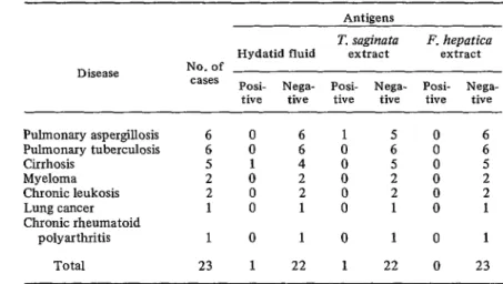
TABLE 3 -Specificity of electiosyneresis in the analysis of 23 concentrateda sera from non-hydatid disease cases.
Antigens
Disease
T. sagina ta F. hepatica Hydatid fluid extract extract No. of
cases Posi- Nega- Posi- Nega- Posi- Nega- tive tive tive tive tive tive Pulmonary aspergillosis
Pulmonary tuberculosis Cirrhosis
Myeloma Chronic leukosis Lung cancer Chronic rheumatoid
polyarthritis Total
6 0 6 1 5 0 6
6 0 6 0 6 0 6
5 1 4 5 0 5
2 0 2 i 2 0 2
2 0 2 2 0 2
1 0 1 1 0 1
1 0 1 0 1 0 1
23 1 22 1 22 0 23
aSera were concentrated to one-third of original volume by lyophiliza- tion.
from a healthy individual in whom no evidence of concomitant hydatidosis was found (Table 4). If these two cases are considered positive, the non-specificity index for the ES test results would still be less than 4.4 per cent.
Sensitivity and Parasite Location
Some of the sera from hydatidosis patients that showed a positive ES reaction to hydatid fluid also possessed antibodies against various fractions of the T. saginata extract and against
Like IEP, ES showed greater sensitivity for hepatic cysts than for pulmonary cysts (Table 6). Neither test yielded positive results for cysts with cerebral or renal locations.
Cyst Condition
L one component of the F. hepatica antigen Examination of each cyst’s condition mosaic (see Table 5). However, the number of showed that those which had recently ruptured crossed reactions with T. saginata declined produced the highest percentage of reactive sera significantly for the group of sera from hyda- (Table 7). The percentage of positive sera was tidosis patients that showed more than three decidedly lower among patients with hyaline
precipitation bands. cysts.
TABLE $-The specificity of electrosyneresis indicated by analysis of 22 concentrateda sera from blood bank donors.
Antigens No. of
cases Hydatid fluid
T. .saginataf, F. hepatica
extract extract
Posi- Nega- Posi- Nega- Posi- Nega-
tive tive tive tive tive tive
22 1 21 1 21 0 22
46 PAHO BULLETIN . Vol. VII, No. 4, I973 ,
TABLE S-Reactivity of hydatidosis patients’ sera shown positive by electro- syneresis. The sera are grouped according to the number of bands generated when they were tested against various groups of antigens.
Positive to Positive to Positive to
No. of hydatid fluid T. saginata extract F. hepatica extract bands Concen- Unconcen- Concen- Unconcen- Concen- Unconcen-
trated a trated trated trated trated trated 1 2 3 4 5 6 I 8 or more
Total 6 10 11 9 7 3 0 1 41 9 K 6 2 1 i 43 21 14 6 1 1 0 1 0 44 18 4 4 0 1 0 0 0 27 1 0 0 0 0 0 0 '0 1 aSera were concentrated to one-third of original volume by lyophiliza- tion.
TABLE 6-Results of electrosyneresis and immunoelectrophoresis with hydatid antigen in relation to location of the cysts.
Location No. of cases
Electrosyneresis Immunoelectrophoresis Positive Negative Positive Negative Lungs Liver Heart Spleen Multiple Bone Brain Peritoneum Kidneys Thymus Total
37 23
16 15
2 2 2 2 2 1 1 1 66 2 1 2 2 0 1 0 1 47 14 1 0 1 0 0 2 0 1 0 19 26 15 2 1 2 2 0 1 0 1 11 1 0 1 0 0 2 0 1 0
50 16
1
TABLE ‘I-Results of electrosyneresis and immunoelectrophoresis with hydatid antigen in relation to the condition of the cysts.
.A
State of No. of the cysts cases
Eiectrosyneresis Immunoelectrophoresis Positive Negative Positive Negative
Hyalin 22 10 12 10 12
Infected 3 2 1 3 0
Recently ruptured 1.5 14 1 14 1
Residual 5 3 2 4 1
Data unavailable 21 18 3 19 2
Torres, et al. . ELECTROSYNERESIS IN THE DIAGNOSIS OF HYDATIDOSIS 47
Discussion
The experiments reported here demonstrate that ES can be used as a reproducible, simple, and rapid method for detecting precipitating antibodies against hydatid fluid, without con- suming the relatively large quantity of reagents needed for IEP. Given the physical arrange- ments actually used, one could process 54 serum samples per hour, since three slides could undergo electrophoresis at the same time.
The level of ES specificity was found to be very satisfactory, since only one serum from a healthy donor and one from a patient with non-hydatid disease reacted to the hydatid fluid. The index of non-specificity would thus be 4.4 per cent, similar to that for hemag- glutination (I). However, the non-specific pre- cipitation bands produced by these sera (Plate 2) are very different from those of the hydatid serum group (Plate l), and this appears to offer a basis for reducing the index to an insignificant level without much difficulty. However, the appreciable number of precipitating systems formed when reactive hydatid sera contact
Taenia suginata extract (Table 5) corroborates the evidence of Capron, et al. (13) that there are antigen fractions common to this organism and E. granulosus (see Figure 4), and should alert us to the possibility of crossed reactions. Lack of sera from patients with Taenia cestodes
or F. kepatica has made it difficult to explore this point more fully.
The overall synsitivity of ES when concen- trated sera were used was 71.2 per cent, a proportion slightly inferior to that attained by immunoelectrophoresis (75.7 per cent) for the same group of patients. This result was proba- bly influenced by the fact that the two tech- niques were not used simultaneously and that some of the sera used for ES had been subjected to repeated freezing and thawing. The rate of positive results with ES when unconcen- trated sera were used was 65.1 per cent; and here the number of precipitation bands formed (Plate 3) declined significantly.
The higher rate of positive results observed in cases of hepatic hydatidosis than in cases of pulmonary hydatidosis confirms earlier findings obtained by other serologic tests (I, 2, 14, 1.5).
Our results also agree with the conclusions of earlier investigations (2, 3, 16) concerning the influence of the cyst’s condition on the sensitivity of immunologic tests. In our study, 12 of the 19 sera that did not show a positive ES reaction to hydatid fluid were from patients with hyaline cysts, and two others were from patients with parasitic residues (Table 7).
Our observation of complex precipitates in some slides 60 minutes after electrophoretic migration indicates that adjustment of the concentrations of reagents and the distances
48 PAHO BULLETIN . voz. VII, No. 4, 1973
between them could make this method faster than any precipitation test now used for diag- nosing hydatidosis.
Our results and the findings of Castagnari and Sorice (12) indicate that ES should be used in diagnosing this zoonosis, since it is more economical, specific, and rapid than the double gel diffusion used by Guisantes and Yarzdbal (17) for detecting the disease.
The fact that our variety of electrosyneresis is less sensitive than that used by Castagnari and Sorice (12) could be due to differences in the
layout on the slide and to a possible denaturing of the sera.
With regard to specificity, these authors do not provide information on the possible effects of other helminthiases on the results of the test. Therefore, in view of proven cross-reactions, the technique will have to be evaluated with sera from cases of teniasis and distomatosis before it can be regularly used for diagnosing hydatidosis in areas where other helminthiases h are found.
ACKNOWLEDGMENTS
The authors wish to thank Drs. Victor M. American Zoonoses Center for their advice and u Varela-Diaz and Emiho A. Coltorti of the Pan assistance in carrying out this study.
SUMMARY This paper points out the advantages of the new technique of electrosyneresis (ES) in im- munologic diagnosis of human hydatidosis. The technique was applied in studying 66 unconcen- trated and triply concentrated sera from pa- tients with surgically confirmed hydatidosis, as well as 55 reference sera, all of which had been analyzed by immunoelectrophoresis.
ES, which is simple, rapid, and reproducible, showed an overall sensitivity of 71.2 per cent for sera concentrated to one-third of their initial volume. However, this sensitivity de-
creased significantly when unconcentrated sera were used. A higher rate of positive results was obtained for cases of hepatic hydatidosis than for cases of pulmonary hydatidosis; in addition, the technique’s sensitivity was found to be related to the biological state of the cysts. The ES results also showed a low degree of non- specificity (4.4 per cent), comparable to that of the indirect hemagglutination test. Immuno-
electrophoresis showed slightly higher sensi- 4 tivity (75.7 per cent), with respect to the same
group of sera.
REFERENCES (I) Kagan, LG. “A Review of Serological Tests
for the Diagnosis of Hydatid Disease.” Bull WHO 39: 25-37, 1968.
(2) Capron, A., L. A. Yarzabal, A. Vemes, and J. Fruit. “Le diagnostic immunologlque de l’echinococcose humaine. (Bilan personnel a propos de 400 observations).” Path Biol (Paris) 18: 357-365, 1970.
(3) Yarzabal, L. A., and A. Capron. “Aportes de la mmunoelectroforesis al diagnostic0 inmu- nol&lco de la hidatidosis.” T&ax (Monte- video) 20: 168-174, 1971.
(4) Bussard, A. “Descriptions d’une technique combinant simultanement l’electrophorese
et la precipitation immunologique darts un .L gel: lUectrosyn8rese.” Biochim Siophys
Acta 34: 258-260, 1959.
(5) Rageth, H. W., and M. Weintraub. “Immuno- Osmophoresis, a Rapid and Sensitive Method for Evaluating Viruses.” Science 144: 1023-1024, 1964.
(6) Dodin, A., and E. R. Brygoo. “Technique h modifiee d’electrosynerese. Son application
a l’identification rapide de germes, de toxines, d’antig&res et d’anticorps.” C R Sot Biol (Paris) 162: 1443-1447, 1969. (7) Prince, A. M., and K. Burke. “Serum
Torres, et al. . ELECTROSYNERESIS IN THE DIAGNOSIS OF HYhATIDOSIS 49
by High Voltage Immunoelectro- osmophoresis.” Science 169: 593-595, 1970.
(8) ShuIman, N. R. “Hepatitis-Associated Anti- gen.” Am J Med 49: 669-692, 1970. (9) Culliford, B. .I. “Precipitin Reactions in
Forensic Problems.” Nature 201: 1092-1093, 1964.
(10) Capron, A., A. Vernes, and J. Biguet. “Le diagnostic immuno~lectrophorktique de l’hydatidose.” In Le kyste hydatique du 4 foie. Lyon, Simep, 1967.
(11) Sorice, F., and L. Castagnari. “L’immuno- elettroforesi nella diagnostica deu’idatidosi umana.” Boll Ist Sieroter Milan 48: 44-51, 1969.
(12) Castagnari, L., and F. Sorice. “L’immuno- precipitazione elettxoforetica (crossed-over electrophoresis) nella diagnosi dell’idatidosi umana, I.” Boll Ist Sieroter Milan 50: 99-106, 1971.
(13) Capron, A., J. Biguet, A. Vernes, and D.
Afchain. “Structure antigenique des hel- minthes. Aspects immunologiques des rela- tions h8te-parasite.” Path Biol (Paris) 16: 121-138, 1968.
(14) Kagan, I. G., J. J. Osimani, J. C. Varela, and D. Allain. “Evaluation of Intradermal and Serologic Tests for the Diagnosis of Hy&tid Disease.” Am J Trop Med Hyg 15: 172-179, 1966.
(15) Williams, J. F., M. V. P&ez Esandi, and R. Oriol. “Evaluation of Purified Lipoprotein Antigens of Echinococcus granubsus in the Immunodiagnosis of Human Infection.” Am J Trop Med Hyg 20: 575-579, 1971. (16) Yar&baI, L. A. La inmunoelectroforesis en la hidatidosis. Montevideo, Facultad de Medi- cina, 1969.