1. INTRODUCTION
Rhinosinusitis (RS) is characterized by inflammation of nasal and paranasal sinuses mucosa, which is one of the most prevalent upper airways affections, leading to high social cost. Owing to its high prevalence, RS is re-cognized and treated by many medical professionals in addition to otorhinolaryngologists, from general practitio-ners who work with primary health care to pediatricians, pneumologists and allergologists.
After the publication of I Brazilian Consensus on rhinosinusitis in 1999, the term RS was preferred over sinusitis, which was more commonly used. This choice followed the global trend because there is almost no case of paranasal sinuses inflammation without affection to nasal mucosa.
RS is a consequence of infectious, viral, bacterial or fungal processes and may be associated with allergy, nasosinusal polyposis and mucosa vasomotor dysfunction. However, when we use the isolated term RS, we are nor-mally referring to bacterial infectious cases. The remaining types are followed by the main term, reason why it is preferred to use viral RS, fungal RS or allergic RS.
Viral RS is the most prevalent form. It is estimated that adults have on average 2 to 5 episodes of cold per year and children have 6 to 10. However, it is difficult to precisely define this prevalence because most patients who have flu or cold do not go to the doctor. Out of viral episodes, about 0.5% to 10% progress to bacterial infec-tions, which explains the high prevalence of this affection in the general population. As to chronic rhinosinusitis (CRS), it is estimated that 14% of the population in the United States have the disease. In Canada, the prevalence is 3.4% in men and 5.7% in women, whereas a Korean study showed prevalence of 1.01%. These differences are due to different methods used by epidemiological studies. The North-American study was carried out with interviews using standardized questionnaires, the Canadian study included subjects with confirmed diagnosis of CRS, and the Korean one included patients submitted to nasoendos-copy and had mucopurulent secretion in middle meatus. Brazil does not have statistics on prevalence and incidence of RS. There is still controversy on the topic, especially concerning chronic presentations. Acute rhinosinusitis (ARS) is infectious by nature, whereas CRS is considered multifactorial. There is increasing evidence that CRS is an immune and inflammatory response from the host in addition to the initial infection. Obstruction of drainage ostia of paranasal sinuses seems to be less important for the pathophysiology than in the acute cases.
Another major question that remains concerns CRS pathogenesis associated with nasal polyposis (NP). Why
do some patients with RS develop polyps whereas others do not? Are they different diseases? Is CRS to be mana-ged clinically or surgically? Considering its multifactorial nature, is RS a disease or a syndrome?
These and other issues are discussed by the pre-sent publication, which will also try to define practical guidelines to managing RS by all medical professionals who see these patients.
2. DEFINITION
RS is defined as mucosal inflammatory process of nose and paranasal sinuses characterized by:
• two or more of the following symptoms: nasal obstruction, anterior or posterior rhinorrhea, facial pain or pressure, reduction or loss of olfaction;
• one of more of the following endoscopic findin-gs: polyps, mucopurulent secretion from middle meatus, mucosa obstructive edema of middle meatus;
• and/or mucosa affection of ostiomeatal complex (OMC) or paranasal sinuses visualized by computed to-mography scan (CT).
3. CLASSIFICATION
The most common classification of RS is based on duration of symptoms and frequency of onset.
• acute rhinosinusitis (ARS): symptoms last for up to 4 weeks;
• subacute rhinosinusitis (SRS): symptoms last for over 4 weeks and less than 12 weeks;
• chronic rhinosinusitis (CRS): symptoms last for more than 12 weeks;
• recurrent rhinosinusitis (RRS): four or more epi-sodes of ARS within one year, with complete resolution of symptoms between them;
• chronic rhinosinusitis with periods of agudization (CRSA): lasts longer than 12 weeks with mild symptoms and periods of intensification.
Severity of Rhinosinusitis Symptoms
0____________________________________________10 absence of symptoms most troublesome ever
Another practical method to assess severity of di-sease is to use visual analog scale (VAS).
Chart 1 - Classification of rhinosinusitis
• Acute/ intermittent Rhinosinusitis Symptoms for up to 12 weeks
• Symptoms for up to 12 weeks
Symptoms for more than 12 weeks - CRS without NP
- CRS with NP
• Recurrent acute Rhinosinusitis 4 or more annual episo-des of ARS
• Mild Rhinosinusitis VAS scores 0 to 4
• Rinossinusite moderada/acentuada VAS scores 5 to 10
4. PREDISPOSING AND ASSOCIATED FACTORS Etiopathogenesis and pathophysiology of RS are associated with multiple factors that may be either local or systemic. Awareness of such factors is essential for appropriately managing and controlling the disease. Any factor that obstructs sinusal ostia (causing difficulty to drain and to oxygenate them), mucociliary transport (MCT) dysfunction and patient immune deficit, resulting in pathogen growth, may predispose the patient to RS.
4.1. ACUTE RHINOSINUSITIS
Viral upper airways infections (UAI)
Many studies have shown that UAI - cold, flu - lead to inflammatory impairment of paranasal sinuses. Bacterial or purulent ARS develop as complications in 0.5% to 10% of UAI. The main mechanisms through whi-ch viral infection predisposes to bacterial infection are: nasal epithelial damage (high virulence pathogens such as influenza and adenovirus), increase in adherence of potential pathogenic bacteria to rhinopharynx, increase in production of histamine, bradykinin and many cytoki-nes, and suppressant effect of virus to the functions of neutrophils, macrophages and leukocytes.
Allergic Rhinitis
Coexistence of allergic rhinitis and RS, both in adults and children, have been documented by many studies. High prevalence of allergic rhinitis in patients with ARS has also been demonstrated. Allergic rhinitis is shown as a predisposing factor to RS because it causes nasal mucosa edema, especially around drainage ostia, which lead to sinusal hypoventilation and retention of secretion, favoring colonization of nasosinusal mucosa by virus and bacteria. Other implied mechanisms are release of mediators by mast cells and exposure of ligation sites of Streptococcus pneumoniae by inflammatory mediators secreted by eosinophils. The higher frequency of allergic rhinitis in patients with acute maxillary RS is a given, but the number of previous episodes of RS among allergic and non-allergic subjects does not show significant differences. Thus, despite the suspicion that allergic rhinitis has a key role in the genesis of RS, the literature is still controver-sial and lacks sufficient evidence (prospective studies) to confirm its real role as predisposing factor of RS.
Smoking
A Canadian study has demonstrated increase in incidence of RS in smoking patients, but another one from Korea did not confirm these results. The topic is still controversial.
There is still controversy on the classification of RRS with and without polyposis in the same group. Is RRS with polyposis a continuum of RRS without polyposis or are they different diseases? Histologically, NP causes frequent epithelial damage, thickened basal membrane, edematous or fibrotic stroma, with reduced number of vessels and glands. There is inflammatory infiltrate with predominance of eosinophils. RRS with polyposis is characterized by thickened basal membrane, edematous and sometimes fibrotic stroma, with reduced number of vessels and glan-ds. There is inflammatory infiltrate with predominance of eosinophils. RRS without polyposis is characterized by thickened basal membrane, globous cell hyperplasia, subepithelial edema, fibrosis and mononuclear infiltrate. Another difference between the diseases is at molecular level. Without polyposis, there is polarization of T-helper 1 lymphocytes (Th1), with high levels of interferon (INF) and beta transforming growth factor (TGF-ß). Nasal polyps have Th2 polarization, with high concentrations of inter-leukin 5 (IL-5) and immunoglobulin E (IgE).
Structural affections
Anatomical anomalies to nasal septum (septal deviation) and/or middle meatus structures (middle bullous concha, uncinate process and ethmoidal bulla hypertrophy, paradoxical middle concha and presence of Haller cells) are referred as causes of narrowing of sinus drainage paths, characterized as RS predisposing factors. However, there are few studies that show the prevalence of these diseases among healthy people and patients with ARS or CRS. Evidence does not lead to the conclusion that anatomical affections play a role in the origin and progression of infectious RS.
Foreign body
Presence of foreign bodies in the nasal cavity may provide accumulation of secretion and consequent bacterial superinfection. Despite being more common in children, it may also occur in adults, including after nasal or dental surgical procedures. Suspicion should be inves-tigated in presence of RS with unilateral fetid secretion in patients with compatible clinical history.
Barotrauma
In cases of sinusal barotrauma (plane, diving) there is accumulation of blood in the sinuses in addition to in-flammatory process resulting from mucosa lesion. These combined factors may lead to subsequent bacterial RS.
4.2. CHRONIC RHINOSINUSITIS
4.2.1 Factors associated with chronic rhinosinusi-tis without polyposis
Mucociliary transport (MCT) affection
Ciliary function has an important role in paranasal sinuses clearance, preventing chronic inflammation. Se-condary ciliary dyskinesias are observed in patients with CRS, but they are probably reversible. Primary ciliary dyskinesias, such as in Kartagener’s syndrome or cystic fibrosis (CF) patients, who have increased mucus viscosity, leads to chronic RS presentations.
Allergy
Despite the association observed by some authors between CRS and allergy, the role of allergy in CRS pathophysiology is uncertain. Karlsson et al. did not observe increase in incidence of RS in pollen seasons and Hinriksdottir et al. did not identify differences in prevalence of CRS in patients with and without allergic rhinitis. Nasosinusal radiological affections observed in allergic patients should be carefully interpreted given that 24.7% to 49.2% of CT scans performed in subjects without symptomatology may be abnormal.
Asthma
Rhinosinusitis and asthma are frequently coexis-tent, but their interrelation is not very well understood. It is known that clinical and/or surgical treatment of CRS reduces the need to take asthma medication.
Gastroesophageal reflux disease (GERD)
There are few studies about the influence of acid reflux on pathogenesis of bacterial RS, but owing to the potential impact on ciliary activity, reflux should be considered as a potential predisposing factor until new studies are completed.
Immune status
The presence of congenital or acquired immuno-deficiency may favor the onset of CRS. There are studies indicating high rate of immunodeficient patients who have difficult to treat CRS. Common variable immunodeficiency may be diagnosed in 10% of these patients, whereas se-lective immunoglobulin A (IgA) deficit may be detected in 6%. Some authors have observed that RS may be consi-dered among the most prevalent diseases in subjects with acquired immunodeficiency syndrome (AIDS). Therefore, immune tests should be part of diagnostic investigation in patients with CRS.
Genetic factors
Genetic affections have not been associated with CRS to present.
Pregnancy
The incidence of RS among pregnant women is approximately 1.5%. During pregnancy, women may have nasal congestion. However, the correlation between ges-tational rhinitis and development of RS is not clear.
Local factors
Despite the fact that many studies have shown anatomical variations in patients with CRS, none has correlated CT scan as predisposing factor to RS. In addi-tion, other studies have shown the presence of similar anatomical affections in CT studies of subjects with RS and in controls.
Microorganisms
Environmental factors
Some studies have shown that smoke from cigaret-tes and low income may be associated with CRS. However, there are no convincing studies that associate pollutants and toxins such as ozone to CRS.
Iatrogenic factors
Iatrogenia that may occur during nasosinusal endoscopic surgeries may predispose to CRS episodes, such as the accidental perforation of a new ostium in an attempt to access the natural ostium of maxillary sinus, promoting the phenomenon of recirculation or, in cases of mucoceles.
4.2.2 Factors associated with chronic rhinosinusi-tis with nasal polyposis
Allergy
It is known that 0.5-4.5% of patients with allergic rhinitis have NP, and approximately 25% of the patients with NP are allergic. Recently, it has been concluded that there is increase in eosinophil and total IgE in nasal polyps, associated with negative skin allergic tests. Thus, allergy may be associated and exacerbate symptoms of CRS with NP, but it is not the cause of polyposis.
Asthma
It is known that asthma is reported in 26% of pa-tients with NP, whereas 7% of papa-tients with asthma have NP. Late asthma development is associated with nasal polyps in 10-15% of the patients. In approximately 69% of the patients with NP and asthma, asthma preceded the presentation, whereas polyps appeared 9 to 13 ye-ars later. However, not all patients with NP have lower airway affections.
Aspirin sensitivity
It is known that among all patients with aspirin (AAS) intolerance, 36-96% have NP. Normally, these pa-tients do not have atopy and prevalence increases after the age of 40 years.
Genetic factors
Some studies have suggested the existence of as-sociated hereditary factor to NP. This is explained by the fact that patients who have RRS with NP have increase incidence of NP in the family. Rugina et al. observed that more than half of 224 patients with NP had positive family history. Some studies have shown the association between HLA-A74, HLA-DR7-DQA1*0201, HLA-DR7-DRB1*0202 and HLA-DRB1*03 and *04.
Environmental factors
The role of environmental factors in pathophysio-logy of CRS has not been established yet.
5. INFLAMMATORY MECHANISMS IN RHINOSINUSITIS
The term CRS comprises a heterogeneous and bro-ad group of diseases that affect the nose and paranasal sinuses involving cells and chemical mediators that lead to formation of chronic inflammatory process. It is still not clear whether CRS results from RRS, which lead to formation of nasal polyps, or whether they have different physiopathogenesis and develop independently.
NP and CRS are frequently classified as one single disease owing to the fact that it is very difficult to diffe-rentiate them. Currently, NP is considered as a subgroup of CRS. As not all subjects with CRS develop nasal polyps, it is suggested that there are different physiopathogenic mechanisms and properties in the mucosa of NP and CRS subjects.
5.1. INFLAMMATORY MECHANISMS IN ACUTE RHI-NOSINUSITIS
Sinusal mucosa of patients with ARS is seldom submitted to histopathological analysis unless in cases of complications that result in emergency surgeries. As a consequence, there are relatively few studies with cytokines and inflammatory mediators in ARS. They show inconsistent results, especially in studies made in animal models or with cadavers, when compared to CRS, in which there is increase in IL-5 mediator.
Inflammatory mediators
In one of the first studies performed with 10 patients operated on due to complications, samples of maxillary sinus mucosa showed significant increase in protein concentration of IL-8 compared to 7 controls. Similar results (not significant) were obtained from IL-1ß and IL-6, whereas other cytokines such as GM-CSF, IL-5 and IL-4 were not increased. A recent study has shown that IL-8 and TNF-alpha and total proteins increased in nasal lavage with patients with ARS, compared to controls and patients with allergic rhinitis.
constantly synthesized at the nasal mucosa. The cytoki-ne pattern found in ARS reminds us of the viral rhinitis naturally acquired.
Inflammatory process triggering factors
Environmental factors such as allergens, virus or air pollutants stimulate mucous epithelium inducing local inflammation of sinusal mucosa. In narrow channels, the inflammation causes approximation of mucosa surfaces, inducing ostial obstruction and accumulation of sinus secretion that drain at the site. Bacteria find the condi-tions to proliferate within the paranasal sinuses. There is metaplasia, with reduction of quantity and quality of cilia responsible for movement of secretion and particles of fragmented material to the outer part of paranasal sinuses, creating additional inflammation.
Local factors that reduce MCT of paranasal sinuses may contribute to the development of ARS. During acute viral rhinitis, about 80% of the patients have reduction of permeability of maxillary sinus ostium. Therefore, ostia are considered key to inflammatory response of parana-sal sinuses. Sinunaparana-sal MCT acts as a mechanical cleaning system and removes secretion and its contents from the sinuses and nose towards the rhinopharynx. MCT in many sinuses is directed to the ostium, and it is not modified with the surgical creation of an accessory opening within the sinus. When there is obstruction, there is a convenient pathological environmental to bacterial growth. Tissue congestion worsens as the immune system respond to infection. pH becomes acid and it favors anaerobiosis.
When mucosa and cilia are damaged, there is the opportunity of transforming the process into chronic. Os-tium obliteration induces to development of intrasinusal negative pressure owing to reabsorption of air into the si-nusal cavity. Thus, obstruction gives rise to a vicious cycle of ciliary dysfunction, retention of secretions, lymphatic drainage obstruction, edema and mucosa hyperplasia, which generates chronic disease. Regardless of the etio-logical factor, if the ostium is obliterated, ventilation and transport of secretions become inefficient and promote sinusitis. This theory is supported by the investigation of patients with RRS, which have ostia smaller than normal subjects.
5.2. INFLAMMATORY MECHANISMS OF CHRONIC RHINOSINUSITIS
Inflammatory mechanisms of CRS without forma-tion of polyps are described based on histopathology, inflammatory pattern, cytokine profile, and remodeling process.
Histopathology
Histopathological abnormalities of nasosinusal mucosa of CRS are well documented and are characte-rized by significant affections of ciliary pseudo-stratified columnar epithelium: goblet cell hyperplasia, cilia loss, epithelial metaplasia, subepithelial edema, mononuclear cell infiltrate, basal membrane thickness, submucous gland hyperplasia, and presence of fibrosis.
Goldwyn et al., in a quantitative study of inflam-matory cells in CRS, observed increase in number of lymphocytes, neutrophils and eosinophils when compa-red to control group. Berger et al. found two different groups in a histopathological and immunohistochemical study of subjects with CRS. The first study showed ma-croscopic edema of sinusal mucosa and mima-croscopically it showed polypoid mucosa, massive edema, with many eosinophils infiltrated into the lamina propria. Next to the mucosa, the mucous was filled with inflammatory cells, including eosinophils. In some samples, the epithelium was intact, in others there was complete desquamation or low epithelium comprising one single layer of cells. Basal membrane had considerable thickness and lamina propria showed lymphocytes, plasmocytes, in addition to many eosinophils. In the second group, the mucosa had less edema, and microscopically the submucosa showed ma-rked hyperplasia of serous mucous glands, which formed continuous layers and occupied wide regions of lamina propria, without connective tissue interposition.
Ethmoidal bone assessment in CRS showed his-topathological affections, such as bone remodeling. It is suggested that nasosinusal mucosa inflammatory process is involved in the stimulation of osteoblastic and osteo-clastic activity.
Lee et al. have also observed osteitis and bone re-modeling in subjects with CRS and suggested that the bone below nasosinusal mucosa is involved in the pathogenesis of persistent inflammation found in CRS.
The comparison of inflammation cells and collagen deposits in nasosinusal mucosa of adults and children with CRS showed that the number of T lymphocytes, eo-sinophils, basophils and subepithelial collagen deposits is increased in both groups when compared to normal mucosa. The number of mast cells is higher in children, whereas the number of eosinophils, neutrophils and colla-gen deposit in submucous layer is greater in adults.
Sinusal mucosa of children with CRS has higher density of lymphocytes in the submucosa, less density of eosinophils, finer and intact epithelium, thinner basal membrane and fewer submucosa glands than sinusal mucosa of adults with CRS.
ethmoidal mucosa of subjects with CRS and NP compa-red to patients without NP. Basal membrane thickness, goblet cell and submucous gland hyperplasia, number of neutrophils, lymphocytes and mast cells did not show any difference between the groups. The authors assumed that CRS with NP and without NP are different diseases, with different etiologies and discrepant management approaches.
Immunohistochemical studies have demonstrated that the number of eosinophils, activated T lymphocytes and plasmocytes are increased in CRS, but when compa-red to nasosinusal mucosa of subjects with NP, we can observe greater amount of these inflammatory cells. As to neutrophils and macrophages, there was no statistical difference between CRS with and without NP.
Inflammatory pattern, cytokine profile, remode-ling
Interleukin 8 (IL-8) has power activity in chemotaxis and activation of neutrophils and expression of messenger RNA are enhanced in CRS, which is a findings suggesting contribution of neutrophils to CRS pathogenesis.
Upon studying the concentration of IL-3, IL-4, IL-5, IL-8 and GM-CSF, we observed increase in IL-3 in CRS and IL-8 in the mucosa of ARS. IL-3 is responsible for local defense and repair of chronically inflamed mucosa, because it regulates many cell populations and indirectly contributes to fibrosis and mucosa thickness.
Concentration of many cytokines such as IL-1, IL-6, IL-8, TNF-alpha, IL-3, GM-CSF, ICAM-1, neutrophil markers (MPO) and eosinophilic cationic protein (ECP) are increased in CRS. VCAM-1, a molecule involved in selective recruiting of eosinophils and IL-5, key cytokine to survival and activation of eosinophil, is not increased in CRS. Such cytokines and the profile of mediators are similar to that of viral rhinitis and ARS, except for the small amount of ECP.
The study about type of inflammation of nasosi-nusal mucosa in subjects with NP and CRS without NP showed quantitative and qualitative differences. Authors suggested a subdivision in the classification of CRS into two different diseases. Whereas in NP the predominant type of inflammation is eosinophilic, IL-5 and albumin (inflammatory marker of vascular permeability), in CRS without NP lymphocytes and neutrophils are the pre-dominant cells. In this study, concentration of IL-8 was similar in both groups.
The correlation between clinical parameters and molecular, cellular and histological markers shows that the presence of polyps (clinical parameters) and tissue eosi-nophilia (histological marker) is correlated with severity of CRS. Concentration of cysteinyl-leukotriene-4 (molecular marker) is increased in all groups, representing a general
marker of inflammation that does not show correlation with disease severity.
In the study by Lee et al. they observed increase in messenger RNA that codified gene MUC8 and marked expression of protein MUC8 in sinusal mucosa of subjects with CRS. It is suggested that MUC8 may be related with the mechanism of overproduction of mucus in CRS.
Overproduction of mucus in CRS is followed by histopathological affections, such as presence of goblet cell and submucosa gland hyperplasia. Kim et al. de-monstrated that expression of messenger RNA to codify mucus MUC5AC and MUC5B is increased, respectively, in the cytoplasm of goblet cells and in mucosa cells of submucous glands.
There is high concentration of TGF-ß in CRS in comparison to NP. TGF-ß in extracellular matrix is related with fibrosis and may differentiate CRS from NP.
Metalloproteinase matrix (MMP) cells are endo-peptidases capable of degrading extracellular matrix and are regulated by cell inhibitors (TIMPs). In CRS and NP, there is increase of MMP-9 and TIMP-1, whereas MMP-7 is increased only in NP. CRS and NP have different cha-racteristics of MMP-9, MMP-7 and TIMP-1, suggesting that regulation of enzymes is related with tissue remodeling observed in both diseases.
Bhattacharyya identified aerobic and anaerobic bacteria in affected and non-affected paranasal sinuses of subjects with CRS. This finding questioned the etiological role of bacteria in CRS and suggested that other agents or factors may be involved in the pathogenesis of CRS.
Watelet et al. demonstrated that the concentration of MMP-9 in nasal secretion and in extracellular matrix, after sinusal surgery, is increased. Inflammatory cells represent the major source of MMP-9 increase, which is related with low quality of healing.
The study of biomarkers in nasal secretion of sub-jects with ARS and CRS with and without NP shows that, regardless of type of RS, all biomarkers are increased in relation to the control group. Nasal IL-5 and IgE are spe-cific markers of CRS with NP.
Analysis of interleukins IL- 4, IL-6, IL-8, IL-11 and TGF-ß in CRS with and without NP shows that IL-6, IL-8 and IL-11 are non-specific markers of sinusal inflammation and are present in CRS with and without NP. However, in cases of CRS with NP, there is increase in transcription of TGF-ß in response to use of IL-4, which suggests the participation of IL-4 in the mechanism of stromal prolife-ration, in the formation of nasal polyp.
subjects and provided a possible explanation to failure in antimicrobial therapy in CRS.
Eicosanoid receptors (leukotrienes and prosta-glandins) are proteins that regulate immunemodulation in respiratory inflammatory diseases. Perez-Novo et al. observed increase in leukotriene receptors in nasal polyps and correlation with eosinophilic inflammation. Recep-tors of prostaglandins EP1 and EP3 are reduced in NP, whereas prostaglandins EP2 and EP4 are increased, both in NP and in CRS without NP. It is suggested that these receptors are not directly related with the pathogenesis of eosinophilic inflammation and may be related with other inflammatory cells.
It is known that innate immune recognition of pa-thogens by nasosinusal mucosa epithelial cells plays an important role in the pathogenesis of CRS. The expression of TLR-9 protein (receptor toll-like) is present both in nor-mal epithelial cells and in CRS, but the level of expression is reduced in CRS with NP. This finding suggested that inappropriate immune response to pathogens via TRL9 in epithelial cells of nasosinusal mucosa may represent the critical point in inflammatory mechanism of CRS.
The presence of fungi may also be related with the pathogenesis of CRS with eosinophilic mucus owing to the fact that surfactant proteins (Sp-D) play a key role in immune response to Aspergillus fumigatus in the lungs. Ooi et al., upon studying healthy and diseased nasosinusal mucosa, showed for the first time the expression of SP-D. In subjects with CRS, expression of SP-D was increased, which is regulated by fungal allergens.
The profile of cytokines in CRS without NP has abundant expression of INF-. (cytokine Th1) and TGF-ß, whereas in CRS without NP there is predominance of IL-5 (cytokine Th2), ECP and IgE. There are higher con-centrations of MPO and IL-8 observed in the nasal polyp of subjects with CF. As a result of these observations, authors have determined the sensitivity and specificity of all cytokines and inflammatory mediators, and revealed that ECP, IgE and EL-5 contribute to differentiate NP from CRS and NP with CF, presenting sensitivity and specificity above 80%. It confirms that eosinophils and their inflam-matory products represent a marked characteristic of NP, when compared to CRS without NP and NP associated with CF. Finally, NP with CF may be differentiated from CRS using MPO IL-8 and MPO, which are increased in NP with CF.
5.3. INFLAMMATORY MECHANISMS OF CHRONIC RHINOSINUSITIS WITH POLYPS AND EOSINOPHI-LIC INFLAMMATION
Many studies have detected the action of
eosinophi-lic mediators in nasal polyp tissues and have demonstrated the different types of cells produced by these mediators. Initial studies by Denburg et al. showed that the culture of epithelial cells of nasal polyps presented powerful eosinophilic stimulating colony activity, as well as simi-lar activity of interleukin-3. Authors have suggested that accumulation of eosinophils in polyps should be in part due to differentiation of stem cells stimulated by factors that derive from mucosa cell populations. Later, other mechanisms such as enhanced synthesis of GM-CSF by epithelial cells, fibroblasts, monocytes and eosinophils were suggested. According to Hamilos et al. polyps of patients with and without allergy have different cytokine profiles. “Allergic” polyps present higher tissue density of GM-CSF, IL-3, IL-4, and IL-5 than controls, and polyps of non-allergic patients have higher tissue density of GM-CSF, IL-3 and IFN. Other studies of proteins in homogenized tissue did not confirm these results. However, authors have found IL-5 at significantly increased levels in nasal polyps when compared to controls and the increase was independent from the existence of allergy. The highest levels of IL-5 were related to patients with non-allergic asthma and intolerance to AAS. Eosinophils were positi-vely stained for IL-5, suggesting an autocrine mechanism of this cytokine in the activation of eosinophils, and strong correlation between concentration of IL-5 and ECP was later demonstrated. The key role of IL-5 was confirmed by the fact that treatment with monoclonal antibodies anti-IL-5 of polypoid tissue with eosinophilic infiltrate resulted in apoptosis of eosinophils and reduc-tion of tissue eosinophilia. The combinareduc-tion of these studies has suggested that increased production of IL-5 influences predominance and activation of eosinophils in nasal polyps, regardless of atopy. However, many other studies have not found differences in amount of cytoki-nes found in polyps of allergic and non-allergic patients. Wagenmann et al. have demonstrated that Th1 and Th2 cytokines are increased in NP, regardless of the results of skin allergic tests. Recently, some authors have studied the regulation of IL-5 receptor, which is divided into two isoforms: soluble (antagonist effect) and transmembrane (signal transducer). In NP, the former is increased and the later is reduced. However, the first study using anti-IL5 antibodies in patients with NP confirmed the role of IL-5 in this disease, showing that local concentration of IL-5, and not its receptor, could predict clinical response to treatment. The study has confirmed previous descriptive and in vitro studies suggesting that anti-IL5 antibodies represent a new approach in NP management.
of mRNA, eotaxin and RANTES was increased in allergic and non-allergic nasal polyps, compared to normal nasal mucosa.
Similarly, Jahnsen et al. found enhanced expression of mRNA to eotaxin, eotaxin-2 and MCP-4. Other studies have indicated that eotaxin and not RANTES, in coopera-tion with IL-5, play a key role in chemical attraccoopera-tion and activation of eosinophils in nasal polyp tissue. These data are in accordance with findings of a recent study with 950 allergic and non-allergic patients with NP, who have also suggested that attraction and activation of eosinophils are correlated with enhanced expression of eotaxin genes and not with expression of RANTES. Increased production of eotaxin in nasal polyps was recently confirmed by com-paring it to controls and patients with CRS.
Studies of cell adhesion molecules are relatively scarce. Initial studies by Symon et al. have demonstrated that ICAM-1, E-selectin and P-selectin are well expres-sed by nasal polyp endothelium, whereas expression of VCAM-1 is poor or absent. In an elegant study by Jahnsen et al., using three-color immunofluorescence staining, it was demonstrated that the number of eosinophils and proportion of positive vessels to VCAM-1 was significantly higher in nasal polyps than in the turbinate mucosa of the same patients. Moreover, treatment with glucocorticoids had reduced density of eosinophils and expression of VCAM-1 in polyps. Interaction between VLA-4 in eosi-nophils and VCAM-1 in endothelial cells is essential not only to transendothelial migration of eosinophils, but it may also modify its activation and functions.
Regulation of extracellular matrix
Expression of TGF-ß1 and TGF-ß2, predominantly by eosinophils, and their effects in activities of fibroblasts and pathogenesis of NP were suggested in many different studies. Authors have compared levels of these proteins in homogenized tissue of patients with NP treated or not with oral corticoids and controls. Patients with untreated NP and controls presented significantly higher levels of IL-5, eotaxin, ECP and albumin, and significantly lower levels of TGF-ß1. Conversely, treatment with corticoste-roids significantly reduced concentrations of IL-5, ECP and albumin, and increased concentration of TGF-ß1. These observations suggested that IL-5 and TGF-ß1 are cytokines with contrary activities, with low concentrations of TGF-ß1 in IL-5-rich polyps. Moreover, they support the theory that albumin deposits and other plasma proteins take part in the pathogenic mechanism of forming nasal polyps, caused by reduction of ß1 production. TGF-ß1 is a powerful cytokine that stimulates formation of extracellular matrix, has chemotactic action to fibroblasts, but inhibits synthesis of IL-5 and reduces the effect of
prolongation of eosinophil survival produced by IL-5 and GM-CSF. Edema and formation of pseudocysts with only some areas of fibrosis characterizes NP. Imbalance between metalloproteinases and increase in MMP-7 and MMP-9 in NP has recently been demonstrated, which may contribute to the formation of edema and accumulation of albumin.
Role of enterotoxins in Staphylococcus aureus (SAEs)
Initial studies have shown that tissue concentra-tion of IgE and number of positive cells for IgE may be increased in NP, suggesting the possibility of local pro-duction of IgE. The local propro-duction is a characteristic of NP, and the number of IgE producing plasma cells may be 10 times greater than in controls. The analysis of IgE specific revealed a multiclonal response to nasal polyp tissue, and anti-SAE IgE antibodies in approxima-tely 30% to 50% of the patients and 60% to 80% of those with NP associated with asthma. A recent prospective study confirmed that colonization of middle meatus by Staphylococcus aureus is more frequent in NP (63.6%) than in CRS (27.3%), and it is related with prevalence of anti-classical enterotoxin IgE antibodies (27.8%) in NP and in CRS (5.9%). In cases of NP association, intolerance to AAS and asthma, colonization of middle meatus by Sta-phylococcus aureus reached 87.5%, anti-enterotoxin IgE antibodies were found in 80% of the cases. Classical SAEs, especially TSST-1 and Staphylococcus protein A (SPA), are inducers of synthesis of multiclonal IgE, based on release of IL-4 and expression of CD40 in T cells, and B7.2 in B cells. Moreover, enterotoxins stimulate T cells binding to variable beta chain receptors of these cells, inducing to production of cytokines IL-4 and IL-5, which activates directly the epithelial cells, leading to release of cytokines. SAEs activate antigen presenting cells and enhance their function. Animal models have confirmed the important role of Staphylococcus enterotoxins in upper airways affections, with SAEs inducing eosinophilic inflammatory responses in sensitized mice with their application in upper and lower airways.
Thus, Staphylococcus enterotoxins induce eosino-philic inflammatory reaction in addition to multiclonal IgE synthesis with high tissue concentrations of total IgE suggesting that SAEs are at least modifiers of NP disease. Similar findings were recently reported for asthma, which is known to be associated with NP, in chronic obstructive pulmonary disease (COPD), defining a correlation betwe-en upper and lower airways.
Conclusion
differentiate it from CRS with NP. Many factors contribute to activation of these mechanisms: mucociliary system dysfunction, viral infections, bacterial or fungal infection, allergy, mucosa edema, obstruction caused by anatomical variations of nasal cavity or paranasal sinuses, environ-mental factors and cell receptors of the subject.
Despite the overlapping of affections found in CRS and NP, most studies about histopathological characte-ristics of CRS describe the main findings as: goblet cell hyperplasia, thickened basal membrane, submucous gland hyperplasia in stroma, chronic inflammatory cell infiltrate, with predominance of lymphocytes and neutrophils.
We have observed some disagreeing issues con-cerning type of inflammatory cells. Goldwyn et al. e Van Zele et al. have demonstrated increase in lymphocytes, eosinophils, plasmocytes and neutrophils in CRS, whereas Poelzehl et al. have not observed statistical difference in quantity of lymphocytes and neutrophils between the groups.
In general, chronic inflammatory process of CRS without NP shows predominance of lymphoplasmocytic characteristic with increase in secretory cells, but in CRS with NP the main characteristic is in the presence of eo-sinophil with stromal edema.
The bone tissue that supports the nasosinusal mu-cosa in CRS also shows abnormalities such as remodeling and neosteogenesis, suggesting the involvement in the pathogenesis of CRS.
Concerning the profile of cytokines and inflam-matory mediators, it is observed that in CRS there is a standard type Th1 and increase in inflammatory mediators related with neutrophils, plasmocytes and lymphocytes such as INF-, TGF-ß, IL-8, MPO. Conversely, Th2 standard is predominant in NP and key inflammatory mediators are related with presence of eosinophils such as ECP, IL-5, IgE. According to Van Zele et al., to differentiate entities based on such small numbers and concentrations is at least intriguing. Authors have suggested that studies with larger samples of patients may increase the sensitivity and specificity of these markers.
The concept of airway remodeling refers to the anatomical consequence of the chronic inflammatory action on the airways and it is spread to other affections resulting from the inflammatory process itself, such as epithelial hyperplasia, increased deposit of extracellular matrix, degradation and accumulation of plasma proteins, in addition to lack of repair appropriate to chronic lesion. It is believed that the understanding of pathogenic mecha-nisms involved in remodeling of the airways may support classification and management of CRS.
Inflammatory mechanisms are activated and main-tained by a number of factors such as infection, irritant
agents, pollutants (physical and chemical agents) and the site where there is these inflammation and their con-sequent structural changes (histopathological) and func-tional changes (inflammatory mediators and cytokines) need genetic predisposing. Recent studies have shown that innate immune recognition of pathogens by epithe-lial nasosinusal mucosa cells plays an important role in pathogenesis of CRS. The expression of TLR9 protein is present both in normal epithelial cells and in CRS, but the level of expression is reduced in CRS with NP. This finding suggests that there is inappropriate immune response to pathogens via TRL9 in nasosinusal mucosa epithelial cells and it may represent the critical point to the inflammatory mechanism of CRS.
6. DIAGNOSIS
6.1. SIGNALS AND SYMPTOMS
6.1.1 Acute rhinosinusitis
Most cases of RS are viral. In some cases, there may be bacterial superinfection and more rarely RS starts from the beginning as a bacterial affection. The suspicion of bacterial RS after viral episode should be considered if symptoms remain for over 10 days, or if there is worsening of symptoms after 5 days. However, there is no specific symptom for etiological diagnosis of RS (viral x bacterial). The most frequently observed symptoms are:
Nasal obstruction and facial congestion: Despite the fact that they are non-specific symptoms, they are pre-sent in most of the patients with ARS, in viral or bacterial presentations, in addition to being present also in allergic episodes. Assessment of nasal obstruction is subjective and it varies from subject to subject but there are tests that try to assess objectively these symptoms. Objective tests that have better correlation with clinical complaints are those that measure flow (rhinomanometry, nasal peak flow), and not those that measure area (acoustic rhinometry).
Facial pain or pressure: Pain may be present in viral and bacterial RS. In viral episodes it tends to be diffuse, but it is very intense. Pain caused by bacterial RS is classically characterized by weight, it is non-pulsatile and worsens when the head is moved forward. There may be referred dental pain, which worsens with mastication. Despite the popular belief that headache and sinusitis episodes are related, this symptom is frequent but not specific to diagnosis. Studies that tried to relate the pain to objective findings of infection (aspirate, CT scan) or to the affected sinus had no convincing results.
Hyposmia or anosmia: Olfaction abnormalities in bacterial RS may occur by nasal obstruction, hindering the access of odor particles to olfaction areas or the influence of purulent secretions present in nasal cavity (cacosmia). Moreover, both viral and bacterial infections may cause direct lesions on the olfactory epithelium.
Others: Other symptoms that may be present in bacterial RS episodes are: ear fullness, caused by drainage of secretions in the pharyngeal ostium region of auditory tube. Cough (dry or productive) because of secretions draining posteriorly through the rhinopharynx. Laryngeal, pharyngeal and tracheal irritation causing sore throat and hoarseness, in addition to other distant symptoms such as lower airways affections, fever, dizziness and malaise, which depends on severity of infection and predisposition of each patient.
Subjective assessment of these symptoms should take into account their intensity, duration and to what extent they affect patients’ quality of life. To scientific studies, assessment of intensity is normally made using questionnaires and it may include terms such as mild, moderate and severe, numeric or visual analog scale. To assess duration, we may use the terms symptomatic or asymptomatic in a specific period of time (hours per day, days of the week, etc).
Signals
The most suggestive signs of bacterial RS (Chart 2) are:
Upon inspection and palpation: Periorbital edema, without hyperemia or infectious signs that would lead to suspicion of complication. Halitosis, caused by presence of purulent secretions in nasal fossa and drained by rhi-nopharynx. Pain upon facial palpation corresponding to sinus region (maxillary, frontal and ethmoidal).
Anterior rhinoscopy: Presence of edema and nasal concha hyperemia. After vasoconstriction (not necessarily) we may observe presence of secretion in middle meatus region or nasal fossa and the characteristic of the secre-tion may suggest possible etiology (viral or bacterial). Drainage of secretion may also be visualized through anterior rhinoscopy.
Oroscopy: Posterior drainage of mucopurulent secretion is suggestive of bacterial RS. Hyperemia of posterior oropharyngeal wall may be present in viral and bacterial episodes.
Despite the fact that it is an initial evaluation, we should bear in mind that even in experienced hands, clinical examination has sensitivity and specificity of 69% and 79%, respectively, which is many times may require use of diagnostic tools.
6.1.2 Chronic rhinosinusitis
The main differential between episodes of ARS and CRS is duration of symptoms (> 12 weeks). Despite the fact that most symptoms found in patients with CRS are similar to those found in ARS, some characteristics are different and should be pointed out. However, it is important to bear in mind that in cases of agudization of chronic cases, we may find the same symptoms of acute cases.
In CRS, the presence of nasal obstruction and con-gestion is less frequent, and when present it is normally associated with other factors, such as septal deviations, allergic rhinitis and others.
Rhinorrhea tends to be less abundant in chronic cases and it may be characteristically aqueous, mucoid or mucopurulent. It may be evident by the nostril or be perceived only as retronasal drainage.
Cough is a common symptom, specially in chil-dren and it is normally non-productive. Sometimes, it may be the only symptom present in cases of CRS. It has periods of exacerbation at night and it is associated with retronasal rhinorrhea that causes secondary inflamma-tion in the pharynx. Cough may also result from release of inflammatory mediators in the inflamed nasosinusal mucosa, which stimulates tracheobronchial mucosa and nasopulmonary reflexes.
Facial pain is an infrequent symptom in chronic Chart 2 - Suggestive signs of bacterial rhinosinusitis.
• Periorbital edema, without hyperemia or infectious signs that would lead to suspicion of complication.
• Halitosis caused by presence of purulent secretion.
• Pain upon facial palpation corresponding to sinus region (maxillary, frontal and ethmoidal).
• Secretion in the region of middle meatus or nasal fossa.
• Posterior drainage of mucopurulent secretion.
pictures and when present it suggests episodes of rea-gudization.
Olfactory affections may occur primarily due to the presence pf pathological secretions or destruction of olfactory epithelium due to prolonged infectious pre-sentation.
6.1.3 Chronic rhinosinusitis with polyposis
In cases of CRS associated with polyposis, symp-toms are very similar to those cases without the asso-ciation. However, depending on the amount of polyps present in nasal cavity, the symptom of nasal obstruction may be exuberant. Moreover, patients who have ethmoi-dal polyposis may present constant nasal congestion and facial pressure.
Polyposis cases normally have high level of olfac-tory affections as well, especially anosmia and hypos-mia. It occurs because the polyps obstruct the passage of odors dissolved in the air to the regions of olfactory epithelium.
6.2 EXAMS
Diagnosis and management of RS are difficult if ba-sed exclusively on clinical history. Thus, objective exams are becoming increasingly more necessary to precisely determine presence of RS. The two most used objectives methods of assessment by ENT physicians are nasal en-doscopy and paranasal sinuses CT scan.
Rhinoscopy: It is an exam routinely used before and after application of topical vasoconstrictor over the nasal mucosa. Anterior rhinoscopy provides definition of nasal mucosa aspect, especially at the level of inferior concha and nasal septum, as well as the presence and aspect of secretions inside the nasal cavity. The exam is not appropriate to carefully assess the middle meatus and the upper and posterior regions of the nose.
Endoscopy Nasal endoscopy enables the exami-nation of all portions of nasal cavity and the detailed macroscopic analysis of nasal and sinusal mucosa, if the patient had been previously operated on. To help identi-fy erythema, edema, polyps, crusts, synechia, scars, and nasal mucus the aspect and presence of mucous pus or clearly purulent secretion in any region of the nasal cavity or rhinopharynx is helpful. Semi-quantitative scores may be applied to abnormalities noticed in endoscopy to stage polyposis, for example.
It is a mandatory exam to assess and manage pa-tients with persistent, recurrent or chronic symptoms. In addition to supporting diagnosis, the technique provides material for bacteriological exams collected using
non-invasive methods. However, it is important to point out that normal endoscopic test does not exclude RS.
Imaging: Despite the fact that RS may be diagnosed by most patients based only on clinical history and physi-cal examination (endoscopy), patients with persistent or recurrent disease normally require imaging exams.
• Simple x-ray - It is a technique that is getting less attention from otorhinolaryngologists. In acute cases, sim-ple X-ray is dispensable, and clinical history and physical otorhinolaryngological exam are enough. If required, they should be performed at orthostatic position. In recurrent or chronic cases, it does not correctly assess middle me-atus, OMC, frontal recess, sphenoethmoidal recess, nor the upper 2/3 region of the nasal cavity.
• Computed Tomography scan (CT) - it is the imaging of choice to assess RS. It is specially indicated in cases of poor response to clinical treatment, in recurrent or chronic cases, in the presence of complications and for surgical planning. Traditionally, only coronal and axial sections are ordered. As a result of top generation multislice tomographers, 3D reconstructions at coronal and axial sections provide careful analysis of anatomy of all paranasal sinuses and their drainage ports, in addition to the possibility of saggital visualization.
Despite high sensitivity, the specificity of affections at CT scan and other imaging tests should be carefully interpreted. In many cases, differentiation between mu-cosa thickness, presence of secretions and presence of fibrous scars, for example, is difficult to make. Another factor to be considered is that not always is there corre-lation between CT and clinical findings with trans and/ or postoperative findings.
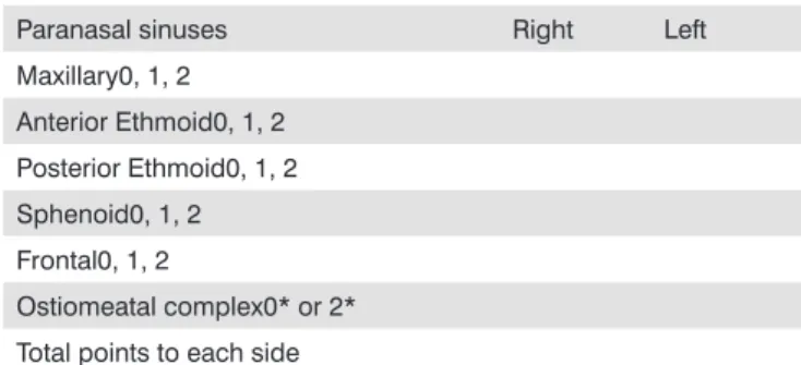
Moreover, CT scan is used as standard method for RS staging. There are many staging systems and most are based on presence and quantity of inflammatory disease in the paranasal sinuses. The most widely accepted system to classify RS is Lund-McKay (Table 1).
Table 1 - Lund-McKay staging system.
Paranasal sinuses Right Left
Maxillary0, 1, 2
Anterior Ethmoid0, 1, 2
Posterior Ethmoid0, 1, 2
Sphenoid0, 1, 2
Frontal0, 1, 2
Ostiomeatal complex0* or 2*
Total points to each side
0 = no abnormalities; 1 = partial opacification; 2 = total opacifica-tion
One of the problems of Lund-McKay classification is the broad spectrum of patients that may be classified as grade 1. Partial opacification ranges from 10% to 90% of the affected sinus. A patient with 10% (grade 1) that improves with clinical or surgical treatment may go to grade 0 (no abnormalities). However, a patient with 90% opacification (also grade 1) that improves 70% with clini-cal or surgiclini-cal treatment goes down to 20% opacification and, thus, despite the substantial improvement, it is still classified within the same group, grade 1 (partial opaci-fication). Therefore, Meltzer et al. proposed a modified Lund-McKay staging system. It is basically the same sys-tem, but grade 1 is divided into 3 subgroups 1A, (1-33% opacification), 1B (34-66% opacification) and 1C (67-99% opacification).
It is important to emphasize that the exam should be ideally requested at acute stages of the disease (except if there is suspicion of complications).
• Magnetic resonance imaging (MRI) - MRI provi-des important information about mucosa and other soft tissues. It is superior to CT in showing spreading of naso-sinusal processes beyond the limits of paranasal sinuses, such as into the orbits and intracranial compartment. The technique is used to diagnose and stage tumors and can differentiate infectious inflammatory disease by bacteria and virus from fungal diseases.
• Others - Both transilumination and ultrasound are not widely used by otorhinolaryngologists because of the high rate of false positive and false negative results, considered to have low specificity and low sensitivity.
Bacteriology: To determine the microbiology of RS and/or its response to treatment, samples of sinusal secretion should be collected without contaminating the normal respiratory or oral flora. Bacteriology is indicated preferentially in cases of recurrence or in chronic cases or in cases that do not respond to conventional treatment (for ex. immunodepressed patients, among others). The two most widely used techniques are maxillary sinus punc-ture and endoscopy. Maxillary sinus puncpunc-ture provides aspiration of secretions that may be performed through the canine fossa or inferior meatus. Nasal endoscopy provides staining of microswab of the middle meatus or even collection of aspiration material, which is less invasive and morbid than a puncture. A recent metaa-nalysis has shown accuracy of 87% in culture of middle meatus assisted by endoscopy in relation to puncture and aspiration through the canine fossa in cases of acute maxillary RS. Quantitative analysis is important because it shows the likelihood that the revealed organism is the agent responsible for the local infection and not simply that contamination increases if density of bacteria is high (≥103 - 104 cfu/mL).
Nasal cytology and biopsy: The presence of eosi-nophils in nasal secretion may indicate the presence of allergy, whereas presence of neutrophils indicate infec-tious process. However, nasal cytology is not normally used for diagnosis of RS and in isolation it can not diag-nose allergic rhinitis. Anatomic pathological exam may be indicated to exclude the presence of neoplasm, vasculitis or autoimmune diseases (Wegener’s granulomatosis, no-dous poliarteritis, recurrent polychondritis) and to study nasal polyps.
Mucociliary function
• Mucociliary clearance: For general assessment of mucociliary clearance we may use saccharin or radioiso-tope. Despite the fact that assessment using radioisotopes is more objective, saccharin test is more used because of its simplicity, safety and low cost. However, abnormal saccharin test results (> 30 minutes) do not differentiate between primary or secondary ciliary dysfunction.
• Others: Assessment of frequency of ciliary bea-ting with microscopy with phase contrast or other culture techniques and assessment of ciliary ultrastructure using transmission electron microscopy or scan microscopy are also used to have more detailed definition of architecture and ciliary function and may take to more specific diag-nosis, such as primary ciliary dyskinesia.
Specific tests of nasal permeability:
Rhinomanometry (airflow verification) and acoustic rhinometry (verification of area and nasal volume) quanti-fy the magnitude of “nasal obstruction” symptom at a given time, but they do not contribute to diagnosis of RS.
Olfaction: Olfaction may be qualitatively and/ or quantitatively checked. However, these tests are not used for diagnosis of RS, but rather to determine olfaction function itself and to follow up its response to employed treatments, both clinical and/or surgical. The most popular test, especially in North America, is the one from Univer-sity of Pennsylvania, named UniverUniver-sity of Pennsylvania Smell Identification Test - UPSIT (Sensonics, Inc.) They are plates soaked with different micro-encapsulated odors that the subject scratches and smells.
Others test olfaction threshold, presenting to pa-tients serial dilutions of specific pure odors, such as for example, pm carbinol. There are many other tests and exams including assessment of olfactory evoked poten-tials, but their review goes beyond the objectives of the present guidelines.
bacterial infection. Others are chlorine level in sweat (CF), total and specific immunoglobulins (immunodeficiency, RS allergic to fungi), complement (CH50, CH100), anti-neutrophil cytoplasmatic antibodies (c-ANCA, Wegener’s granulomatosis), angiotensin converting enzyme (sarcoi-dosis), among others.
7. IMPACT OF RHINOSINUSITIS ON QUALITY OF LIFE
To study quality of life in patients with RS, it is im-portant to differentiate three basic concepts: health, health status and quality of life. Health is defined as physical, mental and social wellbeing, according to the World He-alth Organization. HeHe-alth status refers to physical, social and emotional limitations and disabilities of the patient that may be determined by physicians and health-related professionals. Quality of life is assessed based on personal experience and reflects other circumstances of patients’ life in addition to health status.
To assess quality of life, we use validated question-naires with health measurements of physical, mental and social aspect, which show impact of the disease, mana-gement or environment on patient’s life. These question-naires are important in the development of health policies for the area of prophylaxis and/or disease treatment.
Pre-defined questionnaires may assess general and specific aspects of diseases. This concept was mentioned by Lembcke, who stated that the best quality of life me-asurement is the one that intends to prolong life, relieve stress, restore function and prevent disability.
Non-specific questionnaires to assess impact on quality of life
Medical Outcomes Study Short Form 36 (SF-36)
It is the most widely used and better validated questionnaire available. It may be applied to any disease, including RS, gathering general information (non-specific). It comprises 8 aspects: physical limitation, health status, body pain, limitations of activities, vitality, social limita-tion, mental health and emotional limitation. It has been validated in many languages (France, Spain, German, Australia). It is self-applicable to people over the age of 14. Higher scores indicate better quality of life (0-100). It has been applied in RS to assess pre and postoperative status (evidence level B).
Specific questionnaires
There are specific questionnaires to analyze quality of life in nasosinusal diseases, especially RS and allergic rhinitis. In addition to specific nasosinusal symptoms, it
includes assessment of physical, functional, emotional and social aspects. Some analyze the duration of symptoms, others their severity. These questionnaires check the im-pact of RS on quality of life and efficacy of clinical and surgical treatment. In Brazil, we used translated questio-nnaires because specific RS questioquestio-nnaires have not been validated in Portuguese yet (consistency, applicability and reliability).
The disease-specific questionnaires related with quality of life are:
Rhinosinusitis Outcome Measure (RSOM): 31 ite-ms divided into 7 domains; 20-Item Sinunasal Outcome (SNOT-20) was a modification validated by RSOM-31, to primarily assess RS treatment.
Sinunasal Outcome Test 16 (SNOT 16) and 11 Point Sinunasal Assessment Questionnaire (SNAQ-11) are other questionnaires modified from RSOM that address quality of life of people with RS.
Chronic Sinusitis Survey (CSS): 6 items assessing severity and duration of symptoms and treatment (medica-tion), validated, indicated to assess CRS (high sensitivity to clinical changes after long periods); higher scores indicate better quality of life.
Rhinosinusitis Disability Index (RSDI): 30 items about specific nasosinusal symptoms and functional li-mitation, similar to RSOM 31; does not allow indication of most important symptom; has some general questions similar to SF-36.
The Chronic Rhinosinusitis Type Specific Questio-nnaire: 3 forms (nasosinusal symptoms before and after treatment and clinical classification of RS); it is time-demanding.
Rhinoconjunctivitis quality of life questionnaire (RQLQ): it is validated but specifically designed to assess allergic rhinitis and conjunctivitis, with no relevance to RS.
Rhinosinusitis Quality of Life Survey (Rhino QoL): validated, it has 17 items (frequency, disturbance and impact of symptoms); applicable to CRS but not consis-tent for ARS.
Sinus and Nasal Quality of Life Survey (SN-5): 5 items (nasosinusal infection, nasal obstruction, allergic symptoms, emotional stress and limitation of activities); it is indicated for children with persistent nasosinusal symptoms; validated.
Results
• General
CRS has worse impact on quality of life when com-pared to rheumatoid arthritis, insulin-dependent diabetes and chronic obstructive pulmonary disease (COPD), with worst impact on body pain and social limitation, according to Gliklich and Metson. SF-36 showed significant diffe-rences in 8 domains, when compared to normal subjects (evidence level B).
There is variation in the impact of RS on quality of life in different populations, probably due to cultural influences. In Taiwan, it has been demonstrated that wo-men were more impacted by it than by migraine or initial stage breast cancer. Application of SF-36 in the Taiwanese population showed worse scores than the results of the questionnaire in the American population studied by Gliklich and Metson, with high deterioration of emotional aspect, even though it did not cause physical limitation (evidence level B).
The study analyzed the impact of depression in patients with CRS. Depressive patients had pain and nega-tive impact on physical activity. These patients presented poorer response to surgical treatment via endoscopic approach.
• Specific
Assessment by SNOT-20 showed that higher impact symptoms on quality of life of patients with RS are: thick nasal secretion, posterior discharge, fatigue, poor sleep quality, and tiredness upon waking up. Damm showed that CRS influenced quality of life of 94% of the patients and 74% of them characterized it as severe or intolerable owing to nasal obstruction, posterior discharge, heada-che, hyposmia and/or dry syndrome (evidence level B). In children, the most impacting symptoms on quality of life are nasal obstruction, sinusal infection, medication use, emotional stress, allergic symptoms and limited activities.
Studies using TyPE and SF-36 have shown that RS associated with asthma and allergy cause greater impact on vitality and overall health perception of patients than isolated rhinosinusitis.
Radenne et al. used questionnaire SF-36 to com-pare allergic rhinitis and RS with NP. Both reduce quality of life, but NP has greater impact, especially on vitality, general health and pain. Mental health is more affected than physical health and there is change to emotional health. Association with asthma reduces vitality, pain and physical function, but in isolation, NP has more impact than asthma.
NP associated with bronchiectasia does not worsen SF-36 scores, according to Guilemani et al; therefore, it does not cause additional impact on quality of life.
A prospective randomized study involving patients with CRS and NP has compared 3-month clinical treatment with macrolide and nasosinusal endoscopic surgery. Du-ring 3, 6, 9 and 12-month follow-up they analyzed nasal symptoms, SNOT-20, SF-36, expired nitric oxide, acoustic rhinometry, saccharin test and nasal endoscopy. Ninety patients were randomized and there were 40 surgical cases and 38 clinical cases to be analyzed in the end. There was significant improvement in all objective and subjective parameters in both groups, without differences between treatment except for nasal volume (rhinometry) that was greater in operated cases. The study conformed the reliability of subjective measurement of quality of life with objective parameters (evidence level B).
Many studies have confirmed quality of life im-provement in patients with CRS and NP submitted to endoscopic surgery, with 3-year follow-up. Damm et al. demonstrated improvement in 85% quality of life and the best clinical improvement occurred in 76.4% of the cases, whereas hyposmia was the symptom that persisted the most after surgery. A study using isolated RS has de-monstrated that endoscopic surgery caused less impact on symptoms of hyposmia, fever, dental pain, halitosis and cough. Moreover, there was reduction in use of antihista-minic (AH) and antibiotics and increase in use of topical corticoids. Radenne et al. noticed that nasal endoscopic surgery in massive nasal polyposis was associated with asthma and improved nasal obstruction and quality of life, reducing the need to use asthma medication, but did not modify objective pulmonary parameters. Application of SF-36 and CSS to assess the benefit of nasosinusal en-doscopy surgery at 3, 6, and 12-month follow-up showed significant improvement in quality of life, but there was no difference during the follow-up. Patients that had lowest preoperative scores maintained the lowest postoperative scores, showing the low impact of surgery when the pa-tient has few symptoms.
Quality of life of osteoplastic surgery with obli-teration of frontal sinus improved in only 48.7% of the patients. Patients were assessed after the treatment with conventional sinusal surgery (Dencker access), using SF-36 and McGill Pain Questionnaire-Dutch Language Version (MPQ) and there was improvement of pain and physical limitation, but the other 6 domains remained unaltered.
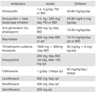
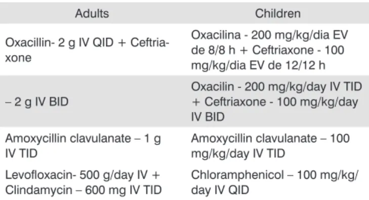
Table 2 - Acute rhinosinusitis.
Antibiotics Adults Children
Amoxycillin 1.5- 4 g/day TID
or BID 45-90 mg/kg/day
Amoxycillin + beta lactamase inhibitor
1.5- 4g / 250 mg/ day TID or BID
45-90 mg/6.4 mg/ kg/day
2nd generation Ce-phalosporin
500 mg-1g /day
BID 15-30 mg/kg/day
Macrolides 500 mg /day BID or qd
10-15 mg/kg/day- qd or BID
Trimethoprim-sulfame-thoxazole
1600 mg + 320mg /day BID
30 mg/kg + 6 mg/ kg/day
Doxycycline
200 mg /day BID, 1st day, after 100 mg qd
Ceftriaxone 1 g/day / 5days qd 50 mg/kg/day / 5days
Levofloxacin 500 mg /day qd
Moxifloxacin 400 mg /day qd
Gemifloxacin 320 mg qd
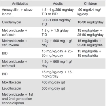
not used antibiotics in the past 4-6 weeks include: amoxycillin, amoxycillin-beta lactamase inhibitors, second-generation cephalosporin (axetil cefuroxime, cefprozil, cefaclor). Trimethoprim-sulfamethoxazole, doxycycline, and new macrolides (azythromycin, cla-rithromycin, or roxithromycin) may be considered for patients with allergy to beta-lactamic antibiotics, but there is estimated treatment failure of 20 to 25% of the cases (Table 2).
Assessment of quality of life in RS is a way of quantifying disease impact and efficacy of treatment on patient’s life. However, these questionnaires have to be validated in the Brazilian population and new randomized multicenter studies have to be carried out to transform them into important instruments to determine the best management of patients with RS.
8. TREATMENT
8.1. ANTIMICROBIAL TREATMENT IN RHINOSINU-SITIS
8.1.1 Acute Rhinosinusitis
The main purpose of using antibiotics in ARS is to eradicate bacteria from the infection site, making the affected sinus restore its normal status, reducing symp-toms and, preventing complications to avoid a chronic process.
Antimicrobial treatment of both acute and chronic RS is normally empirically based, supported by microbio-logical data (cultures and sensitivity to in vitro antimicro-bials) and studies published in the literature. Especially in acute maxillary sinusitis, antibiotic therapy has shown efficacy in moderate to severe cases, reducing the time required to resolve symptoms. In previously healthy pa-tients with mild ARS general and supportive therapeutic measurements may suffice to quickly solve the sympto-matology without requiring use of antibiotics.
In ARS in adults and children, the most common etiological agents, which amount to over 70% of the cases, are Streptococcus pneumoniae and Haemophilus influen-zae; less frequently, there is also Moraxella catarrhalis, Staphylococcus aureus and Streptococcus beta hemolytic. Thus, antimicrobial treatment should necessarily be effecti-ve against pneumococcus and Haemophilus influenzae.
Use of antibiotics, especially in RS, has been object of literature review and comparative studies using the many antimicrobials available and amoxycillin has shown the equal efficacy.
In bacterial RS, the selection of antibiotic should take into account severity of the disease, its progression and exposure to recent antibiotic therapy. Patients are divided into two categories: those with mild symptoms who had not used antibiotics for the past 4 to 6 weeks and those with mild symptoms but who had used antibiotics in the past 4 to 6 weeks, with or without moderate to severe disease regardless of previous antibiotic use.
The recommendation of initial therapy in adults with mild disease who need antibiotic therapy and had
The recommendation of initial therapy in adults with mild disease who had used antibiotics for the past 4-6 weeks, adults with moderate-severe disease, regardless of recent antibiotic use, include: high doses of amoxycillin-clavulanate, respiratory fluoroquinolones: levofloxacin, moxifloxacin and gemifloxacin. Ceftriaxone 1 g/day IM or IV for five days.
The recommendation of initial therapy in children with mild disease who had not used antibiotics in the past 4-6 weeks include: amoxycillin, amoxycillin- beta lactamase inhibitors, second-generation cephalosporin (axetil cefuroxime, cefprozil, cefaclor). Trimethoprim-sul-famethoxazole, macrolides (azythromycin, clarithromycin and roxithromycin) may be considered if the patient has allergy to beta-lactamic agents. It is important to bear in mind that the latter has limited action over most patho-gens, leading to possible treatment failure (Table 3).