Article
J. Braz. Chem. Soc., Vol. 22, No. 3, 583-591, 2011. Printed in Brazil - ©2011 Sociedade Brasileira de Química
0103 - 5053 $6.00+0.00
A
*e-mail: [email protected], [email protected]
Enzyme Kinetics, Structural Analysis and Molecular Modeling Studies on a Series
of
Schistosoma mansoni
PNP Inhibitors
Matheus P. Postigo,a Renata Krogh,a Marcela F. Terni,a Humberto M. Pereira,a
Glaucius Oliva,a Marcelo S. Castilho*,b and Adriano D. Andricopulo*,a
aLaboratório de Química Medicinal e Computacional, Instituto de Física de São Carlos,
Universidade de São Paulo, 13560-970 São Carlos-SP, Brazil
bLaboratório de Bioinformática e Modelagem Molecular da Faculdade de Farmácia,
Universidade Federal da Bahia,40170-290 Salvador-BA, Brazil
A enzima purina nucleosídeo fosforilase do parasita Schistosoma mansoni (SmPNP) é um
alvo molecular atrativo para o desenvolvimento de candidatos a novos fármacos para o tratamento da esquistossomose, doença tropical negligenciada que afeta mais de 200 milhões de pessoas em todo mundo. No presente trabalho, estudos de cinética enzimática foram conduzidos para a determinação da potência e do mecanismo de inibição de uma série de inibidores da enzima
SmPNP. Além das investigações bioquímicas, estudos cristalográicos e de modelagem molecular
revelaram importantes bases moleculares para a ainidade de ligação frente à enzima alvo, levando ao desenvolvimento de relações entre a estrutura e atividade (SAR).
The enzyme purine nucleoside phosphorylase from Schistosoma mansoni (SmPNP) is an
attractive molecular target for the development of novel drugs against schistosomiasis, a neglected tropical disease that affects about 200 million people worldwide. In the present work, enzyme kinetic studies were carried out in order to determine the potency and mechanism of inhibition of a series of SmPNP inhibitors. In addition to the biochemical investigations, crystallographic and
molecular modeling studies revealed important molecular features for binding afinity towards the target enzyme, leading to the development of structure-activity relationships (SAR).
Keywords:neglected tropical diseases,schistosomiasis, enzyme inhibition, crystal structure, binding afinity
Introduction
Neglected tropical diseases are responsible for millions of deaths and disabilities every year. Schistosomiasis is a major cause of illness, morbidity and death worldwide. Currently, there are about 200 million people infected,
with more than 650 million living in endemic areas.1
Praziquantel is the only effective drug available for the treatment of schistosomiasis, however, less than 20% of those in need currently receive the drug. Additionally, praziquantel has been in use for more than 20 years and
signiicant resistance to the chemotherapy has emerged.2
Therefore, this scenario highlights the urgent need for the
development of new drugs to treat the disease.2,3
Enzymes are attractive biological targets for
small-molecule drug discovery.4,5 Purine nucleoside phosphorylase
(PNP, EC 2.4.2.1) is a key enzyme in the purine salvage pathway which has been primarily studied as a target for the treatment of T-cell proliferative diseases, such as T-cell leukemias or lymphomas, organ transplant rejection, rheumatoid arthritis, psoriasis, and some other
autoimmune diseases.6,7 In fact, two inhibitors discovered
at BioCryst Pharmaceuticals are currently undergoing
clinical trials: (i) BCX1777 (forodesine) for the treatment
of cutaneous T-cell lymphoma, chronic lymphocytic leukemia and acute lymphoblastic leukemia; and
(ii) BCX4208 for the treatment of gout.8 More recently, it
has been suggested that PNP inhibitors could also be used for the therapy of parasitic tropical diseases, such as malaria
and schistosomiasis.9-14 The parasite Schistosoma mansoni,
the de novo pathway for purine biosynthesis and depends
entirely on the salvage pathway for its purine requirements
for synthesis of RNA and DNA.7,11,13 Taking into account
that PNP from S. mansoni (SmPNP) is an important
component of the purine salvage pathway and has been identiied as an attractive drug target, the use of selective inhibitors can cause purine starvation, leading to death of
the parasite.7,13,14
In the present work, we have evaluated a series of
guanine derivatives against SmPNP in order to determine
their in vitro potency, afinity and mechanism of inhibition.
In addition, crystallographic and molecular modeling studies were performed to identify the interaction proile of the inhibitors, as well as key structural elements responsible for the experimental properties.
Experimental
SmPNP, data set and biochemical assays
SmPNP was expressed and purified as described
previously.9,11 Xanthine oxidase was of the best grade
available from Sigma Aldrich and was used without further puriication. All other reagents and solvents were obtained commercially from Sigma Aldrich and were of the highest
purity available. The data set of inhibitors (1-20, Table 1)
employed in this work, consisting of a series of guanine, 9-substituted-guanine, 9-substituted-9-deazaguanine and 8,9-disubstituted-9-deazaguanine derivatives, were synthesized by scientists at BioCryst Pharmaceuticals Inc. and are the gift of that organization. Kinetic measurements were carried out spectrophotometrically with the aid of a Cary100 UV-Vis spectrophotometer,
using a standard coupled assay as previously described.13-16
The reaction mixture contained 5 nmol L-1SmPNP (as the
monomer), 50 mmol L-1 phosphate buffer (KPO
4, pH 7.4),
10 μmol L-1 inosine, and xanthine oxidase 40 milliunits
mL-1. Uric acid formation was monitored at 293 nm, in
triplicate at 25 oC (extinction coeficient for uric acid,
ε293 = 12.9 L mmol-1 cm-1).17 The percentage of inhibition
was calculated according to the following equation:
% of Inhibition = 100 × (1 − Vi / V0)
where, Vi and V0 are the initial velocities (enzyme
activities) determined in the presence and in the absence
of inhibitor, respectively. Compound 3, a known SmPNP
inhibitor, was used as a positive control for enzyme
inhibition.13 Values of IC
50 (concentration of compound
required for 50% inhibition of SmPNP) for the whole
series of inhibitors were independently determined by
making rate measurements for at least six inhibitor
concentrations. The type of inhibition and Ki (inhibitor
constant) values were determined for a subset of potent inhibitors under the same experimental conditions for three different inhibitor concentrations at ive varying
substrate concentrations (5.0, 7.5, 10, 15 and 20 μmol L-1).
All kinetic parameters were determined from the collected data by nonlinear regression employing the SigmaPlot enzyme kinetics module. The values represent means of
at least three individual experiments. Values of IC50 for
inhibitors 3, 7, 13, 18 and 20 measured at 10 µmol L-1
inosine are in good agreement with those previously
described,13,14 whereas comparable values are not available
for the other inhibitors of the data set.
Molecular modeling studies
The analyses, calculations and visualizations were performed using the programs SYBYL 8.0 (Tripos Inc., St. Louis, USA) and Pymol 0.99 (DeLano Scientiic, Palo Alto, USA). Molecular docking and scoring protocols, as implemented in FlexX (BioSolveIT GmbH, Sankt Augustin, Germany) and GOLD 4.12 (Cambridge Crystallographic Data Centre, Cambridge, U.K.), were employed to search for reasonable binding
poses of the ligands within the SmPNP binding pocket.
The 3D structures of all SmPNP inhibitors used in the
modeling studies were constructed using CONCORD and standard geometric parameters of the molecular modeling software package SYBYL 8.0 running on Red Hat Enterprise Linux workstations. Although GOLD assigns atoms potential according to a pre-established internal function, all 3D structures were energetically minimized through atom-centered partial charge AM1-ESP calculations as implemented in MOPAC 6.0 before docking runs. This strategy aimed at avoiding possible bond length or bond angles distortions present in any molecule. The X-ray crystallographic coordinates for
SmPNP in complex with acetate used in the docking
of all amino acid residues within a sphere radius of 6.5 Å (FlexX) or 10.0 Å (GOLD) centered on the oxygen atom of Tyr202. Default parameters were employed to generate the poses, which were then ranked by FlexX and GOLDSCORE scoring functions. Next, the best ranked conformation of each ligand was submitted to the web-based interface DrugScore ONLINE to rescore
the proposed binding modes.18 Only poses that rank well
in both function and that were consistent with kinetic studies were considered further.
Crystallization and soaking of SmPNP
The highly puriied SmPNP enzyme was crystallized as
previously described.11-13 Next, crystals grown at 4 °C in
20% PEG 1500, 15 mmol L-1 sodium acetate buffer (pH 4.9
or 5.0) and 20% glycerol (buffer A) were soaked for
48-96 h in solutions containing 10 mmol L-1 of compound 7
(Table 1), dissolved in 1:9 DMSO/buffer A.
X-ray data collection and structure reinement
X-ray diffraction data were collected at cryogenic temperatures (100 K ) at W01B-MX-2 beamline, equipped with a Marmosaic 225 CCD scanner, of the LNLS (National Laboratory of Synchrotron Light, in Campinas - Brazil).
The crystals of SmPNP in complex with compound 7
(SmPNP-7) diffracted up to 2.30 Å. The data were indexed
and integrated using the program MOSFLM and scaled
using the program SCALA from the CCP4 suite.19-22
Next, the starting phases were determined by molecular
replacement using SmPNP in complex with acetate
(PDB ID 1TD1) as the search model, as available in the
program Molrep.21 The model reinement was carried out
with Refmac22 and PHENIX23 using σ
a-weighted 2Fo-Fc
and Fo-Fc electron density maps. The compounds were automatically placed in the electron density using the “Find
Ligand” routine of Coot.24 Additional manual building
and placement of water molecules were performed with the Coot and PHENIX software. During all steps of the reinement, 5% of the data were used to calculate the free
R-factor25 and thus avoid overitting. All inal models
were validated using PROCHECK.26 The coordinates and
structures factors of SmPNP-7 complex are deposited in
the PDB under ID 3DJF.
Results and Discussion
Considering the key role of PNP in the purine salvage
pathway of the S. mansoni parasite, this enzyme has been
selected as a potential target for the chemotherapeutic
treatment of schistosomiasis. Despite the significant amount of structure-activity relationship (SAR) studies
available for inhibitors of the mammalian PNPs,15,27 similar
investigations for SmPNP remain outstanding, and would
be essentially important for the design of new inhibitors.
The difference of about 6-fold in the KM
(Michaelis-Menten constant) values between human and parasite PNPs
(human PNP = 41 µmol L-1, SmPNP = 6.4 µmol L-1) for the
natural substrate (inosine) indicates that binding afinity depends on speciic molecular interactions between small-molecule ligand and protein. As a matter of fact, the irst
SmPNP inhibitors were only recently characterized in the
literature.13,14 In an effort to expand our studies, enzyme
kinetic, crystallographic and molecular modeling studies were carried out to investigate the fundamental chemical and structural requirements involved in the inhibition of
the catalytic activity of SmPNP by a series of guanine
derivatives. In this context, a series of twenty compounds (Table 1) was experimentally evaluated to determine their
in vitro potency (IC50) and to provide new SAR information.
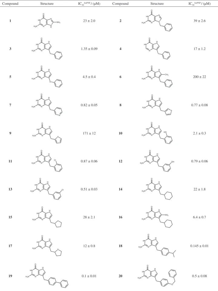
As can be seen, the two guanine derivatives of
the data set (compounds 1 and 2) showed moderate
inhibitory potency against SmPNP (IC50 values of 39 and
23 μM, respectively). These results contrast with that of
compound 3 (IC50 = 1.35 μM), a 9-benzyl-9-deazaguanine
derivative, which is approximately 30-fold more potent than
the structurally related guanine analog 2. This conirms
previous studies showing that 9-deazaguanine analogs are
more potent than the corresponding guanine analogs.6,28,29
A hypoxanthine analog of the data set (compound 4,
IC50 = 17 μM), which is structurally related to compounds
2 and 3, is 12-fold less potent than compound 3, and about
2-fold more potent than the guanine analog 2. These results
highlight the importance of the 9-deazaguanine scaffold
in the development of selective inhibitors of SmPNP,
indicating that stereochemical features in the ribose binding
site might play a pivotal role in SmPNP inhibition.
In order to extend our SAR studies, we have evaluated
some 9-deazaguanines (5-20) possessing a variety of
substituents in the 9-position. On the basis of structure
3, we investigated the bioisosteric replacement of the
methylene group (benzyl group) at position 9 of the purine
scaffold with an oxygen atom, leading to compound 5.
However, this structural modiication led a 3-fold decrease in potency. The substitution of the hydrogen at position 8 of the purine ring with a methyl group afforded a very
weak inhibitor of SmPNP (compound 6, IC50 = 200 μM).
On the other hand, the presence of heteroaromatic
moieties at position 9 of the purine ring (7 and 8) in place
of the traditional phenyl group improved potency against
Table 1. Inhibitory potency of a series of SmPNP inhibitors
Compound Structure IC50SmPNP / (μM) Compound Structure IC50SmPNP / (μM)
1 HN
N N H N O
H2N
NH2 23 ± 2.0 2
HN
N N N O
H2N 39 ± 2.6
3 HN N H N O
H2N 1.35 ± 0.09 4
HN
N H N O
17 ± 1.2
5 HN N H N O O
H2N 4.5 ± 0.4 6
HN
N H N O
H2N
CH3
200 ± 22
7 HN N H N N O
H2N 0.82 ± 0.05 8
HN N H N O H2N S
0.77 ± 0.08
9 HN N H N O H2N S
171 ± 12 10
HN N H N O H2N
HO 2.1 ± 0.3
11 HN N H N O
H2N
Cl
0.87 ± 0.06 12
HN
N H N O
H2N
OH 0.79 ± 0.06
13 HN N H N O
H2N Cl 0.51 ± 0.03 14
HN
N H N O
H2N 22 ± 1.8
15 HN N H N O
H2N 28 ± 2.1 16
HN
N H N O
H2N
NH2
6.4 ± 0.7
17 HN N H N O H2N S
12 ± 0.8 18
HN
N H N O
H2N 0.145 ± 0.01
19 HN N H N O
H2N 0.1 ± 0.01 20
HN N H N O H2N O
and 770 nM, respectively). Our SAR data show that the replacement of aromatic (phenyl, pyridine, thiophene) to non-aromatic moieties led to new compounds with
decreased inhibitory potency (14, IC50 = 22 μM; 15,
IC50 = 28 μM; 16, IC50 = 6.4 μM; and 17, IC50 = 12 μM)
when compared with those of compounds 7 and 8. The
substitution on the phenyl group of compound 3 was
also considered, employing a series of 9-deazaguanines
having the phenyl group substituted by hydrophilic (10,
IC50 = 2.1 μM and 12,IC50 = 0.79 μM) and hydrophobic
(11,IC50 = 0.87 μM and 13,IC50 = 0.51 μM) substituents.
As can be seen in Table 1, the presence of the ortho- or
meta-chlorine (11 or 13) or meta-hydroxyl (12) led to
a slight increase in potency, which was not observed
for compound 10 (IC50 = 2.1 μM). On the other hand,
substituents in the para position of the phenyl ring led
to the most potent inhibitors of this series, compounds
18 (IC50 = 0.14 μM) and 19 (IC50 = 0.10 μM), suggesting
that bulkier hydrophobic groups would be important for
biological activity. The other para-substituted inhibitor
of the series also conirms this tendency (compound 20,
IC50 = 0.5 μM).
Although the SAR information gathered from this series is valuable and revealed several structural aspects related to the inhibitory potency, the underlying explanation for these results would require more in-depth investigation. Two things are especially important. First, the evaluation
of the mechanism of SmPNP inhibition associated to the
determination of the afinity of the inhibitors. Second, the structural analysis of the binding mode of the inhibitors into
the SmPNP active site. In order to explore the mechanism
of inhibition in more detail, Ki values and the type of
inhibition with respect to the physiological substrate were determined employing a subset of inhibitors as shown in Table 2 and Figures 1 and 2. The Lineweaver-Burk double-reciprocal plots show the intercepts of the lines
Figure 1. Competitive inhibitory proile of the SmPNP inhibitors 7 (A), 13 (B), 18 (C) and 19 (D). Kinetic data was collected in the presence of increasing
concentrations of inhibitor. Panel A 0.8 µmol L-1 (), 1.6 µmol L-1 (q), 3.0 µmol L-1 (r). Panel B 0.8 µmol L-1 (), 1.6 µmol L-1 (q), 3.0 µmol L-1 (r).
Panel C 150 nmol L-1 (), 300 nmol L-1 (q) and 600 nmol L-1 (r). Panel D 200 nmol L-1 (), 400 nmol L-1 (q) and 800 nmol L-1 (r). In all panels the
absence of inhibitor is depicted by ().
Table 2. Ki values for a subset of potent SmPNP inhibitors
Compound Ki / (μM) IC50 / Ki
7 0.451 1.8
13 0.120 4.2
18 0.038 3.9
(obtained at three different inhibitor concentrations)
converging at the y-axis (1/Vmax), whereas the slope
(KM/Vmax) and the x-axis intercepts (-1/KM) vary with
inhibitor concentration (Figure 1). Consequently, the Vmax
values remain constant, whereas the apparent values of KM
(KMapp, deined as K
M(1 + [I]/Ki)) increase with increasing
inhibitor concentrations. This behavior is consistent with a mutually exclusive binding mode between inhibitor and
substrate, thus indicating that the inhibition of SmPNP was
found to be competitive for the inhibitors 7, 13, 18 and 19.
A similar behavior was observed for the other compounds of the data set under the same experimental conditions (results not shown).
The determination of the inhibitor constant Ki is
particularly relevant in the present case, since the IC50
values were obtained at only one concentration of substrate.
For competitive inhibitors, IC50 values increase as the
concentration of the substrate increases. As shown in Figure 2, the intersection on the x-axis of the graphs of
KMapp/V
maxappversus inhibitor concentration indicates – Ki.
The Ki values are collected in Table 2.
The data of Table 2 show that the values of IC50/Ki vary
from 1.4 to 4.2, which supports the competitive mechanism
of inhibition. Despite the strong evidences that all of 9-deazaguanine inhibitors bind to the active site of the enzyme, the kinetic data does not provide insights into the binding proile of the inhibitors (in the substrate pocket of
SmPNP). Therefore, crystallization and X-ray studies were
carried out to complement the kinetic indings. Extensive soaking experiments afforded the crystallographic structure
of SmPNP in complex with the nanomolar inhibitor 7
(SmPNP-7) at 2.3 Å (Figure 3 and Table 3).
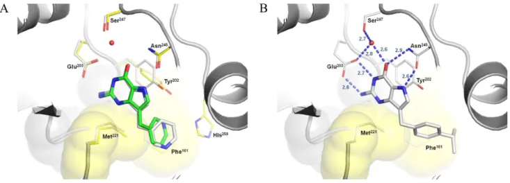
As expected, the purine moiety shows a conserved H-bonding network to Glu203 and Asn245, in agreement
with previous studies for the bovine and human PNPs.27-29
As can be seen in Figure 3, an extra H-bonding to a crystallographic water is found, although this feature could be also present in other 6-oxo purine inhibitors with
moderate potency, such as compound 14 (IC50 = 22 μM).
As a consequence, the relative high potency of compound
7 (IC50 = 0.82 μM) might be a result of a more complex
binding network that incorporates particular interactions of the 3-pyridine substituent at position 9 of the purine ring. Accordingly, this moiety not only H-bonds to Tyr202 and His259, but also reaches a hydrophobic pocket lined by Met221 and Phe161. These experimental results led us to
investigate the binding mode of other potent 9-deazaguanine inhibitors using different molecular docking strategies. In this context, FlexX and GOLD 4.12 were employed to
cross-dock compound 7 into the SmPNP active site (PDB
ID 1TD1). The best FlexX solution shows a RMSD of 1.29Å from the crystallographic coordinates of compound
7 (Figure 4A). Similar results were obtained with GOLD
4.12. These initial results prompted us to further explore the docking tools to provide additional SAR insights within this series.
The binding modes of the 9-deazaguanine derivatives shown in Figure 4B highlight the most important intermolecular interactions involved in ligand binding. The H-bonding pattern between the deazaguanine moiety and the side chains of Glu203 and Asn245 is fundamental, where the catalytic residue Asn245 donates a hydrogen bond to the carbonyl oxygen at the position 6, and accepts a hydrogen bond from the N-H group at position 7 of
the purine ring. Similarly, the residue Glu203accepts
hydrogen bonds from the NH2 and NH groups at positions
2 and 1, respectively. The π-π interaction between the
electron rich guanine ring and the side chain of Tyr202 might also be important for inhibitor binding. In addition, the side chain of Phe161 from the adjacent subunit generates a hydrophobic surface near the site entrance (Figure 4), which provides a suitable interaction region for hydrophobic moieties at position 9 of the purine ring,
such as the para-phenyl and para-isopropyl benzyl of
compounds 19 (IC50 = 0.1 μM) and 18 (IC50 = 0.14 μM),
respectively. On the other hand, more bulk groups,
such as the para-cresyl benzyl ether of compound 20
(IC50 = 0.5 μM), becomes more exposed to solvent and
led a decrease in potency.
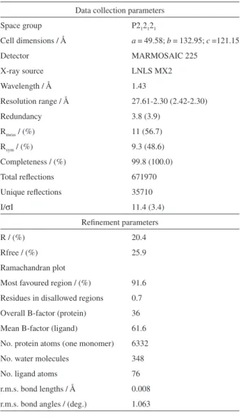
Table 3. Full data collection and reinement statistics for the SmPNP-7 crystallographic complex
Data collection parameters Space group P212121
Cell dimensions / Å a = 49.58; b = 132.95; c =121.15
Detector MARMOSAIC 225
X-ray source LNLS MX2
Wavelength / Å 1.43
Resolution range / Å 27.61-2.30 (2.42-2.30)
Redundancy 3.8 (3.9)
Rmeas / (%) 11 (56.7)
Rsym / (%) 9.3 (48.6)
Completeness / (%) 99.8 (100.0) Total relections 671970 Unique relections 35710
I/σI 11.4 (3.4)
Reinement parameters
R / (%) 20.4
Rfree / (%) 25.9
Ramachandran plot
Most favoured region / (%) 91.6 Residues in disallowed regions 0.7 Overall B-factor (protein) 36 Mean B-factor (ligand) 61.6 No. protein atoms (one monomer) 6332 No. water molecules 348 No. ligand atoms 76 r.m.s. bond lengths / Å 0.008 r.m.s. bond angles / (deg.) 1.063
Figure 3. Crystallographic structure of compound 7 in complex with SmPNP. A) Electron density (Fo–Fc map), contoured at 3.0δ, where the inhibitor was it. B) Binding proile of compound 7 in the active site of SmPNP. Selected residues from the active site are highlighted in yellow, whereas the protein
Conclusions
The development of new therapeutic approaches to combat neglected tropical diseases continues to be one of the most important scientiic and public health challenges facing humankind today. The identiication of novel small-molecule compounds that modulate speciic biological targets is of great pharmaceutical interest, as this complex
task requires the use of different drug design technologies.5,30
The integration of enzyme kinetics, structural analysis and molecular modeling studies provided important insights into the molecular basis underlying ligand binding afinity
and SmPNP inhibition. The information gathered in this
work should be useful in the design of new inhibitors having improved potency.
Acknowledgments
We gratefully acknowledge inancial support from FAPESP (São Paulo Research Foundation), FAPESB (Fundação de Amparo à Pesquisa do Estado da Bahia) and CNPq (National Council for Scientiic and Technological Development), Brazil. We are also grateful to BioCryst Pharmaceuticals, Inc. for the gift of the inhibitors employed in this work.
References
1. Hotez, P.; Molyneux, D. H.; Fenwich, A.; Kumaresan, J.; Sachs, S. E.; Sachs, J. D.; Savioli, L.; N. Engl. J. Med. 2007,
357, 1018.
2. Sabra, A. N.; Botros, S. S.; J. Parasitol. 2008, 94, 537. 3. Global Forum for Health Research; The 10/90 Report on
Health Research, 2003-2004, Geneva, 2004. http://www.
globalforumhealth.org/Media-Publications/Publications/10-90-Report-2003-2004
4. Guido, R. V. C.; Oliva, G.; Andricopulo, A. D.; Curr. Med. Chem. 2008, 15, 37.
5. Andricopulo, A. D.; Salum, L. B.; Abraham, D. J.; Curr. Top. Med. Chem. 2009, 9, 771; Cardoso, C. L.; Lima, V. V.; Zottis, A.; Oliva, G.; Andricopulo, A.; Wainer, I. W.; Moaddel, R.; Cass, Q. B.; J. Chromatogr., A 2006, 1120, 151.
6. Montgomery, J. A.; Med. Res. Rev. 1993, 13, 209.
7. Bzowska, A.; Kulikowska, E.; Shugar, D.; Pharmacol. Ther.
2000, 88, 349.
8. http://www.biocryst.com/clinical_pipeline, acessed in August 2010.
9. Pereira, H. M.; Cleasby, A.; Pena, S. D.; Franco, G. R.; Garratt, R. C.; Acta Crystallogr., Sect. D: Biol. Crystallogr. 2003, 59, 1096.
10. Kicska, G. A.; Tyler, P. C.; Evans, G. B.; Furneaux, R. H.; Schramm, V. L.; Kim, K. J.; Mol. Biol. 2002, 277, 3226.
11. Pereira, H. D.; Franco, G. R.; Cleasby, A.; Garratt, R. C.;
J. Mol. Biol. 2005, 353, 584.
12. Shi, W.; Ting, L. M.; Kicska, G. A.; Lewandowicz, A.; Tyler, P. C.; Evans, G. B.; Furneaux, R. H.; Kim, K.; Almo, S. C.; Schramm, V. L.; J. Biol. Chem. 2004, 279, 18103.
13. Castilho, M. S.; Postigo, M. P.; Pereira, H. M.; Oliva, G.; Andricopulo, A. D.; Bioorg. Med. Chem. 2010, 18, 1421. 14. Postigo, M. P.; Guido, R. V. C.; Oliva, G.; Castilho, M. S.; Pitta,
I. R.; Albuquerquer, J. F. C.; Andricopulo, A .D.; J. Chem. Inf. Model. 2010, 50, 1693.
15. Farutin, V.; Masterson, L.; Andricopulo, A. D.; Cheng, J.; Riley, B.; Hakimi, R.; Frazer, J. W.; Cordes, E. H.; J. Med. Chem.
1999, 42, 2422.
16. Andricopulo, A. D.; Yunes, R. A.; Chem. Pharm. Bull. 2001, 49, 10.
17. Kim, B. K.; Cha, S.; Parks, R. E., Jr.; J. Biol. Chem. 1968, 243,
1771.
Figure 4. Proposed binding mode of SmPNP inhibitors according to FlexX. A) Overlay of the best solution found for compound 7 onto its crystallographic
pose (RMSD = 1.28 Å). B) Interaction proile of compound 18 into the active site of SmPNP. Selected residues from the SmPNP-7 complex (yellow) and
18. Gohlke, H.; Hendlich, M.; Klebe, G.; J. Mol. Biol. 2000, 295, 337; http://pc1664.pharmazie.uni-marburg.de/drugscore/index.php, accessed in June 2009.
19. Leslie, A. G. W.; Acta Crystallogr., Sect. D: Biol. Crystallogr.
1999, 55, 1696.
20. Collaborative Computational Project, Number 4, The CCP4 suite: programs for protein crystallography; Acta Crystallogr., Sect. D 1994, 50, 760.
21. Vagin, A.; Teplyakov, A.; Acta Crystallogr. 2000, 56, 1622. 22. Murshudov, G. N.; Vagin, A.; Dodson, E. J.; Acta Crystallogr.
1997, 53, 240.
23. Adams, P. D.; Grosse-Kunstleve, R. W.; Hung, L.; Ioerger, T. R.; McCoy, A. J.; Moriarty, N. W.; Read, R. J.; Sacchettini, J. C.; Sauter, N. K.; Terwilliger, T. C.; Acta Crystallogr. Sect D: Biol. Crystallogr. 2002, 58, 1948.
24. Emsley, P.; Cowtan, K.; Acta Crystallogr., Sect. D: Biol. Crystallogr. 2004, 60, 2126.
25. Brunger, A. T.; Acta Crystallogr., Section D:Biol. Crystallogr.
1993, 49, 24.
26. Laskowski, R. A.; Moss, D. S.; Thornton, J. M.; J. Mol. Biol.
1993, 231, 1049.
27. Castilho, M. S.; Postigo, M. P.; Paula, C. B. V.; Montanari, C. A.; Oliva, G.; Andricopulo, A. D.; Bioorg. Med. Chem. 2006,
14, 516.
28. Shewach, D. S.; Chern, J. W.; Pillote, K. E.; Townsend, L. B.; Daddona, P. E.; Cancer Res. 1986, 46, 519; Woo, P. W.; Kostlan,
C. R.; Sircar, J. C.; Dong, M. K.; Gilbertsen, R. B.; J. Med. Chem. 1992, 35, 1451.
29. Secrist-3rd, J. A.; Niwas, S.; Rose, J. D.; Babu, Y. S.; Bugg, C. E.; Erion, M. D.; Guida, W. C.; Ealick, S. E.; Montgomery, J. A.; J. Med. Chem. 1993, 36, 1847.
30. Castilho, M. S.; Guido, R. V. C.; Andricopulo, A. D.; Bioorg. Med. Chem. 2007, 15, 6242; Andricopulo, A. D.; Yunes, R. A.; Nunes, R. J.; Savi, A. O. S.; Correa, R.; Cruz, A. B.; Cechinel, V.;
Quim. Nova1998, 21, 573; Salum, L. B.; Polikarpov, I.; Andricopulo, A. D.; J. Chem. Inf. Model. 2008, 48, 2243; Lima,
E. O.; Queiroz, E. F.; Andricopulo, A. D.; Nunes, R. J.; Yunes, R. A.; Correa, R.; Cechinel, V.; Bol. Soc. Chil. Quim. 1999, 44,
185. Honorio, K. M.; Garratt R. C.; Polikatpov, I., Andricopulo, A. D.; J. Mol. Graphics Modell. 2007, 25, 921.
Submitted: September 10, 2010
Published online: December 21, 2010