Article
0103 - 5053 $6.00+0.00*e-mail: [email protected]
In Vitro
Inhibition of Linoleic Acid Peroxidation by Primary S-Nitrosothiols
Fernanda I. Simplicio, Amedea B. Seabra, Gabriela F. P. de Souza and Marcelo G. de Oliveira*
Instituto de Química, Universidade Estadual de Campinas, CP 6154, 13083-970 Campinas-SP, Brazil
O óxido nítrico (•NO) é um antioxidante efetivo na inibição da peroxidação lipídica e circula
in vivo principalmente como S-nitrosotióis primários (RSNOs). Neste trabalho, a peroxidação in
vitro de comicelas do ácido linileico-SDS (LA-SDS) catalisada por lipoxigenase de soja (SLO) e
íons FeII foi monitorada na presença e na ausência de três RSNOs: S-nitrosocisteína,
S-nitroso-N-acetilcisteína e S-nitrosoglutationa. Medidas cinéticas baseadas na formação de duplas ligações conjugadas e adutos luorescentes de lisina-La oxidado mostraram que os RSNOs são antioxidantes mais potentes que seus tióis livres (RSHs) correspondentes em condições equimolares. Esses resultados são consistentes com o bloqueio da peroxidação de LA-SDS por RSNOs através da inativação dos radicais peroxila/alcoxila (LOO•/LO•), levando a produtos nitrogenados do LA
oxidado, os quais liberam •NO. Esses resultados indicam que os RSNOs endógenos podem
desempenhar um papel importante no bloqueio da peroxidação lipídica in vivo, através da inativação primária de radicais alcoxila/peroxila bem como de hidroperóxidos lipídicos pré-formados.
Nitric oxide (•NO) is an effective chain-breaking antioxidant in the inhibition of lipid
peroxidation and circulates in vivo mainly as primary S-nitrosothiols (RSNOs). In this work, the in
vitro peroxidation of linoleic acid-SDS comicelles (LA-SDS) catalyzed by soybean lipoxygenase
(SLO) and FeII ions was monitored in the presence and absence of three primary RSNOs:
S-nitrosocysteine, S-nitroso-N-acetylcysteyne and S-nitrosoglutathione. Kinetic measurements based on the formation of conjugated double bonds and luorescent oxidized LA-lysine adducts, showed that RSNOs are more potent antioxidants than their corresponding free thiols (RSHs) in equimolar conditions. These results are consistent with the blocking of LA-SDS peroxidation by RSNOs through the inactivation of peroxyl/alkoxyl (LOO•/LO•) radicals, leading to
nitrogen-containing products of oxidized LA, which release free •NO. These results indicate that endogenous
RSNOs may play a major role in the blocking of lipid peroxidation in vivo, through the primary inactivation of alkoxyl/peroxyl radicals and also of preformed lipid hydroperoxides.
Keywords: nitric oxide, S-nitrosothiols, lipid peroxidation, linoleic acid, lipoxygenase
Introduction
An increasing amount of evidence has demonstrated that oxidative and nitrosative stresses, play a fundamental role in atherosclerosis and in other diseases associated with lipid peroxidation (LPO).1-6 In these cases, it is assumed
that free radicals which normally play an essential role in metabolic processes, are released from the active site of enzymes, triggering a cascade of deleterious effects on cells.7 These effects involve the interaction of free radicals
with metal or organic redox centers and the promotion of irreversible oxidation reactions beyond the normal catalytic
cycles. Once formed, free radicals are also capable of initiating other radical reactions, which may become self-sustaining through the regeneration of propagating radicals which are involved in the oxidation of lipids in humans representing a key event in the atherosclerotic process. This conclusion is reinforced by the fact that both primary and secondary lipid oxidation products are found in human atherosclerotic lesions.8,9
Under normal physiological conditions, endothelium-derived nitric oxide (nitrogen monoxide, •NO) has multiple
physiological functions in humans, like the regulation of vascular tone in both the systemic and renal circulation,10,11
of the proliferation and migration of smooth muscle cells.13
In addition to the actions related to the mediation of signal transduction, via stimulation of guanylate cyclase-mediated cGMP synthesis, NO has been shown to exert several antiatherogenic properties assigned to its ability to react directly with free radicals, blocking the propagation of radical reactions. This protective effect has already been observed in model lipid systems,14,15 low-density lipoproteins
(LDL)16-18 and cells19,20 and is supported by several in vitro
studies which have demonstrated the formation of nitrogen-containing products of polyunsaturated fatty acids (PUFA), including alkylnitrites (RONO), alkylnitrates (ROONO and RONO2), alkylepoxynitrite (R(O)NO2), alkylnitrohydroxy (RNO2OH), and nitrolipids (RNO2), when PUFAs are oxidized in the presence of •NO. Such products have already
been characterized in other works by mass spectrometry and can be taken as markers of the in vivo pro-oxidant and/or antioxidant actions of •NO.14,15,21-28 These results stimulate
new therapeutic approaches for treating lipid peroxidation-related diseases by enhancing •NO synthesis and/or activity
by administration of L-arginine and antioxidants.29 As an
alternative strategy, compounds that act as NO donors could be administrated as exogenous NO sources, as already demonstrated for the treatment of hepatic steatosis via oral administration of the S-nitrosothiol (RSNO) S-nitroso-N-acetylcysteine (SNAC).3-6 RSNOs are peptides or proteins
carrying the S-NO moiety and were shown to occur in the plasma and cells of mammals where they have the same physiological functions of free •NO like vasodilation,30,31
inhibition of platelet activation and aggregation32,33 and
post-translational modification of protein function.34,35
S-nitrosoglutathione (GSNO), S-nitrosoalbumin and S-nitrosohemoglobin, for example, have been considered to be •NO carriers and donors in humans and are the focus
of several studies both in vivo and in vitro.36 Other RSNOs,
like S-nitrosocysteine (CySNO) have also been described.37
What classiies a RSNO as primary is the fact that the sulfur atom of its SNO moiety is bound to a primary carbon atom (Figure 1), while in S-nitroso-N-acetylpenicillamine (SNAP), which is widely used in experimental studies, the sulfur atom is bound to a tertiary carbon atom, making it a
“tertiary” RSNO. Evaluating the antioxidant properties of primary RSNOs may have an additional relevance, once the lability of •NO in primary and tertiary RSNOs can be
different.37-39 In any case, one of the important characteristics
of having •NO carried as RSNOs is its preservation from
inactivation caused by reaction with oxygen, leading to nitrite (NO2–) and further to nitrate (NO
3
–),40 two of the main end
products of •NO metabolism. Although several exogenous •NO sources (which are not found endogenously) have been
used as antioxidants in LPO studies like organic nitrites and NONOates,41 only one work has reported the protective role
of primary RSNOs in blocking LPO reactions.42
In this work, the in vitro peroxidation of linoleic acid SDS comicelles (LA-SDS) catalyzed by soybean lipoxygenase (SLO) and by FeII ions was monitored in the presence and
absence of three primary RSNOs: CySNO, SNAC and GSNO and of their corresponding free thiols (RSHs), at 37 ºC. Kinetic data showed that RSNOs can block LA peroxidation much more eficiently than RSHs, by inactivating alkoxyl/peroxyl (LO•/LOO•) radicals and LA hydroperoxides, (LOOH =
13-hydroperoxy-octadecadienoic acid, 13-HPODE) through nitration and transnitrosation reactions. These results suggest that endogenous primary RSNOs may play a major role in blocking lipid peroxidation in vivo.
Experimental
Reagents
Ascorbic acid, cysteine (CySH), ferrous sulfate (FeSO4), copper sulfate (CuSO4), glutathione (g-Glu-Cys-Glu, GSH), linoleic acid (LA), L-lysine monohydrochloride (Lys), malonaldehyde bis(dimethyl acetal) (MDA), N-acetyl-L-cysteine (NAC), phosphate buffer saline (PBS, pH 7.4), sodium dodecil sulfate (SDS), sodium nitrite (NaNO2), soybean lipoxygenase (SLO), hydrochoric acid (HCl), mercury chloride (HgCl2), tert-butyl hydroperoxide (tBOOH, C
4 H10 O2) (Sigma/Aldrich, St. Louis, MO) and
sulfanilamide (Merck, Germay), were used as received. All the experiments were carried out using analytical grade water from a Millipore Milli-Q gradient iltration system.
Synthesis of GSNO, CySNO and SNAC in aqueous solution
Aqueous GSNO solution was prepared by the reaction of GSH with sodium nitrite in acidic medium as described elsewhere.37,43 GSNO was obtained as stable reddish
crystals in the pure form and was dried by freeze-drying. Freshly prepared GSNO solutions in PBS were used in the experiments. S-nitroso-N-acetylcysteine (SNAC) and S-nitrosocysteine (CySNO) cannot be precipitated from solution and stored as dry solids because of their high solubility in water. Therefore, aqueous SNAC or CySNO solutions were synthesized through the equimolar reaction of NAC or CyS, respectively, with NaNO2 in acidiied aqueous solution. Stock acidic SNAC and CySNO solutions freshly prepared, were diluted in PBS and used immediately.
Characterization of linoleic acid peroxidation
Linoleic acid (LA) peroxidation was induced through the addition of SLO to aqueous LA dispersions (final concentration 19 mmol L-1) in SDS solution (0.01 mol L-1)
(LA-SDS comicelles). Each LA dispersion was transferred to a quartz cuvette, blowed with O2 for 2 min and SLO (inal concentration 56 nmol L-1) was added to the cuvette with
a syringe to start the peroxidation reaction. Peroxidation reactions were monitored in the absence or presence of CySH, NAC and GSH (inal concentrations 560 mmol L-1) and of
their corresponding RSNOs, CySNO, SNAC and GSNO, respectively (inal concentrations 56 mmol L-1), through the
increase in absorbance at 234 nm, due to conjugated diene formation. A Hewlett Packard spectrophotometer, model 8453 (Palo Alto, CA, USA) with a temperature-controlled cuvette holder was used to monitor the spectral changes in the range 200-600 nm in the dark at 37 °C, in time intervals of 2 s. Spectra of the solutions were obtained in 1 cm quartz cuvette, under stirring (1,000 r min-1). Each point in the
kinetic curves of absorbance vs. time is the average of two experiments with error bars expressed by their standard errors of the mean (SEM). Statistical differences among the initial rates (Ir) of LA peroxidation, catalyzed by SLO, in the absence and presence of RSHs and their corresponding RSNOs were evaluated using ANOVA followed by Tukey-Kramer multiple test.
Characterization of luorescent MDA-lysine adduct
To characterize the emission spectrum of the luorescent adduct formed in the reaction of oxidized linoleic acid (oxLA) and lysine, an MDA-lysine adduct was prepared as a model adduct by reacting MDA with L-lysine in equimolar condition (inal concentration 0.25 mol L-1)
in PBS solution at room temperature and the emission spectrum was obtained in the range 375-600 nm, with excitation wavelength of 360 nm.
Spectroluorimetric characterization and monitoring of oxidized LA-lysine adduct formation
LA peroxidation was induced through the addition of aqueous FeSO4 solution (inal concentration 5.0 µmol L-1) to
aqueous LA (inal concentration 1.2 mmol L-1) dispersions
in SDS solution (inal concentration 0.01 mol L-1) in the
absence or presence of GSNO (inal concentrations 5 and 500 mmol L-1) for 2 h in PBS (pH 7.4). After LA oxidation,
lysine solution (inal concentration 1.0 mmol L-1) was
transferred to the dispersions followed by incubation for 48 h. The kinetics of formation of luorescent oxidized LA-lysine adduct (oxLA-Lys) in the reaction between oxLA and Lys during the incubation time was characterized based on the spectral changes in the range 375 to 600 nm and on the emission intensity at 430 nm, under excitation at 360 nm. All the spectroluorimetric measurements were performed using a Perkin-Elmer LS55 spectroluorimeter with a temperature-controlled cuvette holder at 37 ºC.
Infrared characterization of linoleic acid peroxidation
Linoleic acid peroxidation was induced by heating a sample of pure LA at 80 ºC for 4 h under stirring in a glass lask with O2 atmosphere, obtained by continuously blowing O2 from a cylinder into the headspace of the lask. Aliquots of LA were removed from the reaction lask 2 and 4 h after the beginning of the peroxidation reaction. Capillary ilms of non-oxidized and peroxidized LA were obtained between two calcium luoride (CaF2) windows, which were mounted in special sample holder. IR spectra were obtained in the range 4000-1000 cm-1 using an FTIR
Bomem MB-series, model B-100. An IR spectrum of non-oxidized LA was obtained as a control.
Detection of •NO released from nitrogen-containing
products of oxidized LA
Nitric oxide released from nitrogen-containing products of oxLA formed in the peroxidation of LA in the presence of GSNO, was detected using a gas-phase chemiluminescence-based nitric oxide analyzer (NOA, Sievers, Bolder Co, USA). Peroxidation of LA was induced by two different procedures. In the irst, the LA-SDS dispersion in the presence of CuII
(900 mmol L-1) ions was previously blowed with O 2 for
for 30 min at room temperature. In the second, peroxidation of LA was induced by heating a sample of pure LA at 80 ºC for 1 h under stirring in a glass lask with O2 atmosphere, obtained by continuously blowing O2 from a cylinder into the headspace of the lask. After oxidation, oxLA was dispersed in SDS solution (0.01 mol L-1) and
incubated with GSNO, for 30 min at room temperature. In both cases, after incubation, non-reacted excess
GSNO was removed from the solution by adding HgCl2
(inal concentration 29.4 mmol L-1) and allowing GSNO
decomposition to GS-SG and free •NO to proceed for
15 min. In this condition, •NO is quantitatively released
from excess GSNO by mercuric catalysis and is rapidly and quantitatively converted to its stable end product, nitrite (NO2–). Nitrite formed was removed by adding a
10% v/v solution of sulfanilamide (6.0 mmol L-1 in HCl
2 mol L-1), followed by incubation for 15 min. A volume
of 5 mL of aqueous saturated ascorbic acid solution, used as a reducing agent, was added in the glass purge vessel of the NOA. Antifoaming agent was used to prevent foaming caused by injection of the samples. Volumes of 100 mL of the inal nitrogen-containing products of oxLA suspension were injected in the glass purge vessel containing ascorbic acid, through an impermeable septum. Nitrogen gas (Air Liquid, Brazil) was bubbled through the solution and free •NO formed due to the
reduction of nitrogen-containing products of oxLA by ascorbic acid in the glass purge vessel was carried into the reaction chamber for detection. Control curves are for the measurements of samples of water incubated with GSNO 900 mmol L-1 without LA-SDS and CuII for 30 min
at room temperature (control 1), and of water incubated with GSNO 900 mmol L-1 and CuII without LA-SDS for
30 min at room temperature (control 2). In both cases, the incubations were followed by the addition of HgCl2 (inal concentration 29.4 mmol L-1) for promoting GSNO
decomposition to GS-SG and free •NO for 15 min and
by the addition of a 10% v/v solution of sulfanilamide (6.0 mmol L-1 in HCl 2 mol L-1), for 15 min for eliminating
nitritre formed in the reaction between NO and O2.
Reaction between RSNOs and tert-butyl hydroperoxide
The formation of tert-butyl peroxynitrites (tBOONOs)
in the reactions between tert-butyl hydroperoxide (tBOOH) and RSNOs was characterized by following
the decomposition of GSNO, SNAC and CySNO (initial concentrations 1 mmol L-1) upon the addiction of tBOOH
(initial concentration 25 mmol L-1) in basic medium
(pH 12). The decomposition of RSNOs in absence or presence of tBOOH was characterized by following
the spectral changes of RSNOs solutions in the range 220-1100 nm in the dark, in a 1 cm quartz cuvette referenced against air. Kinetic curves of GSNO, SNAC and CySNO decomposition were obtained from the absorption changes at 336 nm in time intervals of 15 s, at 37 ºC, for 8 min. The control experiment was performed by incubating RSNOs with pure water at pH 12, adjusted with NaOH solution. Each point in the kinetic curves of concentration vs. time is the average of two experiments with error bars expressed by their standard errors of the mean (SEM).
Results and Discussion
Kinetic characterization of linoleic acid peroxidation
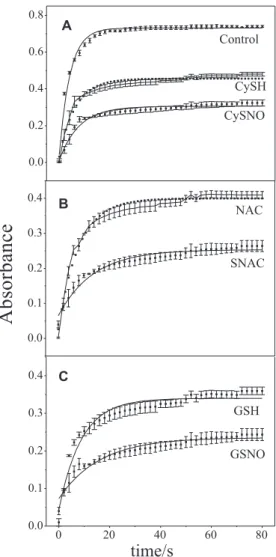
Figure 2 shows the kinetic curves corresponding to the spectral changes monitored at 234 nm in the irst 80 s of LA oxidation by SLO in the presence of RSHs and RSNOs. The band at 234 nm is associated with the formation of conjugated double bonds in LA, as a result of LA peroxidation.35 In this time range, the curves reach
an apparent plateau after ca. 20 s in all cases. The initial rates of reaction (Ir), as well as the height of the plateaus, are decrease signiicantly in the presence of RSHs and RSNOs, compared to the peroxidation of LA alone. As the height of the plateaus can be taken as a measurement of the extent of the peroxidation reaction in its initial phase, this result indicates that RSNOs in concentrations ten times lower than their corresponding RSHs (56 versus
560 mmol L-1) exert a much more extensive blockage of
the peroxidation reaction than the corresponding RSHs in this time range.
The initial rates of LA peroxidation (Ir), extracted from the curves of Figure 2 are shown in the bar graphs of Figure 3 for comparison. It can be seen that the Ir of LA (19 mmol L-1) peroxidation is decreased to about ½ of the
control value in the presence of RSHs (560 mmol L-1),
i.e., at a molar ratio RSH/LA = 29.5, with no signiicant differences among the antioxidant actions of CySH, NAC and GSH. In contrast, the presence of RSNOs at a concentration ten times lower (56 mmol L-1), i.e., at a molar
ratio RSH/LA = 2.9 reduced Ir values to ca. 1/5 of the control value, also with no signiicant differences among the action of CysNO, SNAC and GSNO.
Fluorimetric characterization and monitoring of oxidized LA-lysine adduct formation
Figure 4 shows the emission spectra obtained after LA oxidation catalyzed by FeII ions for 2 h, followed
37 ºC. The peroxidation reactions were performed in the absence (a) and presence of GSNO 5.0 mmol L-1 (b)
and 500.0 mmol L-1 (c). The inset in Figure 4 shows the
emission spectrum obtained after the incubation of MDA with lysine in equimolar concentrations of 0.25 mol L-1,
as a control experiment. Figure 5 shows the kinetic curves of the luorescent oxLA-lysine adduct formation in the reaction between oxidized LA and lysine in conditions (a), (b) and (c) of Figure 4. The curves were based on the spectral changes monitored at 430 nm, during 48 h after lysine addition. It can be observed that the formation of the oxLA-Lys adduct follows a sigmoid pattern with an apparent induction or “lag” phase, which is evident in curves (a) and (b). In curve (c) the reaction is still in an apparent lag phase until 48 h of monitoring, although a small rate of luorophore formation can be detected since the beginning of the reaction.
Figure 2. Kinetic curves of LA (19 mmol L-1) peroxidation catalyzed by
(SLO) (56 nmol L-1), in the absence of RSH or RSNO (Control, A) and
in the presence of CySH 560 mmol L-1 and CySNO 56 mmol L-1 (A);
NAC 560 mmol L-1 and SNAC 56 mmol L-1 (B) and GSH 560 mmol L-1
and GSNO 56 mmol L-1 (C). Absorbance changes were monitored at
234 nm at 37 ºC.
Figure 3. Initial rates (Ir) achieved after ca. 20 s of LA peroxidation, catalyzed by SLO, in the absence and presence of RSHs and their corresponding RSNOs at concentrations ten times lower. [LA] = 19 mmol L-1; [SLO] = 56 nmol L-1; [RSHs] = 560 mmol L-1
and [RSNOs] = 56 mmol L-1. Data extracted from the kinetic curves of
Figure 2. * p < 0.001; ** p < 0.05.
Figure 4. Emission spectra obtained after linoleic acid (LA) oxidation (inal concentration 1.2 mmol L-1) catalyzed by FeII ions (FeSO
4, inal
concentration 5.0 mmol L-1) for 2 h, followed by incubation of the solution
with lysine (Lys) (inal concentration 1.0 mmol L-1) for 48 h at 37 ºC in the
absence (a) and presence of GSNO 5.0 mmol L-1 (b) and 500.0 mmol L-1
(c). Excitation/emission wavelengths 360/430 nm. Inset: Emission spectra of MDA incubated with Lys in equimolar concentrations of 0.25 mol L-1,
used as a control.
Detection of LA peroxidation by infrared spectroscopy
Figure 6 shows the spectral change in the normal infrared region, obtained after 2 and 4 h of heating of pure LA at 80 ºC under O2 atmosphere. The new absorption band with maximum at ca. 1180 cm-1 can be assigned to
the C–O–O vibration of hydroperoxides (LOOH).45
Detection of •NO released from nitrogen-containing
products of oxidized LA
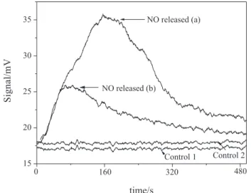
Figure 7 shows the peaks of light emission obtained in the chemiluminescence reaction of free •NO, released
from nitrogen-containing products of oxLA, formed in the reaction between oxLA and GSNO. The two peaks shown were obtained after reduction of nitrogen-containing products of oxLA by ascorbate, according to the procedures described above. Peak (a) was obtained in the reduction of a sample of LA-SDS dispersion oxidized in the presence of CuII ions and GSNO. The same result was observed
when SNAC or CySNO were used in the place of GSNO (data not shown). Peak (b) was obtained in the reduction of a sample of LA-SDS dispersion incubated with GSNO, where pure LA had been previously oxidized by heating under O2 at 80 °C. The detection of free •NO in these cases
shows that nitrogen-containing products of oxLA are formed either when LA is oxidized in aqueous dispersion in the presence of GSNO or when a dispersion of oxLA-SDS is subsequently incubated with GSNO. Control curves are for the measurements of samples of water incubated with GSNO without LA-SDS and CuII (control 1), and of
water incubated with GSNO and CuII without LA-SDS
(control 2), which show that GSNO was completely eliminated through decomposition by HgII ions, followed
by NO2– trapping by sulfanilamide, before injection in the
ascorbic acid soluti
Reaction between RSNOs and tert-butyl hydroperoxide
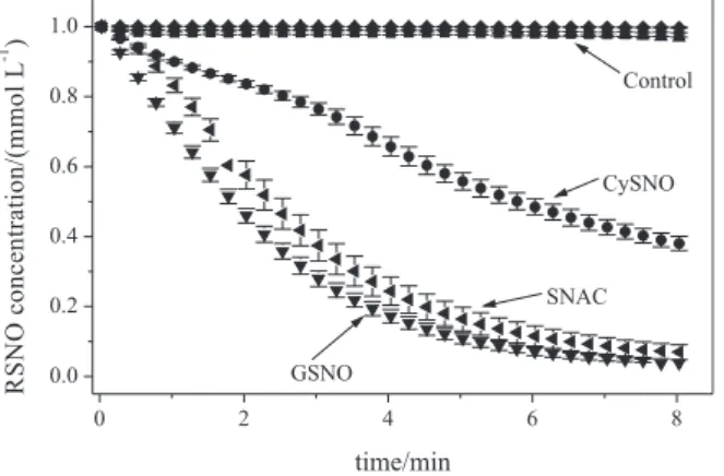
Figure 8 shows the kinetic curves corresponding to the spectral changes due the disappearance of the RSNOs, CySNO, SNAC and GSNO, during their reaction with tert -butyl hydroperoxide (tBOOH) with formation of tert-butyl
peroxynitrites (tBOONOs). Control curves correspond to
the monitoring of RSNOs solutions at the same temperature and pH conditions, but in the absence of tBOOH. It can
be seen that the RSNOs solutions are quite stable in the absence of tBOOH and that their thermal decompositions are negligible in this time range. On the other hand, the presence of tBOOH leads to the fast disappearance of
the absorption bands of the three RSNOs, indicating that they react with tBOOH. The rates of reaction of SNAC
and GSNO are very similar and follow pseudo-irst order kinetics. However, CySNO shows a different kinetic pattern, with an apparent bimodal behaviour. In this case, the rate of reaction is lower and approximately constant up to ca. 3 min and increases after this time, becoming similar to the rates observed in the last 4 min for SNAC and GSNO. In the lipid pool of plasma and cells, polyunsaturated fatty acids (PUFAs) have higher propensity to oxidation due to the fact that bis-allylic methylene hydrogens are more
Figure 6. Infrared spectral changes associated with the formation of hydroperoxides in the oxidation of linoleic acid at 80 ºC under O2 atmosphere for 2 and 4 h.
Figure 7. Light emission peaks obtained in the chemiluminescence reaction of ozone with free NO, released in the reduction of nitrogen-containing products of oxidized linoleic acid (oxLA) formed in the peroxidation reaction of LA in the presence of GSNO (900 µmol L-1).
For details see experimental part. Peak (a) was obtained for the product formed after the incubation of LA with CuII ions (900 µmol L-1) in the
presence of GSNO. Peak (b) was obtained for the product formed after the incubation of LA previously oxidized by heating under O2 with GSNO. Control curves are for the measurements of samples of water incubated with GSNO without LA-SDS and CuII (control 1), and of water incubated
susceptible to hydrogen abstraction by oxidizing radicals than the methylene hydrogens from fully saturated lipids.44
After such initiation process, the rapid reaction between the formed carbon-centered radical and dioxygen (O2) forms a lipid peroxyl radical (LOO•). Propagation occurs via the
reaction between LOO• and intact fatty acid (LH) molecules
forming lipid hydroperoxides (LOOH), which leads to the formation of more LOO• species through the decomposition
of LOOH catalyzed by CuII or FeIII ions either free or in the
form of heme proteins.46-48 In addition, LOOH can lead to
the formation of alkoxyl radicals mediated by FeII. These
processes are represented in Scheme 1.
Linoleic acid (LA), one of the components of LDL particles, is a major unsatured fatty acid in the american diet and is considered to be atherogenic because of its pro-oxidative and pro-inlammatory response through the activation of endothelial cells.49,50 An increase in LA levels
has been reported in the phospholipid fractions of human coronary arteries in cases of sudden cardiac death due to ischemic heart disease.51 Additionally, concentrations of
LA in adipose tissue were positively correlated with the
degree of coronary disease.52 Linoleic acid has a double
bond coniguration with bis-allylic methylene hydrogens (Scheme 1). Due to this characteristic, and due to the reasons mentioned above, LA is an appropriate model compound for LPO studies. The kinetic monitoring of conjugated double bond formation in LA-SDS comicelles catalyzed by SLO (Figure 2) shows that LA is effectively oxidized in aqueous dispersion by dissolved O2. It must be considered that, in this particular condition, SLO is also an appropriate catalyzer as a member of a well known group of enzymes able to induce enzymatic peroxidation of polyunsaturated fatty acids in biological membranes and lipoproteins.53,54 In general, such enzymes contain
an essential iron atom, which is present as FeII in the
inactive enzyme form; enzymatic activation occurs through hydroperoxide-driven oxidation of FeII to FeIII.
Although the kinetics of lipoperoxidation induced by SLO, CuII and FeII ions may be different, the formation
of alkoxyl and peroxyl radicals is a common result in all cases, since hydrogen abstraction by any initiator in the presence of O2 will lead to the formation of these propagating species. The results of all experiments herein presented are in accordance with the inactivation of alkoxyl and peroxyl species by the RSNOs. The expected products in all cases are LOONO and LONO species. In fact, nitrated lipid formation have been shown to occur invivo
as potential “footprint” of the critical role that •NO and/or •NO-derived reactive species play during lipid oxidation
processes.8 The RSNOs used here cannot be considered
classical antioxidants like α-tocopherol (α-TOH) or ascorbic acid. Although α-TOH reacts with LOO• forming
a tocopheroxyl radical (α-TO•) which can scavenge another
LOO• species, allowing two LOO• radicals to be scavenged
by one α-TOH molecule, the primary product of this reaction is LOOH and its accumulation may expose lipids to subsequent oxidation mediated by metal ions.48 A similar
process can be described for other classical antioxidants found in cells, like ascorbic acid and glutathione (GSH).
Figure 8. Kinetic curves corresponding to the spectral changes of CySNO, SNAC and GSNO (initial concentration 1.0 mmolL-1) solutions
in the presence and absence of tBOOH (inal concentration 25 mmol L-1),
monitored at 336 nm for 8 min, at 37 ºC. Control curves correspond to the thermal decomposition of RSNOs solutions in the same temperature and pH conditions but in the absence of tBOOH.
Both are highly susceptible to hydrogen abstraction, generating ascorbyl and thyil radicals and leading to LOOH formation in their primary reactions. In the case of GSH, the fate of the glutathionyl radical (GS•) formed is dimerization,
forming oxidized glutathione (GS-SG), the ration GSH/GS-SG being a well-known marker of oxidative stress.55 The protective actions of RSNOs are primarily
linked to the well-known antioxidant action of •NO as
a radical-chain terminator, which arises from the fact that •NO is itself a free radical. Like its reaction with
superoxide (O2•–) generating peroxynitrite (ONOO–,
k = 6.7 × 10⁹ L mol-1 s-1)56 free •NO may react extremely
fast with LOO• (k = 2 × 10⁹ L mol-1 s-1)48 removing this
chain carrying radical from the reaction scene. Although formation of ONOO– is usually associated with a
pro-oxidant response relected also in the nitration of tyrosine residues,48,57 the deleterious actions of ONOO– have been
shown to depend strongly on the balance between •NO and
O2•–.48 More generally, the balance between oxidant species
and •NO seems to be fundamental in allowing a protective
action of •NO against LPO. Hummel et al.20 for example,
have shown that quite low levels of •NO (ca. 50 nmol L-1)
are enough to suppress FeII-O
2 lipid oxidation in vitro in
cell models.
Although aqueous RSNOs solutions may spontaneous release free •NO in vitro through the homolytic cleavage
of the S–N bond, the chemical stability of RSNOs solutions at low concentrations is high enough to allow these compounds to react directly with other substrates in a bimolecular mechanism. The decomposition of RSNOs with NO release may occur in several hours and days, depending on the experimental condition.19 In the
present work, as shown in Figure 2, the kinetic curves of LA peroxidation were monitored for only 80 s. In this time frame RSNOs are stable and the inhibition of lipid peroxidation can be assigned to the primary reactions between intact RSNOs molecules and oxyl/peroxyl species.
In the case of LA peroxidation, the primary reaction of RSNOs with LO• or LOO• species will lead to the
formation of LOONO or LONO species and not to LOOH or LOH species, as in the case of hydrogen abstraction from classical antioxidants.
As a complementary analysis of the protective action of RSNOs in LPO, one may also consider that linoleate hydroperoxide (LOOH) formed as a primary oxidation product of LA is expected to undergo β-scission generating free aldehydes. It is generally assumed that adducts formed in the conjugation of free aldehydes generated during peroxidation of PUFAs with amino groups on LDL particles are proteins with Schiff bases (containing the -C=N- group). Formation of such adducts is central in the atherosclerotic
process once it neutralizes the cluster of positive charges on the surface of LDL particles, conferring to them a higher anionic electrophoretic mobility and a reduced recognition by the LDL receptor on ibroblasts, while increasing their recognition by macrophages.60 As Schiff
bases have luorescence properties,61,62 it was assumed in
this work that such adducts could be used to characterize the formation of aldehydes from hydroperoxides in the peroxidation of LA. It was found here that the luorescence emission spectra obtained after LA oxidation catalyzed by FeII ions followed by incubation of the solution with Lys,
has exactly the same shape and position of the spectrum obtained in the incubation of MDA with Lys (Figure 4). This result shows that products of LA peroxidation are also reactive toward lysine, forming the same luorescent Schiff base adduct formed in oxidized LDL (like the MDA-lysine adduct). The luorescent adduct identiied in this work was assigned to the reaction between the aldehydes formed from the reduction and b-scission of LOOH, with lysine (Scheme 2). More speciically, the reaction involves the nucleophilic addition between the amino group of lysine and the carbonyl group of the aldehydes, forming hemiaminals, followed by dehydration to generate stable imines. In Scheme 2, these reactions are represented for the two possible aldehydic fragments of LOOH b-scission: 12-oxododecanoic acid and hexanal. Of course, in different oxidative situations several other aldehydic products may form after LA peroxidation, which may also generate luorescent adducts with lysine. In addition to the aldehyde-type lysine adducts, amide-aldehyde-type-lysine adducts have also been described by Kawai and co-workers63,64 as a new class
of protein adducts derived from lipid peroxidation. The dose-dependent reduction in the amounts of aldehyde-type lysine adducts formed in the presence of GSNO 5.0 µmol L-1 and 500.0 µmol L-1 in Figure 4,
compared to the peroxidation reaction performed in the absence of GSNO, relect the protective effect of GSNO in this particular peroxidation condition. This protective action is also relected in the reduction of the rates and increase in the apparent lag phase of the aldehyde-type lysine adducts formation (Figure 5).
The formation of nitrogen-containing products of oxLA, induced by CuII ions and heat, during the peroxidation of
LA in the presence of RSNOs was demonstrated in the present work by reducing these products to free •NO and
hydroperoxides with ascorbic acid, according to procedures already described in other works.14 The reaction involved
can be written as:
2 LOONO/LONO + AscH– + H+→
where AscH– is the ascorbate anion and DHA is
dehydroascorbic acid formed in the oxidation (hydrogen abstraction) of AscH–. Free •NO released in this reaction
was unequivocally detected by chemiluminescence, after its removal from the solution by bubbling with N2 and its reaction with ozone (O3). It must be emphasized here that to avoid any possible interference of •NO released from
excess GSNO, instead of •NO released from nitrated LA,
excess GSNO (which did not reacted with LOO•)was
completely eliminated from the solution by addition of HgII
ions and sulfanilamide. It is know that •NO is quantitatively
liberated from GSNO• by mercuric catalysis, and it is
rapidly and quantitatively converted to its stable solution end-product, nitrite (NO2–) in aerated medium. By adding
sulfanilamide to the solution, NO2– formed is completely
removed, and therefore the •NO signal detected in this
analysis can be attributed solely to •NO released from
nitrogen-containing products of oxLA. This conclusion is supported by the control experiments, which conirmed that GSNO is completely eliminated by the mercuric catalysis/ sulfanilamide procedure. In addition, it was observed that RSNOs react with LA previously oxidized through heating, once incubation of oxLA-SDC comicelles with GSNOs, led also to the formation of nitrogen-containing
products of oxLA, detected by their reduction to •NO with
ascorbate. This result points to the ability of RSNOs to inactivate preformed LA hydroperoxides (LOOH). In this case, the bimolecular reaction is expected to proceed via a transnitrosation mechanism and can be written as:
LOOH + RSNO → LOONO + RSH (2)
It must be noted that the formation of LOOH in the LA oxidized by heating under O2 was proven by observing the appearance of an IR absorption band with maximum around 1180 cm-1, assigned to the C-O-O vibration of
hydroperoxides (LOOH).45 The reactivity of RSNOs against
tert-butyl hydroperoxide (tBOOH) is an additional evidence
for reaction 2. Although the different kinetic behavior of CysNO compared to SNAC and GSNO in Figure 8 is not relected in the initial rates of LA peroxidation in Figure 3, it must be noted that reaction rates in Figure 3 were calculated over the irst 20 s of reaction, while differences in the kinetic behavior among the three RSNOs in Figure 8 begin to appear after 60 s. In addition, reaction rates in Figure 3 relect not only the inactivation of LOOH by RSNOs but mainly of LO• and LOO• species. Therefore, the evidences
of the ability of primary RSNOs to block the propagation of
lipid peroxidation reaction not only by inactivating LO• or
LOO• radicals, but also by inactivating preformed LOOH,
reinforces the potential role of endogenous RSNOs as modulators of peroxidation reactions in vivo. The relevance of this evidence can be better appreciated by considering that the reduction of LOOH to LO• radical by metal ions as
FeII or CuII represents an important via for the propagation of
radical reactions in the lipid peroxidation process. Although SLO, FeII and CuII have chemical peculiarities, it might be
consider that the three compounds lead to the formation of nitrogen containing products of oxLA,8 which can release
NO upon reduction with ascorbic acid. For example, it has been reported that lipid peroxidation of LA by lipoxygenase yielded nitrogen-containing lipid derivatives.8
Thus, if the role of endogenous •NO as an antioxidant
species is linked in vivo to its presence in primary RSNOs, this role must be extended beyond its classical radical chain termination action, to encompass the inactivation of deleterious hydroperoxides also present in the cellular milieu. These multiple protective actions of RSNOs are summarized in Scheme 1 for the case of LA peroxidation and can be generalized for other lipids. These results raise the possibility that primary RSNOs are involved in the formation of nitrogen-containing lipids (and perhaps of nitroalkanes), which all may be natural membrane components and may have biological activities intimately linked with the biological activities of RSNOs.
Acknowledgments
F. I. S. held a studentship from CNPq, project 140702/2003-2. G. F. P. S. holds a studentship from FAPESP, project 07/55877-3. We thank FAPESP and CNPq for inancial support.
References
1. Whitlock, D. R.; Nitric Oxide-Biol. Chem. 2006, 14, 68. 2. Kunsch, C.; Medford R. M.; Circ. Res.1999, 85, 753. 3. de Oliveira, C. P. M. S.; Simplicio, F. I.;. de Lima, V. M. R.;
Yuahasi, K.; Lopasso, F. P.; Alves, V. A. F.; Abdalla, D. S. P.; Carrilho, F. J.; Laurindo, F. R. M.; de Oliveira, M. G.; World J.
Gastroenterol.2006, 12, 1905.
4. de Oliveira, C. P. M. S.; Stefano, J. T.;. de Lima, V. M. R.; Simplicio, F. I.; de Mello, E. S.; de Sá, V. M.; Corrêa-Giannella, M. L.; Alves, V. A. F.; Laurindo, F. R. M.; de Oliveira, M. G.; Giannela-Neto, D.; Carrilho, F. J.; J. Hepatol. 2006, 15, 725. 5. de Oliveira, C. P. M. S.; de Lima, V. M. R.; Simplicio, F. I.;
Soriano, F. G.; de Mello, E. S.; de Souza, H. P.; Alves, V. A.; Laurindo, F. R.; Carrilho, F. J.; de Oliveira, M. G.; J. Am. Coll.
Nutr.2008, 27, 299.
6. de Oliveira, C. P. M. S.; Alves, V. A. F.; de Lima, V. M. R.; Stefano, J. T.; Debbas, V.; de Sá, S. V.; Wakamatsu, A.; Corrêa-Giannella, M. L.; de Mello, E. S.; Havaki, S.; Tiniakos, D. G.; Marinos, E.; de Oliveira, M. G.; Giannella-Neto, D.; Laurindo, F. R.; Caldwell, S.; Carrilho, F. J.; Biochem. Pharmacol. 2007,
74, 290.
7. Girotti, A. W.; J. Lipid Res. 1998, 39, 1529.
8. Rubbo, H.; O`Donnell, V.; Toxicology 2005, 208, 305. 9. Letters, J. M.; Witting, P. K.; Christison, J. K.; Eriksson, A. W.;
Pettersson, K.; Stocker, R.; J. Lipid Res. 1999, 40, 1104. 10. McMackin, C. J.; Vita, J. A.; Methods Enzymol. 2005, 396, 541. 11. Walkowska, A.; Kompanowska-Jezierska, E.; Sadowski, J.;
Kidney Int. 2004, 66, 705.
12. Napoli, C.; Ignarro, L. J.; Nitric Oxide 2001, 5, 88. 13. Cooke, J. P.; Atheroscler. Suppl. 2003, 4, 53.
14. Lima, E.S.; Bonini, M. G.; Augusto, O.; Barbeiro, H. V.; Souza, H. P.; Abdalla, D. S. P.; Free Radic. Biol. Med. 2005, 39, 532. 15. Schopfer, F. J.; Baker, P. R. S.; Giles, G.; Chumley, P.; Batthyany,
C.; Crawford, J.; Patel, R. P.; Hogg, N.; Branchaud, B. P.; Lancaster, J. R.; Freeman, B. A.; J. Biol. Chem.2005, 280, 19289. 16. Hogg, N.; Kalyanaraman, B.; Joseph, J.; Struck, A.;
Parthasarathy, S.; FEBS Lett. 1993, 334, 170.
17. Yamanaka, N.; Oda, O.; Nagao, S.; FEBS Lett. 1996, 398, 53. 18. Rubbo, H.; Trostchansky, A.; Botti, H.; Batthyány, C.; Biol.
Chem. 2002, 283, 547.
19. Kelley, E. E.; Wagner, B. A.; Buettner, G. R.; Burns, C. P.; Arch.
Biochem. Biophys. 1999, 370, 97.
20. Hummel, S. G.; Fischer, A. J.; Martin, S. M.; Schafer, F. Q.; Buettner, G. R.; Free Radic. Biol. Med.2006, 40, 501. 21. Lima, E. S.; Di Mascio, P.; Rubbo, H.; Abdalla, D. S. P.;
Biochemistry2002, 41, 10717.
22. Freeman, B. A.; Baker, P. R. S.; Schopfer, F. J.; Woodcock, S. R.; Napolitano, A.; d’Ischia, M.; J. Biol. Chem. 2008, 283, 15515. 23. Coler, B.; Bloodsworth, A.; Clark, S. R.; Lewis, M. J.; Cross, A.
R.; Freeman, B. A.; O’Donnell, V. B.; Circ. Res. 2002, 91, 375. 24. Schopfer, F. J.; Lin, Y.; Baker, P. R. S.; Cui, T.; Garcia-Barrio,
M.; Zhang, J.; Chen, K.; Chen, Y. E.; Freeman, B. A; Proc. Natl.
Acad. Sci. U.S.A. 2005, 102, 2340.
25. Wright, M. M.; Schopfer, F. J.; Baker, P. R. S.; Vidyasagar, V.; Powell, P.; Chumley, P.; Iles, K. E.;. Freeman, B. A.; Agarwal,
A.; Proc. Natl. Acad. Sci. U.S.A. 2006, 103, 4299.
26. Cui, T.; Schopfer, F. J.; Zhang, J.; Chen, K.; Ichikawa, T.;. Baker, P. R. S.; Batthyany, C.; Chacko, B. K.; Feng, X.; Patel, R. P.; Agarwal, A.; Freeman, B. A.; Chen, Y. E.; J. Biol. Chem.
2006, 281, 35686.
27. Villacorta, L.; Zhang, J.; Garcia-Barrio, M. T.; Chen, X.; Freeman, B. A.; Chen, Y. E.; Cui, T.; Am. J. Physiol.2007, 293, H770. 28. Baker, P. R. S.; Lin, Y.; Schopfer, F. J.; Woodcock, S. R.;
29. Adams, M. R.; McCredie, R.; Jessup, W.; Robinson, J.; Sullivan, D.; Celermajer, D. S.; Atherosclerosis1997, 129, 261. 30. Gladwin, M. T.; Adv. Exp. Med. Biol. 2006, 588, 189. 31. Seabra, A. B.; Fitzpatrick, A.; Paul, J.; de Oliveira, M. G.;
Weller, R.; Brit. J. Dermatol. 2004, 151, 977.
32. Pignatelli, P.; Di Santo, S.; Buchetti, B.; Sanguigni, V.; Brunelli, A.; Violi, F.; FASEB J. 2006, 20, 1082.
33. Marcondes, S.; Cardoso, M. H. M.; Morganti, R. P.; Thomazzi, S. M.; Lilla, S.; Murad, F.; De Nucci, G.; Antunes, E.; Proc.
Natl. Acad. Sci. U. S. A.2006, 103, 3434.
34. Chen, C.; Huang, B.; Han, P. W.; Duan, S. J.; Prog. Biochem.
Biophys. 2006, 33, 609.
35. Batthyany, C.; Schopfer, F. J.; Baker, P. R. S.; Duran, R.; Baker, L. M. S.; Huang, Y.; Cervenansky, C.; Branchaud, B. P.; Freeman, B. A.; J. Biol. Chem. 2006, 281, 20450. 36. Giustarini, D.; Milzani, A.; Colombo, R.; Dalle-Donne, I.; Rossi,
R.; Clin. Chim. Acta2003, 330, 85.
37. de Oliveira, M. G.; Shishido, S. M.; Seabra, A. B.; Morgon, N.
H.; J. Phys. Chem. A2002, 106, 8963.
38. Roy, B.; Dhardemare, A. D.; Fontecave, M.; J. Org. Chem.
1994, 59, 7019.
39. Bainbrigge, N.; Butler, A. R.; Gorbitz, C. H.; J. Chem. Soc.,
Perkin Trans. 21997, 351.
40. Stamler, J. S.; Singel, D. J.; Loscalzo, J.; Science1992, 258, 1898. 41. Nicolescu ,A. C.; Reynolds, J. N.; Barclay, L. R. C.; Thatcher,
G. R. J.; Chem. Res. Toxicol. 2004, 17, 185.
42. Krieger, M.H.; Santos, K.F.R.; Shishido, S.M.; de Oliveira, M.G.; Franchini, K.G.; Spadari-Bratish, R.C.; Wanschel, A. A. C. B.; Estrela, H.F.G.; Santos, L.; Laurindo, F. R. M.; Nitric
Oxide2006, 14, 12.
43. Hart, T. W.; Tetrahedron Lett. 1985, 26, 2013.
44. Hogg, N.; Kalyanaraman, B.; Biophys. Acta1999, 1411, 378. 45. Silvertein, R. M.; Franciz, X.; Spectrometric Identiication of
Organic Compounds, John Wiley & Sons Inc.: New York, 1998.
46. Ohyashiki, T.; Kadoya, A.; Kushida, K.; Chem. Pharm. Bull.
2002, 50, 203.
47. Pinchuk, I.; Lichtenberg, D.; Prog. Lipid Res. 2002, 41, 279. 48. Patel, R. P.; Moellering, D.; Murphy-Ullrich, J.; Jo, H.;
Beckman, J. S.; Darley-Usmar, V. M.; Free Radical Biol. Med.
2000, 28, 1780.
49. Hennig, B.; Lei, W.; Arzuaga, X.; Ghosh, D. D.; Saraswathi, V.; Toborek, M.; J. Nutr. Biochem.2006, 17, 766.
50. Young, V. M.; Toborek, M.; Yang, F.; McClain, C. J.; Hennig,
B.; Metabolism1998, 47, 566.
51. Luostarinen, R.; Boberg, M.; Saldeen, T.; Atherosclerosis1993,
99, 187.
52. Hodgson, J. M.; Wahlqvist, M. L.; Boxall, J. A.; Balazs, N. D.;
Am. J. Clin. Nutr. 1993, 58, 228.
53. Lapenna, D.; Ciofani, G.; Pierdomenico, S. D.; Giamberardino, M. A.; Cuccurullo, F.; Free Radical Biol. Med. 2003, 35, 1203. 54. Brash, A. R.; J. Biol. Chem.1999, 274, 23679.
55. Rahman, I.; Biswas, S. K.; Jimenez, L. A.; Torres, M.; Forman, H. J.; Antioxid. Redox Signal.2005, 7, 42.
56. O’Donnell, V. B.; Eiserich, J. P.; Chumley, P. H.; Jablonsky, M. J.; Krishna, N. R.; Kirk, M.; Barnes, S.; Darley-Usmar, V. M.;
Chem. Res. Toxicol.1999, 12, 83.
57. O`Donnell, V. B.; Freeman, B. A.; Cir. Res.2001, 88, 12. 58. Nakano, E.; Williamson, M. P.; Williams, N. H.; Powers, H. J.;
Biochim. Biophys. Acta2004, 1688, 33.
59. Zhang, Y. H.; Hogg, N.; Proc. Natl. Acad. Sci. U. S. A.2004,
21, 7891.
60. Cominacini, L.; Garbin, U.; Davoli, A.; Micciolo, R.; Bosello, O.; Gaviraghi, G.; Scuro, L. A.; Pastorino, A. M.; J. Lipid Res.
1991, 32, 349.
61. Fruebis, J.; Parthasarathy, S.; Steinberg, D.; Proc. Natl. Acad.
Sci. U. S. A. 1992, 89, 10588.
62. de Oliveira, F. G.; Rossi, C. L.; de Oliveira, M. G.; Saad, M. J. A.; Velloso, L. A.; Cardiovasc. Res.2000, 47, 567.
63. Kawai, Y.; Kato, Y.; Fujii, H.; Makino, Y.; Mori, Y.; Naito, M.; Osawa, T.; J. Lipid Res. 2003, 44, 1124.
64. Kawai, Y.; Fujii, H.; Kato, Y.; Kodama, M.; Naito, M.; Uchida, K.; Osawa, T.; Biochem. Biophys. Res. Commun. 2004, 313, 271.
Submitted: November 16, 2009
Published online: June 23, 2010