www.elsevier.com/locate/bjid
The Brazilian Journal of
INFECTIOUS DISEASES
1413-8670/© 2012 Elsevier Editora Ltda. All rights reserved. A R T I C L E I N F O
Article history: Received 20 July 2011 Accepted 21 August 2011
Keywords: Drug resistance Genes, MDR Mutation
Polymerase chain reaction
* Corresponding author at: Department of Microbiology, School of Medicine, Ahvaz Jundishapur University of Medical Sciences (AJUMS), Ahvaz, 61335, Iran
E-mail address: [email protected] (Hamed Goodarzi) A B S T R A C T
Objective: Isoniazid (INH) and rifampin (RIF) are the most effective first line antibiotics against Mycobacterium tuberculosis. Mutations in several genes determine resistance of M. tuberculosis to INH, with the most common gene target of katG, and resistance to RIF is due to mutation in rpoB gene. The aim of present study was to assess the mutations in the regions related to RIF and INH resistance.
Methods: We characterized 80 clinical isolates of confirmed M. tuberculosis to analyze the most commonly observed INH and RIF mutations. PCR analysis and sequencing were used to detect mutations related to RIF and INH resistance. The multiplex allele-specific-PCR (MAS-PCR) was performed as a comparative assay and for evaluation of this method.
Results: The sequencing of the 250-bp region of katG codon 315, revealed point mutations at 5 different codons in 13.7% of the M. tuberculosis isolates. The sequencing of the 270-bp central region of the rpoB gene revealed point mutations at 7 different codons in 12 (15%) of the M. tuberculosis isolates. The results obtained with MAS-PCR are in accordance with PCR-sequencing with high sensitivity and specificity for katG315, inhA15, and rpoB (531, 516, 526).
Conclusion: The results of this study suggested that molecular techniques can be used as a rapid tool for the identification of drug resistance in clinical isolates of M. tuberculosis. Both DNA sequencing and MAS-PCR yielded high sensitivity for the detection of RIF and INH mutations and detecting multi-drug resistant tuberculosis cases.
© 2012 Elsevier Editora Ltda. All rights reserved.
Original Article
Detection of genomic mutations in
kat
G,
inh
A and
rpo
B genes
of
Mycobacterium tuberculosis
isolates using polymerase chain
reaction and multiplex allele-specific polymerase chain
reaction
Azar Dokht Khosravi
a,b, Hamed Goodarzi
a*, Seyed Mohammad Alavi
b,caDepartment of Microbiology, School of Medicine, Ahvaz Jundishapur University of Medical Sciences (AJUMS), Ahvaz, Iran bInfectious and Tropical Diseases Research Center, AJUMS, Ahvaz, Iran
Introduction
Drug-resistant tuberculosis (TB) is particularly alarming and an important threat to the control of the disease globally.1 There is also much concern that the TB condition
will aggravate with the growing human immunodeficiency virus (HIV) pandemic worldwide, as the host immune system can weaken and become susceptible to TB endogenous reactivation and exogenous re-infection.2 Effective TB
control is challenged by HIV infection and drug-resistant TB, a fatal combination. In the most recent studies on the anti-tuberculosis drug resistance surveillance, a global project has been published by the World Health Organization (WHO) and the International Union Against Tuberculosis and Lung Disease (IUATLD).3 The ratio of multidrug resistance
(MDR), denoting resistance to at least isoniazid (INH) and rifampin (RIF), in new cases ranged from 0 to 22.3%. The highest proportion of MDR-TB reported was 60% among previously treated cases. It has been estimated that 489,139 cases of MDR-TB emerged in 2006, and the global proportion of such resistance among all cases amounted to 4.8%.3
The strategy of directly observed treatment short course (DOTS) is achieving substantial progress in the control of tuberculosis worldwide. However, MDR-TB has emerged as a new challenge, especially in developing countries. This is mainly due to lack of funding to support the treatment of MDR-TB with second line anti-TB drugs.4
The emergence of MDR strains of Mycobacterium tuberculosis (MTB) poses a significant threat to the global control of tuberculosis. Worldwide surveillance has demonstrated that drug-resistant strains are now widespread and reaching alarmingly high-levels in certain countries.5 MDR-TB is a
potentially untreatable, transmissible disease associated with a high mortality.6 Expeditious identification of
antimicrobial susceptibility patterns of MTB is essential for the control of MDR-TB.
INH is the most widely used first-line anti-tuberculosis drug. Mutations in several genes determine resistance of MTB to INH, but the most common mutations are in codon 315 of katG & rpoB gene mutations in M. tuberculosis. katG accounts for 50-95% of INH-resistant clinical isolates. Resistance to INH can also occur by mutations in the promoter region of inhA operon, causing over-expression of inhA, or by mutations at the inhA active site, lowering the inhA affinity to the INH-NAD adduct.7,8 Mutations in inhA or its promoter
region are usually associated with low-level resistance and are less frequent than katG mutations.9,10 RIF is also an important
first-line drug for the treatment of TB. The vast majority of RIF resistance is caused by mutations located in the 81-base pair (bp) region of the rpoB gene. Mutations at positions 531, 526 and 516 are among the most frequent mutations in RIF-resistant strains.11 Most of the frequent point mutations associated with
drug resistance involve the codons screened in the present study, as reported by previous investigations.9,12,13 In this
investigation, we have studied the prevalence of predominant mutations related to INH and RIF in TB patients in Ahvaz, Iran.
Material and methods
M. tuberculosis isolates
A total of 80 clinical MTB isolates were collected from the TB reference laboratory of Khuzestan, Iran, over one year period from February 2010 to February 2011. The isolates were selected from positive cultures on Lowenstein (LJ) medium after 4-6 weeks growth at 37°C.
DNA extraction
For DNA extraction from MTB colonies harvested from the surface of LJ medium, the simple boiling method was used.14
In brief, a few colonies were dissolved in TE (Tris-EDTA) buffer and boiled at 100°C for 15 minutes with subsequent precipitation in a 1200 x g refrigerated centrifuge at 4°C for 3 min. The supernatant containing DNA was used as template for PCR amplification.
PCR assay
For preliminary detection of MTB, the PCR assay was performed using the primers based on the IS6110 gene.15 The primers used
for PCR amplification and detection of mutations in katG 315, inhA 15 and rpoB genes are listed in Table 1. The primers for detecting resistance in RIF were designed to amplify a 270-bp fragment of the rpoB gene, containing the 81-bp hypervariable region comprising positions with the most frequent mutations in RIF-resistant strains (rpoB codons 511, 513, 516, 522, 526, 531, and 533).16,17
The amplification reactions were performed in a final volume of 20 µL, containing 0.2 µg of genomic DNA, 20 pmol of each primer and 10 µL of AmpliTaq Gold master mix (ABI, USA). PCR was performed in an Eppendorf thermal cycler (Germany), using the following condition for amplification of the IS6110 gene: initial denaturation at 95°C for 5 min, one cycle of 95°C for 20 s, 45°C for 360 s and 72°C for 120 s, followed by 30 cycles of 95°C for 20 s, 62°C for 60 s and 72°C for 180 s.
Genes primer sequence
Amplicon size (bp)
IS6110 5’-CTCGTCCAGCGC-CGCTTCGG-3’ 130 5’-CCTGCGAGCGTAGGCGTCGG-3’
rpoB 5-GGTCGGCATGTCGCGGATGG-3’ 270 5-GTATGCGACGGGTGCA-CGTC-3
katG315 5-GGCCC-CGAACCCGAGGCTGC-3 250 5-AACGGGTCCGGG-ATGGTGCCG-3
inhA15 5- CCGCCGATGAGAGCGGTGAGC-3 245 5- CCACTGCTTTGCCGCCACCGC-3
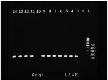
Fig. 1 - Agarose gel electrophoresis of PCR assays for the identification of (a) IS6110 gene. 1, DNA size marker ; 2, negative control; 3-9 and 11-13, positive samples (130bp); 14, positive control.
Fig. 2 - Agarose gel electrophoresis of PCR assays for the identification of (a) katG codon315- inhA- rpoB. 1, DNA size marker; 2-5, katG codon315 (250bp); 6 and 7, inhA (245bp); 8, negative control; 9-11, rpoB (270bp).
Amplification conditions for katG 315 genes were initial denaturation at 95°C for 5 min followed by 30 cycles of denaturation at 95°C for 15 s, annealing at 66°C for 15 s, and extension at 72°C for 15 s, and a final extension at 72°C for 5 min. The PCR amplification for the rpoB was in a similar manner except that the annealing temperature was lowered to 62°C.
Gene sequencing
PCR products were collected and sent for sequencing analysis at Bioneer Company, Korea.
Multiplex allele-specific PCR (MAS-PCR)
MAS-PCR method was used for simultaneous detection of the katG gene codon 315 and rpoB gene codons 516, 526, and 531, and mabA-inhA15. In this assay, in each allele-specific primer, 3′ end was located to pair with the base of the related codons where most point mutations have been found to compare with the wild-type sequences of strain H37Rv. Thus, the wild-type allele-specific fragment was amplified when no mutation existed at a related codon. No allele-specific PCR product was generated when there was a mutation at the targeted codons.
The amount of each pair of primers in MAS-PCR was balanced to achieve acceptable amplification of all target regions. For each MAS-PCR reaction, a standard 35 µL final reaction was used. Each final volume included 8 primers of [rpoB516 (20 pmol in 2 µL), rpoB526 (10 pmol in 1 µL), rpoB531 (30 pmol in 3 µL), RIRm (reverse primer for rpoB531,516,526) (50 pmol in 5 µL), katG OF (10 pmol in 1 µL), katG5R (10 pmol in 1 µL), inhAP-15 (10 pmol in 1 µL), and inhAPF (10 pmol in 1 µL)], 2 µL of genomic DNA (40ng) and 13 µL of AmpliTaq Gold master mix (ABI) (Table 2).18 The cycling parameters included an initial
denaturation at 96°C for 3 min, 27 cycles of 95°C for 50 s, 64°C for 40 s, and 72°C for 1 min, and a final extension at 72°C for 7 min. PCR products were analyzed by electrophoresis on 2.5% agarose gel and visualized with gel documentation.
Target allele-specific primers
Amplicon size (bp)
katG 315 (5R) 5’-ATACGACCTCGATGCCGC 292
katG 315 (OF) 5’-GCAGATGGGGCTGATCTACG
rpoB 516 5’-CAGCTGAGCCAATTCATGGA 218
rpoB 531 5’-CACAAGCGCCGACTGTC 170
rpoB 526 5’-CTGTCGGGGTTGACCCA 185
RIRm 5’-TTGACCCGCGCGTACAC
inhA P15 5’-GCGCGGTCAGTTCCACA 270
inhA PF2 5’-CACCCCGACAACCTATCG
Table 2 - Primers used in MAS-PCR for detection of INH and RIF resistance mutations14
Results
Results of PCR analysis
Results of DNA sequencing
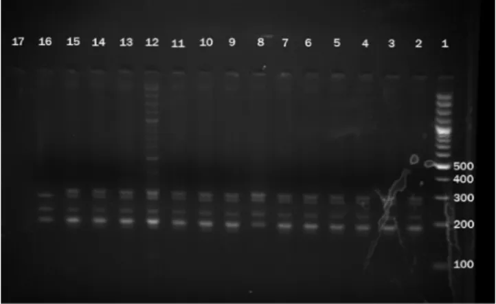
The sequencing of the 250 bp region of katG codon 315 revealed point mutations at 5 different codons in 13.7% of the 80 MTB isolates. The sequencing of the 270 bp central region of the rpoB gene revealed point mutations at 7 different codons in 12 (15%) of the 80 MTB isolates (Table 3). Among rpoB gene point mutations, in 2 isolates, mutation was observed in codon 531, causing TCG to TTG substitution, in 7 isolates mutation was Fig. 3 - Results of MAS-PCR shown by 2.5 % agarose gel electrophoresis. The 292-bp band: the katG codon 315-specific PCR product; the 218-bp band: rpoB codon 516-specific PCR product; the 270-bp band: the
15- promoter region of mabA-inhA–specific PCR product; the 185-bp band: rpoB codon 526-specific PCR product; the 170-bp band: rpoB codon 531-specific PCR product.
rpoB gene rpoB gene
AA (amino acid) Nucleotide AA (amino acid) Nucleotide
Isolate Codon Change Change Isolate Codon Change Change
54 512 Ser→Thr AGC→ACC 4 315 Ser→Thr AGC→ACC
39 527 Lys→Asn AAG→AAC 5 315 Ser→Thr AGC→ACC
38 512 Ser→Thr AGC→ACC 42 312 Ala→Glu GCG→GAG
38 517 Gln→Lu CAG→CCG 17 319 Ala→Val GCG→GTG
37 531 Ser→Lu TCG→TTG 18 328 Trp→Val TGG→GTG
31 512 Ser→Thr AGC→ACC 14 326 Thr→Pro ACG→CCG
42 527 lys→Asn AAG→AAC 13 12 Ala→Glu GCG→GAG
13 527 lys→Asn AAG→AAC 40 326 Thr→Gly ACG→CCG
37 531 Ser→ Lue TCG→TTG 13 315 Ser→Thr AGC→ACC
37 511 Lue→Met CTG→ATG 38 312 Ala→Glu GCG→GAG
18 527 lys→Asn AAG→AAC 59 319 Ala→Val GCG→GTG
50 527 lys→Asn AAG→AAC
52 527 lys→Asn AAG→AAC
Table 3 - Distribution of mutations associated with RIF and INH resistance among MTB isolates
observed in codon 527, causing AAG to AAC and in 3 isolates mutation was observed in codon 512, causing AGC to ACC substitution. One isolate had a mutation in codon 511 and one isolate had two mutations in codons 539 and 541 unexpectedly since these codons are out of the 81 bp region. No mutation was detected in the control RIF sensitive isolate (H37Rv). For INH, the most frequent mutation was observed in katG codons 315 and 312, in which a change from AGC to ACC and GCG to GAG was observed, respectively. No mutation was observed in the inhA promotor region or in the INH sensitive control isolate (H37Rv).
Results of MAS-PCR
Out of 10 isolates that proved to have different mutations in katG gene, 3 isolates which showed mutation in codon 315 by sequencing, were detected by MAS-PCR method. These were accounted for 30% of total katG mutations. The results of PCR-nucleotide sequencing and MAS-PCR assay were identical for the inhA gene (Fig. 3).
Similarly, among 12 isolates that proved to have different mutations in rpoB gene, 2 isolates had a mutation in codon 531 which were also detected by MAS-PCR.
Discussion
In this study, based on PCR and sequencing, the most common mutations related to drug resistance were demonstrated as RIF (15%) and INH (13.7%). The results were not in accordance with a previous report from Ahvaz19
in which the author observed that the most frequent mutations were substitutions in codons 516, 526 or 531 of the rpoB gene, which were found in 25 (83.3%) isolates. Mutations in codon 531 of the rpoB gene were detected in 16 (53.3%) of 30 RIF resistant isolates.
A similar study from the middle-east reported INH resistance in their isolates with all mutations at codon 315 of the katG gene. However, none of the reported mutations was AGC to ACC as found in our study.20
In the study of Yang et al.21 on the simultaneous
detection of INH and RIF in 174 clinical isolates of MTB in Turkey, distinct PCR banding patterns were observed for different mutation profiles and the correlation between MAS-PCR results and DNA sequencing was 99.4%. Other studies used multiplex PCR to detect the mutations in rpoB and katG genes in 20 drug-resistant isolates of MTB from the Southeast of Mexico. Sequencing analysis showed 93% mutations in the rpoB gene; of which 47% exhibited a mutation at 531 (S→L). Fifty-eight percent of their isolates showed mutations in katG; with 52% exhibiting a mutation at 315 (S→T).22 Also, the usefulness of MAS-PCR in detecting
mutations was reported in a study by Mokrousov et al.,23 in
which they were able to detect rpoB mutations at the rate of 82.8%. The reported mutations were in the rpoB 531, 526 and 516 codons. Similarly, in a study by Rathore et al.,24 four
mutations at rpoB codons 526, 531 and 12 were noticed in 16 RIF resistant isolates and 7 mutations were identified in INH resistant isolates in the katG codon 315.
In a study on the detection of katG and inhA gene mutations in MTB by multiplex allele-specific PCR, of 31 INH-resistant isolates, mutations in the katG codon 315 were identified in 64.5% of the isolates by sequencing. From these, 19 were detected by MAS-PCR methods.25
Our findings showed that most of the resistance mutations to INH occurred in codon 315 and this was in agreement with other mentioned studies showing the major involvement of this codon in INH resistance all over the world. However, a variety of involved codons was reported for RIF resistance among MTB isolates in different studies. In our study, the main codon involved in RIF resistance was 527, while similar studies reported several other codons for RIF resistance.
In conclusion, although culture based phenotypic susceptibility test is the gold standard for detecting drug resistance in MTB and no molecular method can yet completely replace it, MAS-PCR provides a rapid screening tool for the majority of mutations occurring in genes related to INH and RIF antibiotics. However, the molecular assays only detected known mutations, which is the most important limitation in detection of drug resistance by such techniques. Based on the other studies, all mutations related to anti-TB drugs are not known yet. Since the prevalence of mutations may vary by geographic area, identification of a resistance-associated mutation can be instructive, but lack of a mutation in the target sequence must be interpreted with caution.
Acknowledgements
This work is part of the MSc. thesis of Hamed Goodarzi approved in Infectious and Tropical Diseases Research Center (No. 88109). Special thanks to research affairs, Ahvaz Jundishapur University of Medical Sciences, Ahvaz, Iran, for the financial support. This work was financially supported by Grant No. 88109, Research affairs, Ahvaz Jundishapur University of Medical Sciences, Ahvaz, Iran.
Conflict of interest
All authors declare to have no conflict of interest.
R E F E R E N C E S
1. Zignol M, Hosseini MS, Wright A, et al. Global incidence of multidrug-resistant tuberculosis. J Infect Dis. 2006;194:479-85. 2. Shamaei M, Marjani M, Chitsaz E, et al. First-line
anti-tuberculosis drug resistance patterns and trends at the national TB referral center in Iran-eight years of surveillance. Int J Infect Dis. 2009;13(5):e 236-40.
3. World Health Organization. Anti-tuberculosis drug resistance in the world. Report no. 4. WHO/HTM/TB/2008.394. Geneva, Switzerland: WHO, 2008. http://www.who.int/tb/ publications/ 2008/drs_report4_26feb08.pdf Accessed July 2009.
4. Mori T. MDR-TB- its characteristics and control in Asia-Pacific rim symposium in USJCMSP 10th international conference on emerging infectious diseases in the Pacific rim. Tuberculosis. 2007;87(Suppl. 1):S5–9.
5. Espinal MA, Laszlo A, Simonsen L, et al. Global trends in resistance to antituberculosis drugs. N Engl J Med. 2001;344:1294–303.
6. Shi R, Otomo K, Yamada K, et al. Temperature-mediated heteroduplex analysis for the detection of drug-resistant gene mutations in clinical isolates of Mycobacterium tuberculosis by denaturing HPLC, SURVEYOR nuclease. Microbes Infect. 2006; 8:128–35.
7. Rozwarski DA, Grant GA, Barton DH, et al. Modification of the NADH of the isoniazid target (InhA) from Mycobacterium tuberculosis. Science. 1998;279:98–102.
8. Banerjee A, Dubnau E, Quemard A, et al. inhA, a gene encoding a target for isoniazid and ethionamide in Mycobacterium tuberculosis. Science. 1994;263:227–30. 9. Zhang Y, Telenti A. Genetics of drug resistance in
Mycobacterium tuberculosis. In: Hatfull G, Jacobs W R, eds. Molecular genetics of mycobacteria. Washington DC, USA: ASM Press, 2000: pp 235–54.
10. Hazbon MH, Brimacombe M, Bobadilla del Valle M, et al. Population genetics study of isoniazid resistance mutations and evolution of multidrug-resistant Mycobacterium tuberculosis. Antimicrob Agents Chemother. 2006;50:2640-9. 11. Kima S-Y, Parkb Y-J, Song E, et al. Evaluation of the
CombiChip Mycobacteriak Drug-Resistance detection DNA chip for identifying mutations associated with resistance to isoniazid and rifampin in Mycobacterium tuberculosis. Diag Microbiol Infect Dis. 2006;54:203–10.
13. Slayden RA, Barry III CE. The genetics and biochemistry of isoniazid resistance in Mycobacterium tuberculosis. Microbes Infect. 2000;2:659–69.
14. Hose KJP, Svastova P, Moravkova M, et al. Methods of mycobacterial DNA isolation from different biological material: A review. Vet Med. 2006;51:180-92.
15. Millera JM, Jenny AL, Payeur JB. Polymerase chain reaction detection of Mycobacterium tuberculosis complex and Mycobacterium avium organisms in formalin-fixed tissues from culture-negative ruminants. Vet Microbiol. 2002;87:15–23. 16. Hwang HY, Chang CH, Chang LL, et al. Characterization of
Rifampin – resistant M. tuberculosis in Taiwan. J Med Microbiol. 2003;52:239-45.
17. Baker LV, Brown TJ, Maxwell O, et al. Molecular analysis of isoniasid – resistant M. tuberculosis isolate from England and Wales reveals the phylogenetic significance of the ahpc – 46A polymorphism. Antimicrob Agents Chemother. 2005;49:1445-64. 18. Mokrousov I, Otten T, Filipenko M, et al. Detection of
isoniazid-resistant Mycobacterium tuberculosis strains by a multiplex allele-specific PCR assay targeting katG codon 315 variation. J Clin Microbiol. 2002;40:2509-12.
19. Doustdar F, Khosravi AD, Farnia P, et al. Mutations in rpoB gene and genotypes of rifampin resistant Mycobacterium tuberculosis isolates in Iran. MDR. 2008;7(2):11-7
20. Ahmad S, Mokaddas E. Contribution of AGC to ACC and other mutations at codon 315 of the katG gene in isoniazid-resistant Mycobacterium tuberculosis isolates from the Middle East. Int J Antimicrob Agents. 2004;23:473-9.
21. Yang Z, Durmaz R, Yang D, et al. Simultaneous detection of isoniazid, rifampin, and ethambutol resistance of
Mycobacterium tuberculosis by a single multiplex allele-specific polymerase chain reaction (PCR) assay. Diag Microbiol Infect Dis. 2005; 53:201-8.
22. Cuevas RZ, Zenteno JC, Cuellar A, et al. Mutations in rpoB and katG genes in Mycobacterium isolates from the Southeast of Mexico. Mem Inst Oswaldo Cruz. 2009;104(3):468-72. 23. Mokrousov I, Otten T, Vyshnevskiy B, et al. Allele-Specific
rpoB PCR assays for detection of rifampin-resistant Mycobacterium tuberculosis in sputum smears. Antimicrob Agents Chemother. 2003;47(7):2231-5.
24. Rathore M, Girish P, Jayalakshmi TK, et al. Rapid detection of multidrug resistant Mycobacterium tuberculosis by real time PCR based assay in Indian population. Rec Res Sci Tech. 2011;3(3):58-62.