Vol.53, n. 2: pp. 353-360, March-April 2010
ISSN 1516-8913 Printed in Brazil BRAZILIAN ARCHIVES OF
BIOLOGY AND TECHNOLOGY
A N I N T E R N A T I O N A L J O U R N A L
New Method for Early Detection of two Random Amplified
Polymorphic DNA (RAPD) Groups of
Staphylococcus aureus
Causing Bovine Mastitis Infection in Paraná State, Brazil
Dicezar Gonçalves1,2
*, Jane Eire Gabriel, Humberto Maciel França Madeira, Guilherme
Schnell e Schühli and Vânia Aparecida Vicente
1Divisão de Bioprocessos e Biotecnologia; Universidade Federal do Paraná; 81531-990; Curitiba - PR - Brasil.
2
Pontifícia Universidade Católica do Paraná; Curitiba - PR - Brasil. 3Universidade Estadual de Ponta Grossa Ponta Grossa - PR - Brasil
ABSTRACT
The aim of this work was to develop a fast and accurate molecular approach to allow early detection of two RAPD groups of S. aureus causing bovine mastitis. Seventy five S. aureus isolates from infected animals were characterized by RAPD. Genomic fragments isolated from the unique bands present in either group were cloned and sequenced. Based on the DNA sequences, specific primers were designed to allow for the simultaneous detection of either group by multiplex PCR of S. aureus DNA isolated from clinical and subclinical bovine mastitis. Results showed that these proposed primers set could be used to detect various clinical and subclinical S. aureus isolates as well as the detection of the microorganism in bulk milk. Their use as a specific method for effective and early diagnostic tool for S. aureus infection in dairy herds is suggested.
Key words:Staphylococcus aureus, molecular markers; subclinical mastitis, clinical mastitis, mastitis detection
*Author for correspondence: [email protected]
INTRODUCTION
The economic importance of the S. aureus causing clinical and subclinical bovine mastitis is largely recognized (Myllys et al, 1997). Clinical mastitis is the single most costly disease of dairy cattle resulting in the reduction of milk yield and quality (Riekerink et al, 2007a). The control of precalving subclinical mastitis by somatic cell count (SCC) is an indicator of intramammary infection (IMI) in bovines (Riekerink et al, 2007b). Udder edema and number of neutrophils in milk (Detilleux, 2004) are also important factors (Compton et al, 2007a). Precalving subclinical mastitis was diagnosed in
18.5% of quarters of the udder (Compton et al, 2007b) and increase in standard deviation log of SCC was associated with the presence of S. aureus
clinical mastitis in bovines (Green et al,(2004). Mastitis persists for long-term and may be detected by Pulsed Field Gel Eletrophoresis (Rabello et al, 2005; Anderson and Lyman, 2006).
Gonçalves, D., et al 354
U.S.A. and classified as either contagious or environmental based upon their primary reservoir and transmission mode (Parker et al, 2007).
S. aureus is a contagious pathogen commonly
transmitted among the cows by contact with infected milk and the infection reach up to 32% of
the herd (Pitkälä et al, 2004). This pathogen is particularly important because it causes mainly subclinical forms of infectious mastitis that are often difficult to detect by the herdsman (Compton et al, 2007b). The evolution of the infection turns difficult its treatment and increases the disease level (Reksen et al, 2006). Upon infection of the secretory tissue, the pathogen diffuses and encysts (Smith, 1977 and Hensen et al, 2000). This condition reinforces the necessity of early diagnosis to prevent the spread of the disease and management of the contaminated milk with S. aureus.
In Paraná State, Brazil, the estimated annual economic loss due to mastitis range from $95.62 to 142.42 per cow (Swinkels et al, 2005). These costs include reduced yield, discarded milk, drug therapy, veterinarian costs, premature culling, and increased labor. Fat and protein production is reduced by 9.0% in the clinical mastitis cases (Hagnestam et al, 2007).
Several biochemical and molecular methods are used in the investigation of S. aureus in bovine infections (Hartstein et al, 1989; Williams et al,
1990; Prevost et al, 1991; Kosteman et al, 1992 and Kapur et al, 1995). Current identification methods are based on microbiological culture of milk and biochemical tests on the isolated bacteria. At present, species identification by standard methods is laborious and takes at least 2 to 3 days to yield results. Other tests are commonly employed in Brazil to detect the occurrence of the subclinical mastitis, as California Mastitis Test, (Schalm and Noorlander, 1957; Riekerink et al, 2007b). However, since specific identification of the causal agent in Brazil is limited to tradicional methods, PCR-based approaches have been developed to identify the mastitis pathogens, which provides a promising alternative for the rapid identification of bacteria. In addition, the sensitivity of PCR-based assays tends to be better than bacterial cultures (Forsman et al, 1997; Kim et al, 2001) allowing the detection of small number of microorganisms.
These methods may be extremely important when rapid and accurate identification of pathogenic bacteria is required. Different PCR-based methods
have been developed for specific and sensitive detection of mastitis pathogens directly in milk. Forsman et al, (1997) identified by PCR test the
Staphylococcus spp. isolates in Russia, Germany and Denmark, but no cross reactivity could be observed investigating staphylococcal reference strains. Riffon et al, (2001) developed molecular probes reacting in PCR with bacterial DNA from bovine milk providing direct and rapid detection of
Escherichia coli, S. aureus, Streptococcus
agalactiae, S. dysgalactiae, S. parauberis and S. uberis, using universal primers in Canada. In this bacterial group, Meiri-Bendek et al, (2002) using 16S subunit of the rRNA genes, developed a specific PCR reaction to detect Streptococcus agalactiae in the milk of subclinical mastitis. Cremonesi et al, (2006) developed a method to extract DNA directly from the main Gram-positive bacteria known to cause bovine mastitis. Some alternative methods for diagnosis were also developed for detection in cows. Puetkes et al, (2001) and Eckersall et al, (2006) developed a multiplex PCR approach to detect the disease directly from milk cows or alternatively from dairy products, using mRNA 23S and genomic DNA. Youngerman et al, (2004) using CXCR2, a chemokine receptor required for neutrophil migration to infections sites and which contains single nucleotide polymorphisms (SNP) within the gene, showed association of CXCR2 genotypes, with subclinical and clinical mastitis. Ramesh et al, (2002) and Graber et al, (2007) obtained high correlation (R= 0.925), with the PCR methods to diagnose S. aureus mastitis. Gillespie and Oliver, (2005), using RT-PCR identified 91.7% of S. aureus in milk and Moon et al, (2007), based on coagulase gene polymorphism, observed relations with enterotoxins-producing by S. aureus strains. The objective of this work was to detect S. aureus
by means of multiplex PCR using a set of specific primers, by previous extraction of DNA, cloning and sequencing of monomorphic bands from isolated S. aureus DNA in order to make a rapid and specific identification of the microorganism.
MATERIALS AND METHODS
Samples were stored in 7.5% NaCl agar manitol at 4º C. They are cultivated in BHI broth at 37º C for 43 h. Genomic DNA was isolated by means of CTAB (cetiltrimetilamonium bromide),
(Dellaporta et al, 1983; Doyle and Doyle 1987; Daneshwar and Sher-Ullah, 2004).
Genetical variability and polymorphism analysis of 75 isolates of S. aureus
To carry out the Random Amplified Polymorphic DNA (RAPD) technique the following primers were used: OPJ 5 (5’CTC CAT GGG G 3’); OPJ 6 (5’ TCG TTC CGC A 3’); Primer 786 (5’ GCG ATC CCC A 3’); Primer 797 (5’ AGC GTC ACT G 3’) and Primer 798 (5’ TGA CCC GCC 3’), based in Myllys et al, (1997) and Pereira et al,
(2002) and OPA 2 (5’ TGC CGA GCT G 3’) (Spolidorio et al, 2003). The DNA amplifications reactions were made according Pereira et al,
(2002). A final volume of 14..9 µL contained 20ng of purified DNA and 1.0 U of Taq DNA polymerase, 3 mM 1-1 MgCl2, 50 mM 1-1 KCl, 10 mM 1-1 Tris-HCl (pH 8.8), 200 µM of Mix dNTP, and 0.5 mM of each primer. The mixture was subjected to a initial denaturation at 94 ºC for 5 min, followed by 45 cycles of 30 s at 94ºC, 30 s at 36ºC and 1 min 30 s at 72ºC. A subsequent final cycle of extension at 72ºC for 1 min was carried out in a thermocycler (PT100 Perkin Elmer). The amplification products were analyzed in 1.6% agarose gel (TBE buffer at 114 mV). Two strains
of S. aureus, ATCC 25923 and ATCC 6538 and
one out-group were also included (Streptococcus
spp.) as references strains. The polymorphism analysis of the isolated strains from the clinical and subclinical cases was carried out under
phenetical methods applying UPGMA
(Unweighted Pair Group Method with Arithmetic Mean) using the similarity coefficient of Jaccard in the NTSYS program, version 2.1 (Numerical Taxonomy and Multivariate Analysis System), (Rohlf, 2000). The support values of the groups were computed by bootstrap (Felsenstein 1986, 1993; Hillis and Bull, 1993).
Isolation of the RAPD genomic DNA fragments Two polymorphic and monomorphic bands were selected, corresponding to both mastitis cases types and the isolation of DNA fragments was due to the similarity in each group. The DNA fragments were cut and purified by the Purelink Quick Gel Extraction Kit (Invitrogen Life).
Fragment cloning in E. coli
Fragments were prepared according to the pGEM® Systems, (Promega Corporation) and cloning in the E. coli TOP10 F1 (Soares et al,
2001). For visualization of the possible insertion fragments in the plasmids, eletrophoresis in agarose gel (0.8 %) was carried out after the treatment with EcoRI restriction enzyme (InvitrogenLife).
AmpliSeq reactions
All the reactions were prepared with the Kit DYEnamicTM ET dye terminator MegaBACETM (Amersham). Universal primers used in the reactions for plasmid amplification were synthesized and purified by means of InvitrogenTM Life, according Murphy et al, (2005): pUC/M 13 Forward Sequencing Primer: 5’ CGT TGT AAA ACG ACG GCA AGT 3’ and pUC/M13 Reverse Sequencing Primer: 5’ TTT CAC ACA GGA AAC AGC TAT GAC 3’. Reaction systems consisted of 3.0 µL mix ET dye terminator; two primers in equal concentration 5-500 mol 1-1; dNTPs in a final concentration of 125 mol l-1, 50-500 mg l-1 of DNA in a final vol. of 7.5µL. All the reactions were cycled at 95ºC for 2 min, followed by 35 cycles at 95ºC for 20 s and 60ºC for 2 min. The PCR product was purified by ammonium acetate 7.5 mol 1-1 and precipitated by ethanol in successive centrifugations (Murphy et al, 2005). Samples were sequenced by electrophoresis in an ABI 377 (Applied Biosystems). The primers were designed considering the sequences obtained.
Sequences edition and primer design
Edited sequences (GenBank: Access AM749799
to S. aureus RAPD clinic fragment and
Gonçalves, D., et al 356
AGC TTC ACG CCG AG 3' with an optimum annealing temperature of 62.72 ºC.
Markers evaluation and protocol development A multiplex PCR according to Vandercan et al,(1995), Pinekter et al, (2001) and Vannuffel et al, (2003) was prepared applying the proper annealing temperature suggested by the software (PerlPrimer) with a final volume of 25.0 µL. The reaction comprised 2.5 µL of PCR buffer minus Mg (final concentration 1X), 1.0 µL of mix dNTP’s (final concentration of 0.2 mM), Primer mix in final concentration of 0.5 µM, 2.5U of Taq, 25.0 ng of genomic DNA (0.5 µl) and 17.5 µL of Milli Q filtered water. The amplifications were preceded with 35 cycles with denaturation of 45s at 94ºC, annealing of 45s at 60ºC and extension of 2 min at 72ºC. The multiplex PCR products were examined with agarose gel 1%.
RESULTS AND DISCUSSION
Strain recognition and DNA isolation
In the present work, 31.25% of the mastitis cases sampled were confirmed as S. aureus infection by morphological and biochemical analysis (Gonçalves et al 2001). Several procedures for the early identification and fast recognition of S. aureus infections are essential for health studies (both animal and human) due to economical
importance in dairy industry. Amplification analysis allowed the identification of two main groups (Fig.1) based on the presence of distinct monomorphic bands. These bands were fully congruent with the identification of the clinic and subclinical strains of S. aureus.
A multiplex PCR according to Vandercan et al, (1995), Pinekter et al, (2001) and Vannuffel et al, (2003) was prepared applying the proper annealing temperature suggested by the software (PearlPrimer) with a final volume of 25.0 µL. The reaction comprised 2.5 µL of PCR buffer minus Mg (final concentration 1X). 1,0 µL of mix dNTP’s (final concentration of 0.2 mM), Primer mix in final concentration of 0.5 µM, 2.5U of Taq, 25.0 ng of genomic DNA (0.5 mL) and 17.5 µL of Milli Q filtered water. The amplifications were proceeded with 35 cycles with denaturation of 45s. The subclinical sample displayed a 950 bp band while the subclinical sample was smaller 800 pb band (Fig. 2).
In addition to its fast performance, the RAPD method assures a high confidence in its diagnosis, being very sensitive to S. aureus studied strains. RAPD data allowed a clear distinction between two clades in the identification of the strains (Fig.1). The identification was fully congruent with the clinical picture noted in the studied infections. Each group of strains might correspond to one cluster of mutually similar genotypes.
Figure 2 - Size and intensity (in maximum absorbance) by samples proceeding from clinical and subclinical mastitis. Bands were eluted with Purelink kit. A star indicates the position of band peak of interest (around of 950 for the subclinical sample and 800 for the clinical).
AmpliSeq reactions
Both inserts were effectively amplified with the proper PCR reaction suggested by the plasmid manufacturer protocol (pGEM® Vector Systems). The results are presented in Fig 3. The two
distinctive monomorphic bands were recovered displaying the same size of the original excised bands (950 bp for the subclinical sample and 800 bp for the clinical) (Fig.3).
Figure 3 - Positively cloned strains in agarose gel (1%). Ld = ladder (1 kbases). The 3 kbases bands correspond to the plasmid pGEM®-T Easy Vector previously digested by EcoRI.
Multiplex PCR
Amplification of the desired regions starting from genomic DNA was always successful using the four designed primers. It was equally effective for single PCR or multiplex PCR reactions carried out
Gonçalves, D., et al 358
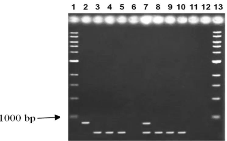
Figure 4 - Multiplex PCR results in 1% agarose gel showing amplifications of genomic DNA. Lanes: 1=Ladder 1 kb; 2=subclinic strain; 3=clinic strain; 4 and 5= samples containing clinic strains, proceeding of illness cases; 6=empty lane; 7=subclinic and clinic strains observed in milk tank with capacity of 5.000L of milk; 8=clinic strain observed in milk tank with capacity of 500L of milk; 9=ATCC 25923; 10=ATCC 6538; 11=Streptococcus mutans; 12=Escherichia coli; 13=Ladder 1 kb.
In this study, 75 field S. aureus were identified using the set of four primers resulting in amplifying the desired fragment and the reference strains ATCC 25923 and ATCC 6538 samples. Also, S. aureus in milk samples collected directly from the milk bulk tank according to Kim et al,
(2001), (for total DNA extraction) were positively detected. To assure the specificity of the primers, the amplification of S. mutans and E. coli was
attempted but the primers failed in their
amplification. The development of a fast diagnosis tool of the subclinical strain was priority. The multiplex PCR approach allowed the detection of both types of strains. The method was able to detect successfully S. aureus contamination even in high-diluted milk samples as the ones taken from a 5000L bulk milk tank. These results suggested another useful application to detect eventual contaminations in the stored milk.
This protocol could be used as a quality inspection tool for producers and in the chain of distribution, contributing to healthy herds and high ranks of quality for the milk producers.
ACKNOWLEDGMENTS
The authors are grateful to Dr. Emanuel M. de Souza, Mr. Helisson Faoro and Mr. Valter Baura (Biochemistry Department to UFPR) for their
technical assistance. Thanks are due to PUCPR and UFPR, for the financial support.
RESUMO
Esta pesquisa objetivou o desenvolvimento de técnica rápida e eficiente para diagnosticar precocemente diferentes linhagens de S. aureus
REFERENCES
Anderson, K. L. and Lyman, R.L. (2006), Long -Term Persistence of Specific Genetic Types of Mastitis-Causing Staphylococcus aureus on Three Dairies. J. Dairy Sci., 89, 4551-4556.
Compton, C.W.R.; Heuer, C.; Parker, K.; McDougall, S. (2007a), Risk Factors for Peripartum Mastitis in Pasture-Grazed Dairy Heifers. J. Dairy Sci., 90, 4171-4180.
Compton, C. W.R.; Heuer, C.; Parker, K.; McDougall, S. (2007b), Epidemiology of Mastitis in Pasture-Grazed Peripartum Dairy Heifers and Its Effects on Productivity. J. Dairy. Sci., 90, 4661-4669.
Cremonesi, P.; Castiglioni, G.; Malferrari, I.B. (2006), Improved method for rapid DNA extraction of mastitis pathogens directly from milk. J. Dairy Sci.,
89, 163-169.
Cullor, J.S. (1992), Mastitis in dairy cows. Does it hinder reproductive performance? Vet. Med., 8, 830-835.
Daneshwar, P. and Sher-Ullah, S.J. (2004), Genomic Extraction From Victoria amazonica. Plant. Mol. Biol. Rep. , 22, 195-196.
Dellaporta, S. L.; Wood, J.; Hicks, J.B. (1983), A plant DNA minipreparation: versión II. Plant Mol Biol Rep., 1, 19-21.
Detilleux, J. C. (2004), Neutrophils in the War against
Staphylococcus aureus: Predator-Prey Models to the Rescue. J. Dairy Sci., 87, 3716-3724.
Doyle, J.J. and Doyle, J.L. (1987), A rapid DNA isolation procedure for small quantities of fresh leaf tissue. Phytochem. Bull., 19, 11-15.
Eckersall, P.D.; Young, F. J.; Nolan, A. M.; Knight, C. H.; Mc Comb, C.; Waterston, M. M.; Hogarth, C. J.; Scott, E. M.; Fitzpatrick, J. L. (2006), Acute Phase Proteins in Bovine Milk in Experimental Model of
Staphylococcus aureus Subclinical Mastitis. J. Dairy Sci ., 89, 1488-1501.
Felsenstein, J. (1986–1993), PHYLIP (Phylogeny Inference Package) version 3.5c. (DNAPARS)-DNA Parsimony Program. Seattle Distributed by the author. Department of Genetics, University of Washington.
Forsmann, P.; Tilsala-Timisjarvi, A.; Alatossava, T. (1997), Identification of staphylococcal and streptococcal causes of bovine mastitis using 16S-23S rRNA. Microbiol., 143, 3491-3500.
Gillespie, B. E. and Oliver, S. P. (2005), Simultaneous Detection of Mastitis Pathogens, Staphylococcus aureus, Streptococcus uberis and Streptococcus agalactiae Multiplex Real-Time Polimerase Chain Reaction. J. Dairy Sci., 88, 3510-3518.
Gonçalves, D. and Kozicki, L .E. (1997), Biochemical and immunological profiles during the parturition period in dairy cows, with and without placental retention and mastitis. Braz. J. Vet. Res. Anim. Sci.,
34 (6), 371-382.
Graber, H. U.; Casey, M. G.; Naskova, J.; Steiner, A.; Schaeen, W. (2007), Development of a Highly Sensitive and Specific Assay to Detect
Staphylococcus aureus in Bovine Mastitic Milk. J. Dairy Sci., 90, 4661-4669.
Green, M. J.; Green, L. E.; Schukken, Y. H.; Bradley, A. J.; Peeler, E. J.; Barkema, H. W.; Haas de, Y.; Collis, V. J.; Medley, G. F. (2004), Somatic Cell Count Distributions During Lactation Predict Clinical Mastitis. J. Dairy Sci., 87, 1256-1264.
Hagnestam, C., Emanuelson, U., Berlung, B. (2007), Yield Losses Associated with Clinical Mastitis Occuring in Different Weeks of Lactation. J. Dairy Sci., 90, 2260-2270.
Hartstein, A.I.; Morthland, V.H.; Eng, S. (1989), Restriction enzymes analysis of plasmid DNA and bacteriophage typing of paired S. aureus blood culture isolates. J. Clin. Microbiol , 27, 1874-1879. Hensen, S.M.; Pavičic, M.J.A.M.P.; Lohuis, J.A.C.M.;
Poutrel, B. (2000), Use of Bovine Primary Mammary Epithelial Cells for the Comparison of Adherence and Invasion Ability of Staphylococcus aureus Strains. J. Dairy Sci., 83 (3), 418-429.
Hillis,D.M and Bull, J.J (1993), An empirical test of bootstrapping as a method for assessing confidence in phylogenetic analysis. Syst. Biol.,42, 17-48.
Kapur,V.; Sischo, W.M.; Green, R.S. (1995), Molecular population genetic analyses of Staph. aureus
recovered from cows. J. Clin. Microbiol., 33, 376-380.
Kim,C.H.; Khan, M.; Morin, D.E.; Hurley, W.L.; Tripathy, M.; Kehrli Jr, M.; Oluocht, A.O.; Kakoma, I. (2001), Optimization of the PCR for detection of
Staph. aureus nuc. Gene in the milk. J. Dairy Sci., 84, 74-83.
Koneman, E.W.; Allen, S.D.; Janda, W.M.; Schreckenberger, P.C.; Wn, W.C. (2001), In Diagnóstico Microbiológico- Texto e Atlas Colorido. 5. ed. MEDSI, 11-16.
Kosteman, J.R.; Edling, T.D.; Lipuma, J.J.; Stull, T.L. (1992), Molecular Epidemiology of pseudomonas cepacia determined by polymerase chain reaction ribotiping. J. Clin. Microbiol., 30, 2004-2087. Marshall, O.J. (2004), PerlPrimer: cross-platform,
graphical primer design for standard, bisulphite and real-time PCR. Bioinformatics, 20, 2471-2472 Meiri-Bendek, I.; Lipkin, E.; Friedman, A.; Leitner, G.;
Saran, A.; Friedman, S.; Kashi, Y. (2002), A PCR-based method for the detection of Streptococcus agalactiae in milk. J. Dairy Sci., 85, 1717-1723. Moon, J. S.; Lee, A.R.; Kang, H. M.; Lee, E. S.; Joo, Y.
Gonçalves, D., et al 360
Murphy, K.M.; Berg, K.D.; Ehleman, J.R. (2005), Sequencing of Genomic DNA by Combined amplification and Cycle Sequencing reaction. Clin. Chem., 51 (1), 35-39.
Myllys, V.; Ridell, J.; Bjorkroth, J.; Biese, I.; Pyorala, S. (1997), Persistence in bovine mastitis of Staph. aureus clones as assessed by random amplified polymorphic DNA analysis, ribotyping and biotyping. Vet. Microbiol., 51, 245-251.
Parker, K.I.; Compton, C.; Anniss, F.M.; Weir, A.; Heuer, C., Mc Dougall, S. (2007), Subclinical and Clinical Mastitis in Heifers Following the Use of a Teat Sealant Precalving. J. Dairy Sci., 90, 207-218. Pereira, M.S.V.; Leal, N.C.; Leal, T.C.A.; Sobreira, M.;
Almeida, A.M.P.; Siqueira-Júnior, J.P.; Campos-Tadaki, G.M.(2002), Typing of human and bovine
Staph. aureus by RAPD and ribotypyng-PCR. Lett. App. Microbiol., 35, 32-36.
Pinekter, P.; Mansell, P.D.; Browning, G.F. (2001), Multiplex Polymerase Chain Reaction Assay for Simultaneous Detection of Staph. aureus and Streptococcal Causes of Bovine Mastitis. J. Dairy Sci., 84, 1140-1148.
Pitkälä, A.; Haveri, M.; Pyörälä, S.; Myllys, V.; Honkanen-Buzalski, T. (2004) Bovine Mastitis in Finland 2001 – Prevalence, Distribution of Bacteria, and Antimicrobial Resistence. J. Dairy Sci., 87, 2433-2441.
Prevost, G.; Pottecher, B.; Dalhet, M. (1991) Pulsed-field gel Electrophoresis as a new epidemiological tool for monitoring methicilin- Resistant Staph. aureus in a intensive care unit. J. Hosp. Infect. (17), 255-259.
Puektes, P.; Mansell, P. D.; Browning, G. F. (2001), Multiplex Polymerase Chain Reaction Assay for simultaneous Detection of Staphylococcus aureus and Streptococcal Causes of Bovine Mastitis. J. Dairy Sci., 84 (5), 1140-1148.
Rabello, R. F., Souza, C.R.V.M., Duarte, R. S., Lopes, R. M.M.; Teixeira, L. M.; Castro, A. C. D. (2005), Characterization of Staphylococcus aureus Isolates Recovered from Bovine Mastitis in Rio de Janeiro, Brazil. J. Dairy Sci.,88, 3211-3219.
Ramesh, A.; Padmapriya, B. P.; Chandrashekar, A.; Varadaraj, M. C. (2002), Application of a convenient DNA extraction method and multiplex PCR for the direct detection of Staphylococcus aureus and
yersinia enterocolitica in milk samples. Mol. Cell. Prob., 16, 307-314.
Reksen, O.; Sølverød, L.; Branscum, A.J.; Østerås (2006), Relationship Between Milk Culture Results and Treatment for Clinical Mastitis or Culling in Norwegian Dairy Cattle. J. Dairy Sci., 89, 2928-2937.
Riekerink, R.G.M.; Barkema, H.W.; Stryhn, H. (2007a), The Effect of Season on Somatic Cell Count
and the Incidence of Clinical Mastitis. J. Dairy Sci.,
90, 2260-2270.
Riekerink, R. G. M.; Barkema, H. W.; Veenstra, W.; Berg, F. E.; Stryhn, H.; Zadoks, R. N. (2007b), Somatic Cell Count During and Between Milkings. J. Dairy Sci , 90, 3733-3741.
Riffon, R.; Sayasaith, K.; Khalil, H.; Dubreuil, P.; Drolet, M.; Lagacé, J. (2001) Development of a rapid and sensitive test for identification of major pathogens in bovine mastitis by PCR. J. Clin. Microbiol., 39 (7), 2584-2589.
Rohlf, F.J. (2000), NTSYS-PC, Numerical taxonomy and multivariate analysis system – Version 2.1. New York, Applied Biostatistic Inc., 44 p.
Schalm, O. W. and Noorlander, D. O. (1957), Experiments and observations leading to development of the California mastitis test. J. Am. Vet. Med. Assoc., 130- 199.
Smith, H. (1977), Microbial Surfaces in Relation to Pathogenicity. Bacteriol. Rev.,41 (2), 475-500. Soares, M. J. S.; Teixeira, L. A.; Nunes, M. R.; Carvalho, M.
C. S.; Ferreira-Carvalho, B. T.; Figueiredo, A. M. S. (2001) Analysis of different molecular methods for typing methicilin-resistent Staphylococcus aureus isolates belonging to the Brazilian epidemic clone. J. Med. Microbiol., 50, 732-742.
Spolidorio, D.M.P.; Hofling, J.F.; Pizzolito, A. C.; Rosa, E. A.; Negrini, T. C.; Spolidorio, L. C. (2003), Genetic polymorphism of Streptococcus mutans in brazilian family members. Braz. J. Microbiol. 34, 3, 213-217.
Swinkels, J.M.; Hogeveen, H.; Zadoks, R.N. (2005), A partial Budget Model to Estimate Economic Benefits of Lactational Treatment of Subclinical
Staphylococcus aureus Mastitis. J. Dairy Sci., 88, 4273-4287.
Vannuffel, P.; Gigi, J.; Ezzedne, H.; Sumer, A.; Daneshwar, P.; Sher-Ullah, S.J. (2003), Isolation of DNA for RAPD Analysis from Dry Leaf Material of Some Hisperis L. Specimens. Plant. Mol. Biol. Rep.,
21, 461-462.
Vandercam, B.; Delmel, M.; Wanters, G.; Gala, J.L. (1995), Especific Detection of Methicilin-Resistant
Staph. Species by Multplex PCR. J. Clin. Microbiol.,
33 (11), 2864-2867.
Williams, J.G.K.; Kubelik, A.R.; Livak, K.J. (1990), DNA polymorphisms amplified by binary primers are useful as genetic markers. Nucl. Ac. Res., 18, 6531-6535.
Youngerman, S. M.; Saxton, A. M.; Oliver, S. P.; Pighetti, G. M. (2004), Association of CXCR2 Polymorphism with Sub clinical and Clinical Mastitis in Dairy Cattle. J. Dairy Sci., 87, 2442-2448.