Ca
2+
signaling in pancreatic acinar cells:
physiology and pathophysiology
O.H. Petersen
MRC Group, Physiological Laboratory, School of Biomedical Sciences, University of Liverpool,
Liverpool, UK
Correspondence to: O.H. Petersen, MRC Group, Physiological Laboratory, School of Biomedical
Sciences, University of Liverpool, Liverpool, UK
E-mail: o.h.petersen@liv.ac.uk
The pancreatic acinar cell is a classical model for studies of secretion and signal transduction mechanisms. Because of the extensive endoplasmic reticulum and the large granular compartment, it has been possible - by direct measurements - to obtain considerable insights into intracellular Ca2+ handling under both normal and pathological conditions. Recent studies have also revealed important characteristics of stimulus-secretion coupling mechanisms in isolated human pancreatic acinar cells. The acinar cells are potentially dangerous because of the high intra-granular concentration of proteases, which become inappropri-ately activated in the human disease acute pancreatitis. This disease is due to toxic Ca2+ signals generated by excessive liberation of Ca2+ from both the endoplasmic reticulum and the secretory granules.
Key words: Ca2+ signaling; Pancreatic acinar cells; Stimulus-secretion coupling; Human acinar cells; Ca2+ entry; Ca2+ extrusion Presented at the IV Miguel R. Covian Symposium, Ribeirão Preto, SP, Brazil, May 23-25, 2008.
Received November 5, 2008. Accepted December 16, 2008
Introduction
The crucial role of changes in the cytosolic Ca2+ con-centration ([Ca2+]
i) for the control of muscle contraction and relaxation was well understood already in the 1960’s (1). However, a proper appreciation of the role of Ca2+ in the regulation of secretion from exocrine glands only be-gan to emerge in the 1970’s (Figure 1). Selinger and colleagues (2) from Israel and Alonso and colleagues (3) from Argentina demonstrated ATP-dependent Ca2+ up-take into microsomal vesicular preparations from salivary gland cells and thereby established that there was a source of intracellular non-mitochondrial Ca2+ from which Ca2+ could in principle be released for signaling purposes. Shortly thereafter, Nielsen and I (4) - working at that time in Copenhagen, Denmark - demonstrated that the physi-ological stimulants of salivary secretion, acetylcholine (ACh) and adrenaline, evoked release of Ca2+ from internal stores in the salivary gland cells and we proposed that the crucial store was in the endoplasmic reticulum (ER). Together with Matthews and Williams (5) in Cambridge, England, I
obtained similar results in studies of the exocrine pan-creas, and Case and Clausen (6) - working at that time in Denmark - also produced the same type of evidence.
After several years of confusion about whether pancre-atic enzyme secretion is Ca2+-dependent or -independent, it became clear that there is an initial phase of stimulant-evoked enzyme secretion that is completely independent of extracellular Ca2+, whereas the following phase of sus-tained secretion depends acutely on the presence of exter-nal Ca2+ (7). The initial phase of stimulant-evoked enzyme secretion follows very closely after the intracellular Ca2+ release (5) and data obtained later, utilizing patch clamp membrane capacitance measurements (8), showed that the exocytotic release has a time course very similar to the time course - during a single Ca2+ spike - of the change in [Ca2+]
i near the apical membrane through which secretion occurs (Figure 2). These findings provide the basis for our current concept of stimulus-secretion coupling in exocrine glands.
mechan-isms underlying physiological and pathological Ca2+ sig-naling in the pancreatic acinar cells, because several such review articles have been published recently (9-11). The purpose of this article is to focus attention on a couple of critical questions, which have received recent attention.
Why is the sustained phase of enzyme
secretion acutely dependent on external Ca
2+?
Considering the existence of large intracellular Ca2+ stores in pancreatic acinar cells and the evidence that Ca2+ is released from such stores upon stimulation with physi-ological stimulants (for example, ACh), it is not immedi-ately obvious why secretion should depend on external Ca2+. One could easily imagine that Ca2+ could be released upon stimulation and then taken up again into the stores when stimulation ceases. However, the pancreatic acinar cell - like all other cells - has Ca2+ pumps in the plasma membrane (plasma membrane Ca2+activated ATPase -PMCA) and these pumps increase the rate of Ca2+ extru-sion as soon as [Ca2+]
i increases from the basal resting level (12). In fact, the rate of PMCA-mediated Ca2+ extru-sion is sufficient to pump out all the Ca2+ released into the cytosol within a few minutes, when the ER is emptied during maximal stimulation (11,12).
Figure 3 shows a simplified diagram in which the major shifts in Ca2+ handling during the transitions from rest to activity and back again to rest are depicted. The initial
sharp rise in [Ca2+]
i (monitored by changes in the Ca2+ -activated Cl- current) is due to opening of Ca2+ release channels (predominantly inositol trisphosphate receptors (IP3Rs) in the ER, but also involving ryanodine receptors and non-ER acidic Ca2+ stores) (for a recent review, see Ref. 11). The subsequent decline in [Ca2+]
i is principally due to PMCA-mediated Ca2+ extrusion, since the Ca2+ pumps in the ER (sarco(endo)plasmic reticulum Ca2+ -activated ATPase - SERCA) have little impact as long as the Ca2+ release channels are open. During the sustained phase of stimulation, when [Ca2+]
i is stable at a moderately elevated level, there is again equilibrium - as in the resting state - with Ca2+ influx from the external solution into the ER matching the release from the ER and extrusion across the plasma membrane, but the Ca2+ fluxes are much higher in the stimulated state than under resting conditions (compare scenarios [1] and [2] in Figure 3). When stimula-tion ceases, the Ca2+ release channels close and now SERCA has a significant impact, causing net Ca2+ reup-take into the stores (scenario [3] in Figure 3). The interest-ing point here is that, whereas [Ca2+]
i returns to the resting level very quickly upon cessation of stimulation, it takes a long time before the ER is replenished with Ca2+ (Figure 3). This reuptake of Ca2+ into the ER can be blocked by the specific SERCA pump inhibitor thapsigargin and is com-pletely dependent on the presence of external Ca2+ (13,14). This is in complete accord with the finding that all the Ca2+ released from the intracellular stores is pumped out of the
Figure 1. Figure 1. Figure 1. Figure 1.
Figure 2. Figure 2.Figure 2. Figure 2.
Figure 2. Basic aspects of pancreatic acinar stimulus-secretion coupling. A, Simultaneous measurements in isolated pancreatic fragments of intracellular Ca2+ release (assessed by fractional efflux of pre-loaded radioactive calcium) and amylase secretion assessed by an on-line fluorescence-based technique (adapted from Ref. 5). B, Ca2+-dependence of acetylcholine (ACh; 1 μ M)-induced amylase secretion from isolated pancreatic fragments. Initially, the fragments were superfused by a Ca2+-free solution, then (during continued ACh stimulation) Ca2+ was added to the inflowing solution. Finally, ACh was removed (adapted from Ref. 7). C, Simultaneous conductance and capacitance measurements from a single isolated pancreatic acinar cell. The changes shown represent a single spike taken from a train of such events elicited by intracellular IP3 infusion. The conductance trace (∆G) refers to the Ca2+-activated Cl- conductance and therefore shows the time course of the [Ca2+]i change. The capacitance trace (∆C) monitors insertion and retrieval of granule membrane (adapted from Ref. 8).
cell by PMCAs and therefore has to re-enter the cell in order to refill the ER (11,13). Finally, after several minutes of active Ca2+ reuptake into the ER we come back to the pre-stimulation resting level (scenario [4] in Figure 3).
How does Ca
2+enter acinar cells during the
sustained phase of stimulation?
After many years of uncertainty and debate about the molecular mechanism responsible for Ca2+ entry second-ary to depletion of intracellular stores, a consensus has now emerged. There is a sensor, STIM1, in the ER mem-brane that monitors [Ca2+] in the ER lumen ([Ca2+]
Lu). When Ca2+ is released from the ER, in response to stimu-lation, [Ca2+]
Lu falls and this triggers translocation of STIM1 to the so-called puncta in the ER membrane. These puncta are localized in parts of the ER very close to the plasma membrane. During ER Ca2+ depletion, STIM1 is localized in the puncta together with the Ca2+ entry channel CRACM1 (also known as Oral1) (15), which is the classical
well-characterized and very Ca2+-selective, Ca2+ release-acti-vated Ca2+ channel (16). We have recently demonstrated STIM1 translocation, triggered by ER Ca2+ depletion, to puncta very close to the plasma membrane and co-local-ized with Oral1 in pancreatic cells (pancreatic cell line, PANC1). Furthermore, we have shown that this transloca-tion does not require ATP (17).
What is physiological stimulation?
In experiments of the type shown in Figures 2B and 3, sustained stimulation with μM concentrations of ACh was used. Is this physiological? Almost certainly not! Prolonged stimulation of this intensity will destroy the acinar cells (18). Unfortunately, we do not have any direct evidence relating to the actual ACh concentrations attained during physiological nerve stimulation near the muscarinic recep-tor sites on the baso-lateral acinar membranes. In order to answer the question about the type of stimulation that could be regarded as physiological, it may therefore be
Relative fluorescence
50
40
30
20
10
0
0.05
0.04
0.03
0.02
0.01
0
A
B
C
Ca2+-free
Ca2+
ACh 10
U/mL
5
0 10 min
10 min ACh
Amylase secretion
45Ca2+
efflux
45
Ca
2+
fractional efflux
3 nS
800 fF
∆G
∆C
better to consider stimulation with the circulating hormone cholecystokinin (CCK). From animal experiments, as well as experiments on man, we have good data about the CCK levels in plasma after a normal meal. They are in the range 1-10 pM (19). When pancreatic acinar cells are exposed to such concentrations of CCK, there is no sustained eleva-tion of [Ca2+]
i, but rather repetitive short-lasting [Ca2+]i spikes. These are mostly local, in the apical granule-containing region, but do occasionally spread out to the whole cell (20,21). In order to obtain a sustained global elevation of [Ca2+]
i, of the type shown in Figure 3, it is necessary to use nM concentrations of CCK, completely outside the physiological range of this hormone (18,19).
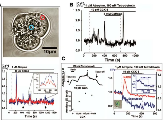
These considerations indicate that physiological stimuli, in general, evoke repetitive, short-lasting and local [Ca2+] i elevations and that sustained (and inevitably global) [Ca2+] i elevations are pathological phenomena. However, the rel-evance of the CCK data described above to human biology and medicine has been disputed, because of a study indicating that human pancreatic acinar cells lack func-tional CCK receptors (22,23). We therefore re-investigated this question, taking advantage of a long-established and well-functioning collaboration with the Division of Surgery and Oncology at the Royal Liverpool Hospital, which enabled us to transfer normal human pancreatic tissue -obtained from patients (with full informed consent) under-going surgery for treatment of pancreatic cancer - very quickly to our laboratory for isolation of acinar cells and cell clusters (24). As shown in Figure 4, these isolated human acinar cells were polarized, exactly like mouse and rat
[1] [Ca 2+] Lu µM 150 0 120 90 60 30 Ca2+ SERCA
[2] [3] [4]
0 50 100 150 200 250
Time (s)
10 µM ACh 10 µM ACh
CCE
Ca2+-activated Cl- current
PMCA IP3R
Ca2+ Ca2+ Ca2+ Ca2+ Ca2+ Ca2+ Ca2+ [1] [2] [3] [4]
0 50 100 150 200 250
Time (s) pA 0 -200 -50 -100 -150
cells, and produced repetitive short-lasting [Ca2+]
i spikes in response to stimulation with physiological (pM) concentra-tions of CCK. These spikes were mixtures of local (in the apical pole) and global [Ca2+]
i transients and - similar to what had originally been found in the mouse acinar cells (20,21), even the global [Ca2+]
i elevations always started in the apical (granular) pole (Figure 4A). These repetitive [Ca2+]
i spikes were able to generate appropriate secretory and metabolic responses, as assessed by several differ-ent methods (Figure 4).
We can therefore conclude that physiological stimuli evoke repetitive [Ca2+]
i spikes, that these spikes often only represent local [Ca2+]
i elevations in the apical granular pole and that global [Ca2+]
i elevations have the character of [Ca2+]
i waves that always begin in the apical pole and then spread out towards the base of the cell.
Why do sustained global [Ca
2+]
i
elevations
cause necrosis?
If sustained stimulation of the type shown in Figure 3 -with almost complete emptying of the ER - is allowed to continue for more than 15-20 min, activation of intracellular trypsin occurs (18,25). This initiates the very dangerous autodigestion process, which causes the often fatal hu-man disease acute pancreatitis (10). Although hyperstimu-lation (stimuhyperstimu-lation of the pancreas with unphysiologically high concentrations of ACh or CCK) does experimentally evoke changes in mouse pancreatic acinar cells that are very similar to those observed in human pathology, it is
Figure 3. Figure 3. Figure 3. Figure 3.
the wrong kind of Ca2+ signal, namely a global sustained elevation of [Ca2+]
i, rather than the physiologically impor-tant repetitive local Ca2+ spikes in the secretory pole that control normal secretion.
Since intracellular trypsin activation is regarded as the most important process initiating acute pancreatitis, it is essential to understand in which intracellular compartment this occurs. Recent work from my laboratory demonstrated that trypsin activation, elicited by a prolonged elevated [Ca2+]
i, occurs in post-exocytotic endocytic vacuoles (30). Taken together with the well-established fact that hyper-stimulation only produces very little secretion and that one of the characteristics of acute pancreatitis is a secretion clear that hyperstimulation of this type is not responsible
for acute pancreatitis. Pancreatitis is typically caused by alcohol abuse or complications arising from biliary disease (gall stones) (10). It turns out that non-oxidative alcohol metabolites, namely fatty acid ethyl esters, and bile acids are capable of eliciting sustained global [Ca2+]
i elevations and necrosis (26-28). This necrosis is Ca2+-dependent, since it can be prevented by incubating the acinar cells with membrane-permeant Ca2+ chelators (27). Although there are no doubt many factors that play a role in the disease development (29), the crucial point is that acute pancreatitis is one of the clearest examples known of [Ca2+]
i toxicity. Basically, acute pancreatitis is caused by Figure 4.
Figure 4.Figure 4. Figure 4.
defect (31), these data lead us to conclude that prolonged global [Ca2+]
i elevation fails to induce sustained secretion. At this point in time, the nature of this secretion defect is not entirely clear. Hyperstimulation clearly elicits insertion of granule membrane into the apical plasma membrane, since endocytosis of a fluorescent extracellular fluid phase marker was observed in our experiments (30). This indicates that hyperstimulation induces formation of a fusion pore large enough to allow molecules of the size of, for example, Texas Red Dextran to be taken up. Since there is little real protein secretion in acute pancreatitis, it would appear that the fusion pore is insufficiently large to allow the passage of large protein molecules. It is now becoming clear that fusion pore expansion is an important part of the secretion process (32) and it is a plausible hypothesis, which still needs to be critically tested, that it is the fusion pore expansion that is defective during conditions leading to acute pancreatitis.
Is the ER the only intracellular store from
which Ca
2+can be released?
The mechanism by which trypsin activation occurs in the granules/vacuoles is not clear. It is not immediately obvious why Ca2+ depletion of the ER should trigger trans-formation of trypsinogen to trypsin inside the granules. In this context, it is important to realize that, although stimu-lant-elicited Ca2+ release from the ER is essential for Ca2+ signal generation and therefore secretion (11), it is not the only store from which Ca2+ can be released. Many years ago, we demonstrated in studies of isolated granules from pancreatic acinar cells that IP3 can liberate Ca2+ stored in these organelles (33) and more recently we have shown, in experiments on permeabilized acinar cells, that high con-centrations of Ca2+-releasing messengers, including IP
3, release Ca2+ not only from the ER, but also from an acid pool specifically localized in the granular area (34). This pool is most likely dominated by the granules. It is impor-tant for pathophysiological considerations that bile acids, which are known to be capable of inducing acute
pancre-atitis, release Ca2+ from the acid pool in the granular area at concentrations that are pathophysiologically relevant (35).
Taken together, these data suggest that release of Ca2+ from the secretory granules may be an important part of the mechanism by which trypsin activation occurs inside the granules. This does not mean that the release from the ER is of no importance. It is clear that sustained elevation of [Ca2+]
i is essential for both trypsin activation and vacuole formation and that this, to a large extent, depends on the ER Ca2+ depletion, which is linked to activation of Ca2+ entry channels in the plasma membrane as explained above. Most likely, trypsin activation depends on both Ca2+ release from the ER, triggering sustained Ca2+ entry that maintains the plateau of elevated [Ca2+]
i, as well as Ca2+ depletion from the granules which, in a way that is still obscure, changes the configuration of the granule matrix (10).
Conclusion
Our understanding of the complex set of processes responsible for Ca2+ signaling in pancreatic acinar cells has improved enormously in recent years. With the help of direct measurements of [Ca2+] in various intracellular com-partments, we now have good data describing the basic movements of Ca2+ in response to stimulation under both normal physiological and pathological conditions. This basic knowledge concerning Ca2+ movements between various well-defined compartments has also helped our understanding of the mechanism by which the important human disease acute pancreatitis is initiated. However, several crucial points require further clarification. Most importantly, we do not at present understand how a sus-tained elevated [Ca2+]
i, in combination with Ca2+ depletion of the ER as well as the secretory granules, triggers the fatal trypsin activation that ultimately kills the acinar cell with devastating results. Progress in this respect would be important for the development of better therapeutic ap-proaches.
References
1. Hasselbach W. Relaxing factor and the relaxation of muscle. Progr Biophys Mol Biol 1964; 14: 167-222.
2. Selinger Z, Naim E, Lasser M. ATP-dependent calcium uptake by microsomal preparations from rat parotid and submaxillary glands. Biochim Biophys Acta 1970; 203: 326-334.
3. Alonso GL, Bazerque PM, Arrigo DM, Tumilasci OR.
Aden-osine triphosphate-dependent calcium uptake by rat sub-maxillary gland microsomes. J Gen Physiol 1971; 58: 340-350.
4. Nielsen SP, Petersen OH. Transport of calcium in the per-fused submandibular gland of the cat. J Physiol 1972; 223: 685-697.
cells: acetylcholine-induced membrane depolarization, cal-cium efflux and amylase release. J Physiol 1973; 234: 689-701.
6. Case RM, Clausen T. The relationship between calcium exchange and enzyme secretion in the isolated rat pan-creas. J Physiol 1973; 235: 75-102.
7. Petersen OH, Ueda N. Pancreatic acinar cells: the role of calcium in stimulus-secretion coupling. J Physiol 1976; 254: 583-606.
8. Maruyama Y, Petersen OH. Delay in granular fusion evoked by repetitive cytosolic Ca2+ spikes in mouse pancreatic acinar cells. Cell Calcium 1994; 16: 419-430.
9. Petersen OH. Ca2+ signalling and Ca2+-activated ion chan-nels in exocrine acinar cells. Cell Calcium 2005; 38: 171-200.
10. Petersen OH, Sutton R. Ca2+ signalling and pancreatitis: effects of alcohol, bile and coffee. Trends Pharmacol Sci 2006; 27: 113-120.
11. Petersen OH, Tepikin AV. Polarized calcium signaling in exocrine gland cells. Annu Rev Physiol 2008; 70: 273-299. 12. Tepikin AV, Voronina SG, Gallacher DV, Petersen OH. Acetylcholine-evoked increase in the cytoplasmic Ca2+ con-centration and Ca2+ extrusion measured simultaneously in single mouse pancreatic acinar cells. J Biol Chem 1992; 267: 3569-3572.
13. Mogami H, Tepikin AV, Petersen OH. Termination of cyto-solic Ca2+ signals: Ca2+ reuptake into intracellular stores is regulated by the free Ca2+ concentration in the store lumen. EMBO J 1998; 17: 435-442.
14. Hofer AM, Landolfi B, Debellis L, Pozzan T, Curci S. Free [Ca2+] dynamics measured in agonist-sensitive stores of single living intact cells: a new look at the refilling process. EMBO J 1998; 17: 1986-1995.
15. Luik RM, Wu MM, Buchanan J, Lewis RS. The elementary unit of store-operated Ca2+ entry: local activation of CRAC channels by STIM1 at ER-plasma membrane junctions. J Cell Biol 2006; 174: 815-825.
16. Parekh AB, Putney JW Jr. Store-operated calcium chan-nels. Physiol Rev 2005; 85: 757-810.
17. Chvanov M, Walsh CM, Haynes LP, Voronina SG, Lur G, Gerasimenko OV, et al. ATP depletion induces transloca-tion of STIM1 to puncta and formatransloca-tion of STIM1-ORAI1 clusters: translocation and re-translocation of STIM1 does not require ATP. Pflügers Arch 2008; 457: 505-517. 18. Raraty M, Ward J, Erdemli G, Vaillant C, Neoptolemos JP,
Sutton R, et al. Calcium-dependent enzyme activation and vacuole formation in the apical granular region of pancreatic acinar cells. Proc Natl Acad Sci U S A 2000; 97: 13126-13131.
19. Walsh JH. Gastrointestinal hormones. In: Johnson LR (Edi-tor), Physiology of the gastrointestinal tract. 2nd edn. New York: Raven Press; 1987.
20. Petersen CC, Toescu EC, Petersen OH. Different patterns of receptor-activated cytoplasmic Ca2+ oscillations in single pancreatic acinar cells: dependence on receptor type, ago-nist concentration and intracellular Ca2+ buffering. EMBO J 1991; 10: 527-533.
21. Thorn P, Lawrie AM, Smith PM, Gallacher DV, Petersen OH. Local and global cytosolic Ca2+ oscillations in exocrine cells evoked by agonists and inositol trisphosphate. Cell 1993; 74: 661-668.
22. Ji B, Bi Y, Simeone D, Mortensen RM, Logsdon CD. Human pancreatic acinar cells lack functional responses to chole-cystokinin and gastrin. Gastroenterology 2001; 121: 1380-1390.
23. Owyang C, Logsdon CD. New insights into neurohormonal regulation of pancreatic secretion. Gastroenterology 2004; 127: 957-969.
24. Murphy JA, Criddle DN, Sherwood M, Chvanov M, Mukherjee R, McLaughlin E, et al. Direct activation of cyto-solic Ca2+ signaling and enzyme secretion by cholecystoki-nin in human pancreatic acinar cells. Gastroenterology 2008; 135: 632-641.
25. Kruger B, Albrecht E, Lerch MM. The role of intracellular calcium signaling in premature protease activation and the onset of pancreatitis. Am J Pathol 2000; 157: 43-50. 26. Voronina S, Longbottom R, Sutton R, Petersen OH, Tepikin
A. Bile acids induce calcium signals in mouse pancreatic acinar cells: implications for bile-induced pancreatic pathol-ogy. J Physiol 2002; 540: 49-55.
27. Criddle DN, Raraty MG, Neoptolemos JP, Tepikin AV, Peter-sen OH, Sutton R. Ethanol toxicity in pancreatic acinar cells: mediation by nonoxidative fatty acid metabolites. Proc Natl Acad Sci U S A 2004; 101: 10738-10743.
28. Criddle DN, Murphy J, Fistetto G, Barrow S, Tepikin AV, Neoptolemos JP, et al. Fatty acid ethyl esters cause pan-creatic calcium toxicity via inositol trisphosphate receptors and loss of ATP synthesis. Gastroenterology 2006; 130: 781-793.
29. Pandol SJ, Saluja AK, Imrie CW, Banks PA. Acute pancre-atitis: bench to the bedside. Gastroenterology 2007; 132: 1127-1151.
30. Sherwood MW, Prior IA, Voronina SG, Barrow SL, Woodsmith JD, Gerasimenko OV, et al. Activation of tryp-sinogen in large endocytic vacuoles of pancreatic acinar cells. Proc Natl Acad Sci U S A 2007; 104: 5674-5679. 31. Saluja AK, Lerch MM, Phillips PA, Dudeja V. Why does
pancreatic overstimulation cause pancreatitis? Annu Rev Physiol 2007; 69: 249-269.
32. Vardjan N, Stenovec M, Jorgacevski J, Kreft M, Zorec R. Elementary properties of spontaneous fusion of peptidergic vesicles: fusion pore gating. J Physiol 2007; 585: 655-661. 33. Gerasimenko OV, Gerasimenko JV, Belan PV, Petersen OH. Inositol trisphosphate and cyclic ADP-ribose-mediated release of Ca2+ from single isolated pancreatic zymogen granules. Cell 1996; 84: 473-480.
34. Gerasimenko JV, Sherwood M, Tepikin AV, Petersen OH, Gerasimenko OV. NAADP, cADPR and IP3 all release Ca2+ from the endoplasmic reticulum and an acidic store in the secretory granule area. J Cell Sci 2006; 119: 226-238. 35. Gerasimenko JV, Flowerdew SE, Voronina SG, Sukhomlin
TK, Tepikin AV, Petersen OH, et al. Bile acids induce Ca2+ release from both the endoplasmic reticulum and acidic intracellular calcium stores through activation of inositol trisphosphate receptors and ryanodine receptors. J Biol Chem 2006; 281: 40154-40163.
36. Petersen OH. Lecture Notes: Human Physiology. 5th edn. New York: Blackwell Scientific Publications; 2007. 37. Michell RH, Kirk CJ, Jones LM, Downes CP, Creba JA. The
Biol Sci 1981; 296: 123-138.
38. Streb H, Irvine RF, Berridge MJ, Schulz I. Release of Ca2+ from a nonmitochondrial intracellular store in pancreatic acinar cells by inositol-1,4,5-trisphosphate. Nature 1983; 306: 67-69.
39. Spät A, Bradford PG, McKinney JS, Rubin RP, Putney JW Jr. A saturable receptor for 32P-inositol-1,4,5-triphosphate in hepatocytes and neutrophils. Nature 1986; 319: 514-516.
40. Furuichi T, Yoshikawa S, Miyawaki A, Wada K, Maeda N, Mikoshiba K. Primary structure and functional expression of the inositol 1,4,5-trisphosphate-binding protein P400. Na-ture 1989; 342: 32-38.