TIMP-1 mediates the inhibitory effect
of interleukin-6 on the proliferation
of a hepatocarcinoma cell line in a
STAT3-dependent manner
1Jiangsu Key Laboratory for Molecular and Medical Biotechnology,
Life Sciences College, Nanjing Normal University, Nanjing, China
2Animal Model Research Center, Nanjing University, Nanjing, China
S.-Y. Guo1, X. Shen1,
J. Yang1, J. Yuan1,
R.-L. Yang1, K. Mao1,
D.-H. Zhao1
and C.-J. Li1,2
Abstract
The tissue inhibitor of metalloproteinases (TIMP)-1 is a multifunc-tional protein which is not only an inhibitor of matrix metalloprotein-ases (MMPs) but also to have a possible “cytokine-like” action. Here, we first compared mRNA expression of TIMP-1 and MMP-9 in BEL-7402 (a hepatocellular carcinoma cell line), L-02 (a normal liver cell line) and QSG-7701 (a cell line derived from peripheral tissue of liver carcinoma) using real-time quantitative RT-PCR. By evaluating the variation of the MMP-9/TIMP-1 ratio as an index of reciprocal changes of the expression of the two genes, we observed that the MMP-9/TIMP-1 ratio was about 13- and 5-fold higher in BEL-7402 than in L-02 and QSG-7701, respectively. Significantly, overexpres-sion of TIMP-1 decreased the MMP-9/TIMP-1 ratio in BEL-7402 and then inhibited the cell growth to 60% and reduced the migration to about 30%. Meanwhile, our data showed that interleukin-6 (IL-6) (100 ng/mL) could also inhibited the cell growth of BEL-7402. Further studies indicated that TIMP-1 mediated the inhibitory effect of IL-6 on BEL-7402 cell proliferation in a STAT3-dependent man-ner, which could further accelerate the expression of the cyclin-dependent kinase inhibitor p21. A dominant negative STAT3 mutant totally abolished IL-6-induced TIMP-1 expression and its biological functions. The present results demonstrate that TIMP-1 may be one of the mediators that regulate the inhibitory effect of IL-6 on BEL-7402 proliferation in which STAT3 signal transduction and p21 up-regula-tion also play important roles.
Correspondence
C.J. Li
Jiangsu Key Laboratory for Molecular and Medical Biotechnology Life Sciences College
Nanjing Normal University Nanjing 210097 China
Fax: +86-25-8359-8812 E-mail: [email protected]
Research supported by the Natural Science Fund of Jiangsu Province (No. 2000SWX000B501).
Received April 24, 2006 Accepted January 19, 2007
Key words
•Tissue inhibitor of
metalloproteinases-1
•Hepatocellular carcinoma •Interleukin-6
•STAT3 •p21
Introduction
Four tissue inhibitors of metalloprotein-ases (TIMP) have been identified; TIMP-1 has been shown to be particularly interest-ing. In addition to its classical role as a broad specific inhibitor of matrix
hepatic stellate cells (HSCs), a key event in the pathophysiology of liver fibrosis (9), is also accompanied by induction of TIMP-1 activity and the expression of its mRNA (10, 11). It was shown that transgenic TIMP-1 inhibits simian virus 40 T antigen-induced hepatocarcinogenesis by impairing hepato-cellular proliferation and tumor angiogene-sis (12). All of these studies suggested that TIMP-1 might be involved in the process of liver pathogenesis.
The pleiotropic cytokine interleukin-6 (IL-6) elicits a wide variety of biological activities in different cell types, including hematopoi-etic and neuronal cells, keratinocytes, osteo-clasts, and vascular endothelial cells. IL-6 also modulates the hepatic expression of acute-phase response genes during inflammation (13). When HSCs are activated, they secrete and respond to a wide range of cytokines and growth factors including IL-6. Furthermore, activated HSCs also secrete and deposit most of the excess extracellular matrix (ECM) in the fibrotic liver through increased secretion of ECM proteins, including 1 and TIMP-2 (14). IL-6 has also been identified as a key protective factor from stress. During hyperoxic lung injury, IL-6 can protect lung cells from apoptosis by inducing the expression of Bcl-2 and TIMP-1 (15).
In the present study, we examined the functional involvement of IL-6 and TIMP-1 during liver carcinogenesis. Using a liver carcinoma cell line, we found that IL-6 was able to increase 1, that IL-6 and TIMP-1 were able to inhibit cell proliferation, and that IL-6 induction of TIMP-1 and p21 ex-pression was STAT3 dependent.
Material and Methods
Antibodies and cytokine
The recombinant human IL-6 was ob-tained from Merck Inc. (Whitehouse Sta-tion, NJ, USA). Rabbit polyclonal antibodies for TIMP-1 and ß-tubulin were from Santa
Cruz Biotechnology, Inc. (Santa Cruz, CA, USA). All other chemicals were from Amersco Inc. (Kaysville, UT, USA).
Plasmids and adenovirus
Dr. David Young (Department of Cell Bi-ology, Harvard Medical School, Boston, MA, USA) kindly provided the reporter plasmid pGL3-TIMP-1, in which the promoter region of human TIMP-1, spanning a fragmentfrom -1714 to +17 bp was inserted before a lu-ciferase reporter gene. The plasmids pcDNA3-TIMP-1 and pEGFP-pcDNA3-TIMP-1 were constructed in our laboratory. The plasmid p21(Waf1) luciferase and the control reporter plasmid pSV-ß-galactosidase were gifts of Dr. Zhi-Ming Yin (Life Sciences College, Nanjing Normal University, Nanjing, China). The cDNA encoding STAT3 and dominant nega-tive STAT3 were gifts from Dr. Xin-Yuan Fu (Department of Pathology, Yale University School of Medicine, New Haven, CT, USA) and were subcloned into expression vector pcDNA3.0 (STAT3wt and pcDNA-STAT3cyf). STAT3cyf is a mutation of Stat3 tyrosine 705 to phenylalanine (Y705F) which causes a reduction of the tyrosine phosphory-lation of wild-type STAT3 and inhibits the action of endogenous STAT3 in transfected cells (16).Dr. Xin-Yuan Fu also provided the M67 reporter plasmid in which STAT3 DNA-binding sites were inserted before a luciferase reporter gene.
adenovi-rus expressing TIMP-1 and GFP (Ad1-TIMP-1). We used a GFP-expressing recombinant adenovirus (Ad1) as a parallel empty control. The adenoviral backbone vector and shuttle vector were kindly provided by Dr. Tong-Chuan He (The Howard Hughes Medi-cal Institute, Baltimore, MD, USA) (17). The simplified system described by He et al. (17) was used for adenovirus generation and amplification and titer determination. Viral particles were purified by cesium chloride density gradient centrifugation.
Recombinant adenovirus infection
Adenoviruses were incubated with cells in a small volume of serum-free medium at 37oC. After adsorption for 2 h, fresh
com-plete growth medium was added and the cells were further incubated for the follow-ing experiments.
Cell culture and transfection
The BEL-7402, QSG-7701 and L-02 cell lines were purchased from the Shanghai In-stitute of Cell Biology, China, and were grown in Dulbecco’s modified Eagle’s me-dium (Gibco, Grand Island, NY, USA) sup-plemented with 10% NCS (HyClone, Hamp-ton, NH, USA), 2 mM L-glutamine, 20 U/ mL penicillin, and 20 mg/mL streptomycin. The cultures were kept in a 5% CO2 and 95%
air humidified incubator at 37ºC.
Transfections were performed using the calcium phosphate method. All promoter-luciferase fusions were co-transfected with the pSV-ß-galactosidase plasmid to estimate transfection efficiency.
Cell growth assay
Subconfluent monolayer cells were trypsinized and plated onto a 6-well plate at a density of 104 cells per well. After
over-night post-plating, the cells were either treated with IL-6 (100 ng/mL), or infected with
Ad1-TIMP-1. The cell numbers were counted with a hemocytometer after trypsinization at different time points.
Real-time quantitative RT-PCR
Total RNA was isolated from cells using Trizol reagent (Invitrogen, Carsbad, CA, USA) according to the protocol of the manu-facturer. Total RNA (2 µg) was used for a reverse transcriptional reaction (Amersham Biosciences, Piscataway, NJ, USA). The primers were synthesized to amplify specif-ic segments of the cDNA sequences of hu-man TIMP-1, MMP-9, and actin; the ß-actin was used as an internal control. The primer sequences for each gene were as follows: TIMP-1 forward primer: 5' TTC CGA CCT CGT CAT CAG GG 3'; TIMP-1 reward primer: 5' ATT CAG GCT ATC TGG GAC CGC 3', MMP-9 forward primer: 5' CCT GGA GAC CTG AGA ACC AAT C 3'; MMP-9 reward primer: 5' GAT TTC GAC TCT CCA CGC ATC 3', actin forward primer: 5' TCC TGT GGC ATC CAC GAA ACT 3'; actin reward primer: 5' GAA GCA TTT GCG GTG GAC GAT 3'.
The qPCR assays were performed with the MyiQ SyBr Green Supermix system (Bio-Rad, Hercules, CA, USA) and 300 nM sense and antisense primers. The cycling conditions were as follows: initial denaturation at 95oC
for 2 min, followed by 40 cycles at 95oC for
30 s, 60oC for 30 s, and 72oC for 30 s. The
expression level of each mRNA was deter-mined relative to the standard curve using the LightCycler computer software (Roche Diag-nostics, Castle Hill, NSW, Australia). The specificity of the SyBr Green assays was con-firmed by melting point analysis and gel elec-trophoresis. The gene expression of ß-actin was used for normalization.
Western blotting analysis
mMMgCl2, 1 mMEGTA, 1.0% NP-40, 1 mM
PMSF, 10 µg/mL leupeptin, and 10 mg/mL aprotinin). Protein concentrations were deter-mined by Bradford assays. Samples of the extract containing equal amounts of total pro-tein were submitted to 10% (w/v) SDS-PAGE and transferred to the PVDF membrane, which was blocked with 3% BSA-blocking buffer and then incubated with polyclonal anti-TIMP-1 antibody diluted anti-TIMP-1:500. The membrane was then incubated with the peroxidase-conjugated secondary antibody for 40 min. The blotting patterns were developed using the ECL sys-tem (Roche, Mannhein, Germany). ß-tubulin was blotted as a control of protein loading.
ELISA
The secreted TIMP-1 protein in culture medium was examined by indirect ELISA. Briefly, the wells of polystyrene microtiter plates (Greiner Bio-One, Maybachstrasse, Frickenhausen, Deutschland) were coated with 150-µL culture medium after 72 h of infection with adenovirus. After overnight incubation at 4oC, the wells were washed three times with
0.1 M PBS containing 0.05% Tween 20, pH 7.4. The coated wells were blocked with 200 µL 2% BSA for 1 h at 37ºC and then incubated with 150 µL polyclonal antibody anti-TIMP-1 diluted 1:1000. After incubation for 2 h at 37ºC, the wells were washed as before and then incubated with 150 µL of horseradish peroxidase-conjugated goat anti-rabbit IgG (1:5000 dilution, Sigma, St. Louis, MO, USA) for 1 h at 37ºC. After washing, o -phenylenedi-amine (Shanghai Chemicals, Shanghai, China) was used as the substrate. The reaction was stopped after 30 min with 50 µL 2 M H2SO4
and absorbance was measured at 490 nm us-ing an ELx800 Microplate Reader (Bio-Tek Instruments, Inc., Winooski, VE, USA).
Luciferase assay
BEL-7402 cells were transfected with the luciferase reporter construct (0.2 µg/each
well) when cells reached about 60% density on 6-well plates. At 24 h after transfection, cells were further stimulated with exogenous IL-6 (100 ng/mL) or infected with Ad1-TIMP-1. Luciferase activities in cell lysates were determined with a luminometer (Lumat LB 9507, Berthold, Germany) 48 h after transfection, using a luciferase assay kit (Pro-mega, WI, USA). Luciferase activity was normalized to ß-galactosidase activity to correcttransfection efficiency. Each experi-ment was performed in triplicate and was repeated at least three times. P values ≤5% were considered to be significant.
Single-cell migration assay
After pEGFP-TIMP-1 transfection, cells were plated onto a grid glass coverslip at low-cell density. In order to trace the move-ment of the cells, randomly selected fields were photographed every hour with a Leica digital camera. pEGFP-TIMP-1-expressing cells were distinguished under a fluores-cence microscope and photographed with a Spot Cool CCD (Diagnostic Instruments, Sterling Heights, MI, USA). All pictures were merged together using the Photoshop software. The migration distance was com-pared between the “Green” cells (TIMP-1-expressing cells) and normal cells.
Motility assay
eight measurements were made at identical marked regions.
Statistical analysis
All experiments were repeated at least three times. Data are reported as means ± standard deviation. Statistical differences between the two groups were evaluated using the paired Student t-test (Statistica, Statsoft Inc., Tulsa, OK, USA). P values less than 0.05 were con-sidered to be significant.
Results
Overexpression of TIMP-1-inhibited cell proliferation of BEL-7402
BEL-7402 is an hepatocellular carcino-ma (HCC) cell line extensively used in the
field of hepatoma research in China, while L-02 is a normal liver cell line used as a control and QSG-7701 is a cell line derived from peripheral tissue of liver carcinoma. Their genetic background, in particular re-garding TIMP-1 expression, remains largely unknown. The result of real-time quantita-tive PCR showed that the expression of TIMP-1 tended to be decreased in BEL-7402 cells compared to normal cell lines, whereas the amount of mRNA of MMP-9 presented an inverse trend. In addition, we evaluated the variation of the MMP-9/TIMP-1 ratio as an index of reciprocal changes of the expression of the two genes and ob-served that the MMP-9/TIMP-1 ratio was significantly higher (about 13- and 5-fold, respectively, P < 0.001) in BEL-7402 than in L-02 and QSG-7701, respectively (Figure 1A). When cells overexpressed TIMP-1 by
in-Figure 1. Overexpression of tis-sue inhibitor of metalloprotein-ases (TIMP-1) could inhibit the proliferation of the hepatocellu-lar carcinoma BEL-7402 cell line. A, Comparison of the MMP-9/TIMP-1 ratio in three different cell lines with recombinant ad-enovirus carrying TIMP-1 (Ad1-TIMP-1) and without (mock) TIMP-1 overexpression. B, De-tection of TIMP-1 overexpres-sion in BEL-7402. Western blot-ting (WB) analysis was pro-cessed with ß-tubulin as internal control. The active protein of TIMP-1 secreted in the culture medium was detected by ELISA. The mock here indicated the cells without any treatment which was an internal control.
fection of recombinant adenovirus carrying the TIMP-1 gene, the MMP-9/TIMP-1 ratio was significantly decreased to almost the same level in BEL-7402, QSG-7701 and L-02 (Fig-ure 1A). Cell growth was inhibited in BEL-7402 and QSG-7701 cell lines (Figure 1C). The number of BEL-7402 cells declined to only about 60% of the number of untreated cells (P < 0.01) after 6-day culture by induc-tion of TIMP-1, but L-02 was not observably affected by TIMP-1 overexpression. Interest-ingly, an obvious growth-inhibitory effect on QSG-7701 (P < 0.01) was also observed. The methyl thiazolyl tetrazolium (MTT) assay dis-played similar results (data not shown).
IL-6 inhibited the proliferation of the hepatocarcinoma cell line BEL-7402 and induced TIMP-1 expression
Increasing evidence has demonstrated that IL-6 plays an important role in regulating cell proliferation. To investigate the role of IL-6 in the HCC cell line, we cultured BEL-7402 cells in a medium containing IL-6 (100 ng/mL) and found that cell proliferation was inhibited. After 6 days of culture, cell numbers decreased to only about 75% of untreated cells (P < 0.05; Figure 2A). This was also confirmed by the MTT assay (data not shown). The results also showed that IL-6 only began to have visible effects after 4 days of treatment, which sug-gests that other factors induced by IL-6 inhib-ited the proliferation of these cells.
To address whether the inhibitory effect of IL-6 on BEL-7402 was accompanied by induction of TIMP-1, we checked the activ-ity of luciferase that was under the control of the TIMP-1 promoter. The data suggested that IL-6 was able to induce TIMP-1 expres-sion. The luciferase activity increased about 9-fold after IL-6 treatment (Figure 2B).
IL-6 induction of TIMP-1 was STAT3 dependent
Two functional binding sites for
activa-Figure 2. Inhibition by interleukin-6 (IL-6) of the proliferation of the hepatocarcinoma cell line BEL-7402 was accompanied by induced tissue inhibitor of metalloproteinases (TIMP-1) expression. A, BEL-7402 cells were collected by trypsinization and resuspended in culture medium after being treated or not with IL-6. The number of cells was counted with a hemocytometer. B, Cells were transfected with the TIMP-1 reporter plasmid and treated or not with IL-6. TIMP-1 expression induced by IL-6 was analyzed according to the luciferase activity of the TIMP-1 reporter construct. The ß-gal reporter plasmid was co-transfected to normalize the transfection efficiency.
tor protein-1 and STAT which responded to IL-6/oncostatin have been identified in the TIMP-1 promoter M (19). Here we exam-ined whether STAT3 signaling was involved in TIMP-1 production induced by IL-6 in BEL-7402. As shown in Figure 3, the TIMP-1 reporter luciferase activity of BEL-7402 cells transfected with wild-type STAT3 ex-pression vector was up-regulated about 4.4-fold compared to control cells, and IL-6 treatment elevated TIMP-1 expression up to 18-fold (P < 0.01) in these cells. The domi-nant negative form of STAT3 totally inhib-ited the expression of TIMP-1 both in IL-6-treated and -unIL-6-treated cells. Thus, we found that the activation of TIMP-1 gene expres-sion by IL-6 in BEL-7402 cells was depend-ent on STAT3 signaling.
p21 was up-regulated by TIMP-1 and IL-6
To elucidate the mechanism of IL-6-in-duced inhibition of cell proliferation in BEL-7402, we checked the p21(Waf1) expression level using a luciferase reporter assay. IL-6 enhanced p21 expression about 3.3-fold com-pared to control cells, in which the transient overexpression of STAT3 augmented the luciferase reporter activity. When the cells
were transfected with STAT3wt and treated with IL-6, they displayed up to 6.7 times more p21 expression than control cells. More-over, dominant negative STAT3 blocked the stimulation of IL-6 to control levels (Figure 4A).
TIMP-1 itself was also able to accelerate p21 expression. The transfection of TIMP-1 resulted in a 5-fold higher expression of p21 than the vehicle vector transfection control and blank control (Figure 4B).
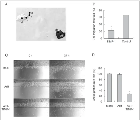
TIMP-1 inhibited the migration of BEL-7402
Both the single-cell migration detection assay and wounding assay showed that cells overexpressing TIMP-1 migrated much slower than control cells did. In Figure 5A, a, b, c, d indicate the cell center at successive time points. The migration route of each cell can be considered to be a→b→c→d ap-proximately, the average motion distance of the transfected cells (green cells) was only about 33% compared to the normal cells (Figure 5B).
The mobility of cell groups was detected by the wounding assay. Confluent monolay-ers of three groups of BEL-7402 cells were wounded and then cultured for 24 h. The
cells were photographed and the migration distance was determined (Figure 5C,D). The data showed that the migration rates of the Ad1-TIMP-1 group and the Ad1 group were 28 and 98%, respectively, of the migration of the mock group. Both experiments indi-cated that TIMP-1 inhibited the migration of BEL-7402 cells.
Discussion
The ECM forms a microenvironment that can modulate cellular behavior by affecting the contact of the cell with the outside world (20,21). Degradation or activation of ECM proteins by proteolysis can change the cellu-lar microenvironment rapidly and irrevers-ibly. The turnover of ECM is delicately regu-lated by a pair of contrary functional mol-ecules: MMP and its tissue inhibitor (TIMP).
MMP produces subtle changes of matrix structure by cleaving and also releasing ma-trix-bound growth factors and thereby con-trolling differentiation.
MMP-9 has been reported to play an important role in the invasion and metastasis of cancers (22). A previous report showed that the MMP-9/TIMP-1 mRNA ratio dif-fered between the stage II-III group and the stage IV group of lung cancer (23). Imbal-ance of the ratio between MMP-9 and TIMP-1 may cause breakdown of the basement membrane and ECM and is an essential step in tumor invasion and metastasis (24). In the present study, BEL-7402 originated from a Chinese patient with HCC which exhibited a high metastasis rate. Real-time PCR data showed that the MMP-9/TIMP-1 ratio was noticeably higher in BEL-7402 than in the normal liver cell line L-02 and QSG-7701,
Figure 5. Inhibitory effect of tis-sue inhibitor of metalloprotein-ases (TIMP-1) on the migration of BEL-7402 cells.A, The cen-ters of cells monitored every hour are shown by a, b, c, d. The cell on the right overexpressed TIMP-1. B, The relative average velocity of single cell migration was calculated for TIMP-1-trans-fected and control cells. C, Abil-ity to migrate affected by TIMP-1 using a wounding assay. Con-fluent monolayers of three groups of BEL-7402 cells were scraped using a tip and then cultured for 24 h. The cells were photo-graphed and the migration dis-tance was determined. The bee-line indicated the same location.
which may be one explanation for the high invasive ability of BEL-7402 compared to the other two lines.
TIMP-1 is a multifunctional protein with MMP-dependent and -independent actions for the regulation of cell death, cell prolif-eration, and angiogenesis (25). After TIMP-1 was initially characterized as a homologue of erythroid potentiating activity factor, it was found to display growth- and survival-promoting activity for a wide range of nor-mal or transformed cells (26,27). Increasing evidence demonstrates a much more com-plex role for TIMP-1 during tumor progres-sion and angiogenesis, in addition to its regu-lation of MMP-mediated ECM degradation. It has been well established that TIMP-1 has anti-apoptotic activity on many different cell types through both MMP-dependent (28,29) and -independent pathways (30,31).
Regarding the role of TIMP-1 in cell proliferation, conflicting data have been re-ported suggesting that TIMP-1 is mitogenic in certain cancer cells and lymphoid cells (2,32) while it inhibits the growth of normal mammary epithelial cells (33). In the present study, TIMP-1 reduced the growth rate of the HCC cell line BEL-7402 by up-regulat-ing the cyclin-dependent kinase inhibitor p21(Waf1) and reducing the MMP-9/TIMP-1 ratio, suggesting that both MMP-9-TIMP-1 balance and TIMP-MMP-9-TIMP-1 expression as a “cy-tokine-like” protein are required for the regu-lation of tumor progression.
Previous studies have shown that IL-6 has a pivotal role in stimulating hepatocytes to produce acutephase proteins cytotoxic to the liver; moreover, IL-6 has been shown to be a cell cycle progression factor for liver cells (34). After 70% partial hepatectomy, liver regeneration is dramatically impaired in IL-6-/- livers. Treatment with IL-6 clearly
accelerated the proliferative response in both IL-6+/+ and IL-6-/- livers (35). Intense
re-search over the past decade has shown that IL-6 could inhibit the proliferation of some cancer cells (36-38). Zeng and Fan (39)
suggested that IL-6 could inhibit the growth of the hepatocarcinoma cell line BEL-7402 by increasing the influx of Ca2+ and
decreas-ing the expression of Bcl-2 in the cell. Acting via gp130, IL-6 activates mul-tiple signaling pathways, including the JAK-STAT, the Src family of protein tyrosine kinases, and phosphatidylinositol 3-kinase. It is also known that IL-6 can induce TIMP-1 expression (40).As indicated in a previous paper, IL-6 inhibits the proliferation of BEL-7402 by promoting apoptosis (39). We found that IL-6 treatment did not inhibit BEL-7402 proliferation until the fourth day of treat-ment, suggesting that there might be another mediator induced by IL-6 which actually exerts the inhibitory effect. Our data showed that TIMP-1 may be the mediator of the inhibitory effects of IL-6, inducing the re-modelingof the ECM. Actually cell growth and proliferation result from a balance be-tween the number of dividing cells and the number of dying cells. And since TIMP-1 has been shown to inhibit apoptosis, our results suggested the presence of a potential downstream pathway regulated by TIMP-1 distinct from the anti-apoptosis pathway.
The present results showed that IL-6 in-duced cell growth inhibition through TIMP-1 production, which was STAT3 dependent. The downstream inhibitory molecule of IL-6 and TIMP-1 was p21. IL-IL-6 could acceler-ate p21 expression through the STAT3 sig-naling pathway which may act by decreas-ing MMP activity.
Acknowledgments
Pa-thology, Yale University School of Medi-cine, New Haven, CT, USA) for providing STAT3 wild-type, dominant negative form constructs and M67 reporter plasmid, and thank Dr. Zhi-Ming Yin (Life Sciences
Col-lege, Nanjing Normal University, Nanjing, China) for providing the p21(Waf1) lu-ciferase plasmid and the control reporter plasmid pSV-ß-galactosidase.
References
1. Denhardt DT, Feng B, Edwards DR, Cocuzzi ET, Malyankar UM. Tissue inhibitor of metalloproteinases (TIMP, aka EPA): structure, control of expression and biological functions. Pharmacol Ther 1993; 59: 329-341.
2. Hayakawa T, Yamashita K, Tanzawa K, Uchijima E, Iwata K. Growth-promoting activity of tissue inhibitor of metalloproteinases-1 (TIMP-1) for a wide range of cells. A possible new growth factor in serum.
FEBS Lett 1992; 298: 29-32.
3. Alexander CM, Howard EW, Bissell MJ, Werb Z. Rescue of mam-mary epithelial cell apoptosis and entactin degradation by a tissue inhibitor of metalloproteinases-1 transgene. J Cell Biol 1996; 135: 1669-1677.
4. Boudreau N, Sympson CJ, Werb Z, Bissell MJ. Suppression of ICE and apoptosis in mammary epithelial cells by extracellular matrix.
Science 1995; 267: 891-893.
5. Guedez L, Courtemanch L, Stetler-Stevenson M. Tissue inhibitor of metalloproteinase (TIMP)-1 induces differentiation and an antiapop-totic phenotype in germinal center B cells. Blood 1998; 92: 1342-1349.
6. Campbell CE, Flenniken AM, Skup D, Williams BR. Identification of a serum- and phorbol ester-responsive element in the murine tissue inhibitor of metalloproteinase gene. J Biol Chem 1991; 266: 7199-7206.
7. Uchijima M, Sato H, Fujii M, Seiki M. Tax proteins of human T-cell leukemia virus type 1 and 2 induce expression of the gene encoding erythroid-potentiating activity (tissue inhibitor of metalloproteinases-1, TIMP-1). J Biol Chem 1994; 269: 14946-14950.
8. Gewert DR, Coulombe B, Castelino M, Skup D, Williams BR. Char-acterization and expression of a murine gene homologous to human EPA/TIMP: a virus-induced gene in the mouse. EMBO J 1987; 6: 651-657.
9. Friedman SL. Seminars in medicine of the Beth Israel Hospital, Boston. The cellular basis of hepatic fibrosis. Mechanisms and treatment strategies. N Engl J Med 1993; 328: 1828-1835. 10. Nieto N, Dominguez-Rosales JA, Fontana L, Salazar A,
Armendariz-Borunda J, Greenwel P, et al. Rat hepatic stellate cells contribute to the acute-phase response with increased expression of alpha1(I) and alpha1(IV) collagens, tissue inhibitor of metalloproteinase-1, and matrix-metalloproteinase-2 messenger RNAs. Hepatology 2001; 33: 597-607.
11. Bahr MJ, Vincent KJ, Arthur MJ, Fowler AV, Smart DE, Wright MC, et al. Control of the tissue inhibitor of metalloproteinases-1 promoter in culture-activated rat hepatic stellate cells: regulation by activator protein-1 DNA binding proteins. Hepatology 1999; 29: 839-848. 12. Martin DC, Sanchez-Sweatman OH, Ho AT, Inderdeo DS, Tsao MS,
Khokha R. Transgenic TIMP-1 inhibits simian virus 40 T antigen-induced hepatocarcinogenesis by impairment of hepatocellular pro-liferation and tumor angiogenesis. Lab Invest 1999; 79: 225-234. 13. Akira S, Taga T, Kishimoto T. Interleukin-6 in biology and medicine.
Adv Immunol 1993; 54: 1-78.
14. Smart DE, Vincent KJ, Arthur MJ, Eickelberg O, Castellazzi M, Mann J, et al. JunD regulates transcription of the tissue inhibitor of metalloproteinases-1 and interleukin-6 genes in activated hepatic stellate cells. J Biol Chem 2001; 276: 24414-24421.
15. Ward NS, Waxman AB, Homer RJ, Mantell LL, Einarsson O, Du Y, et al. Interleukin-6-induced protection in hyperoxic acute lung injury.
Am J Respir Cell Mol Biol 2000; 22: 535-542.
16. Kaptein A, Paillard V, Saunders M. Dominant negative stat3 mutant inhibits interleukin-6-induced Jak-STAT signal transduction. J Biol Chem 1996; 271: 5961-5964.
17. He TC, Zhou S, da Costa LT, Yu J, Kinzler KW, Vogelstein B. A simplified system for generating recombinant adenoviruses. Proc Natl Acad Sci U S A 1998; 95: 2509-2514.
18. Sato Y, Rifkin DB. Inhibition of endothelial cell movement by pericytes and smooth muscle cells: activation of a latent transform-ing growth factor-beta 1-like molecule by plasmin durtransform-ing co-culture.
J Cell Biol 1989; 109: 309-315.
19. Bugno M, Graeve L, Gatsios P, Koj A, Heinrich PC, Travis J, et al. Identification of the interleukin-6/oncostatin M response element in the rat tissue inhibitor of metalloproteinases-1 (TIMP-1) promoter.
Nucleic Acids Res 1995; 23: 5041-5047.
20. Khatiwala CB, Peyton SR, Putnam AJ. Intrinsic mechanical proper-ties of the extracellular matrix affect the behavior of pre-osteoblastic MC3T3-E1 cells. Am J Physiol Cell Physiol 2006; 290: C1640-C1650.
21. Ingber DE. Mechanical signaling and the cellular response to extra-cellular matrix in angiogenesis and cardiovascular physiology. Circ Res 2002; 91: 877-887.
22. Iizasa T, Fujisawa T, Suzuki M, Motohashi S, Yasufuku K, Yasukawa T, et al. Elevated levels of circulating plasma matrix metalloprotein-ase 9 in non-small cell lung cancer patients. Clin Cancer Res 1999; 5: 149-153.
23. Ming SH, Sun TY, Xiao W, Xu XM. Matrix metalloproteinases-2, -9 and tissue inhibitor of metalloproteinase-1 in lung cancer invasion and metastasis. Chin Med J 2005; 118: 69-72.
24. Gomez DE, Alonso DF, Yoshiji H, Thorgeirsson UP. Tissue inhibi-tors of metalloproteinases: structure, regulation and biological func-tions. Eur J Cell Biol 1997; 74: 111-122.
25. Chirco R, Liu XW, Jung KK, Kim HR. Novel functions of TIMPs in cell signaling. Cancer Metastasis Rev 2006; 25: 99-113.
26. Luparello C, Avanzato G, Carella C, Pucci-Minafra I. Tissue inhibitor of metalloprotease (TIMP)-1 and proliferative behaviour of clonal breast cancer cells. Breast Cancer Res Treat 1999; 54: 235-244. 27. Oelmann E, Herbst H, Zuhlsdorf M, Albrecht O, Nolte A, Schmitmann
C, et al. Tissue inhibitor of metalloproteinases 1 is an autocrine and paracrine survival factor, with additional immune-regulatory func-tions, expressed by Hodgkin/Reed-Sternberg cells. Blood 2002; 99: 258-267.
inhibitor of metalloproteinase-1 is mediated via effects on matrix metalloproteinase inhibition: implications for reversibility of liver fi-brosis. J Biol Chem 2002; 277: 11069-11076.
29. Vorotnikova E, Tries M, Braunhut S. Retinoids and TIMP1 prevent radiation-induced apoptosis of capillary endothelial cells. Radiat Res 2004; 161: 174-184.
30. Liu XW, Taube ME, Jung KK, Dong Z, Lee YJ, Roshy S, et al. Tissue inhibitor of metalloproteinase-1 protects human breast epithelial cells from extrinsic cell death: a potential oncogenic activity of tissue inhibitor of metalloproteinase-1. Cancer Res 2005; 65: 898-906. 31. Lambert E, Boudot C, Kadri Z, Soula-Rothhut M, Sowa ML, Mayeux
P, et al. Tissue inhibitor of metalloproteinases-1 signalling pathway leading to erythroid cell survival. Biochem J 2003; 372: 767-774. 32. Bertaux B, Hornebeck W, Eisen AZ, Dubertret L. Growth stimulation
of human keratinocytes by tissue inhibitor of metalloproteinases. J Invest Dermatol 1991; 97: 679-685.
33. Fata JE, Leco KJ, Moorehead RA, Martin DC, Khokha R. Timp-1 is important for epithelial proliferation and branching morphogenesis during mouse mammary development. Dev Biol 1999; 211: 238-254.
34. Streetz KL, Wustefeld T, Klein C, Manns MP, Trautwein C. Media-tors of inflammation and acute phase response in the liver. Cell Mol
Biol (Noisy-le-grand) 2001; 47: 661-673.
35. Michalopoulos GK, DeFrances MC. Liver regeneration. Science
1997; 276: 60-66.
36. Takizawa H, Ohtoshi T, Ohta K, Yamashita N, Hirohata S, Hirai K, et al. Growth inhibition of human lung cancer cell lines by interleukin 6
in vitro: a possible role in tumor growth via an autocrine mechanism.
Cancer Res 1993; 53: 4175-4181.
37. Danforth DN Jr, Sgagias MK. Interleukin-1 alpha and interleukin-6 act additively to inhibit growth of MCF-7 breast cancer cells in vitro.
Cancer Res 1993; 53: 1538-1545.
38. Badache A, Hynes NE. Interleukin 6 inhibits proliferation and, in cooperation with an epidermal growth factor receptor autocrine loop, increases migration of T47D breast cancer cells. Cancer Res 2001; 61: 383-391.
39. Zeng XH, Fan XJ. Apoptosis of liver cancer cells BEL-7402 induced by IL-6 and its calcium signal transduction pathway. Acta Laser Bio Sinica 2001; 10: 55-61.