Effects of septal cholinergic lesion
on rat exploratory behavior in an
open-field
1Faculdade de Filosofia, Ciências e Letras de Ribeirão Preto,
Universidade de São Paulo, Ribeirão Preto, SP, Brasil
2Department of Psychology, Rutgers University, Piscataway, NJ, USA
M.R. Lamprea1,
F.P. Cardenas1,
R. Silveira1, T.J. Walsh2†
and S. Morato1
Abstract
The medial septum participates in the modulation of exploratory behavior triggered by novelty. Also, selective lesions of the cholin-ergic component of the septohippocampal system alter the habituation of rats to an elevated plus-maze without modifying anxiety indices. We investigated the effects of the intraseptal injection of the cholin-ergic immunotoxin 192 IgG-saporin (SAP) on the behavior of rats in an open-field. Thirty-nine male Wistar rats (weight: 194-230 g) were divided into three groups, non-injected controls and rats injected with either saline (0.5 µl) or SAP (237.5 ng/0.5 µl). Twelve days after surgery, the animals were placed in a square open-field (120 cm) and allowed to freely explore for 5 min. After the test, the rats were killed by decapitation and the septum, hippocampus and frontal cortex were removed and assayed for acetylcholinesterase activity. SAP increased acetylcholinesterase activity in the septum, hippocampus and frontal cortex and decreased the total distance run (9.15 ± 1.51 m) in compar-ison to controls (13.49 ± 0.91 m). The time spent in the center and at the periphery was not altered by SAP but the distance run was reduced during the first and second minutes (2.43 ± 0.36 and 1.75 ± 0.34 m) compared to controls (4.18 ± 0.26 and 3.14 ± 0.25 m). SAP-treated rats showed decreased but persistent exploration throughout the session. These results suggest that septohippocampal cholinergic mechanisms contribute to at least two critical processes, one related to the motiva-tion to explore new environments and the other to the acquisimotiva-tion and storage of spatial information (i.e., spatial memory).
Correspondence S. Morato
Faculdade de Filosofia, Ciências e Letras de Ribeirão Preto, USP Av. Bandeirantes, 3900 14040-901 Ribeirão Preto, SP Brasil
Fax: +55-16-633-5668 E-mail: [email protected]
Research partially supported by grants from CNPq (No. 523094/95-7), FAPESP (No. 98/11187-2) and NSF IBN (No. 9514557). M.R. Lamprea and F.P. Cardenas were the recipients of fellowships from CAPES and FAPESP, respectively.
†In memoriam.
Received March 27, 2002 Accepted December 12, 2002
Key words
·Exploratory behavior ·Open-field
·Cholinergic immunotoxin ·Medial septum
·Saporin
·Acetylcholinesterase
Cholinergic neurons in the medial sep-tum receive afferents from a variety of brain-stem and midbrain areas that participate in arousal, motivation, and vegetative function (1). These neurons seem to integrate subcor-tical information and the projections of these neurons to the hippocampus through the sep-tohippocampal cholinergic pathway modu-late the hippocampal responsiveness to its primary cortical input, i.e., the entorhinal
an open-field accessible from the home cage and reduced locomotor activity. Similarly, medial septal lesions have been reported to decrease the exploration of both neutral and novel objects as well as rearing and locomo-tor activity levels in the open-field (6). Addi-tionally, it has been demonstrated that le-sions of the medial septum decrease the total activity in an open-field, especially in the central area, as well as exploratory behavior (7). It is interesting to note that other reports by these investigators (8,9) as well as by others (10) consider increases in the activity in the central area of the open-field to be an index of decreased anxiety. Recently, we reported that intraseptal microinjections of the cholinergic immunotoxin 192 IgG-saporin (SAP) decreased exploratory behav-ior in an elevated plus-maze without affect-ing behaviors related to anxiety. In addition, a minute-by-minute analysis showed a defi-cit in habituation of exploratory behaviors (11).
The present study also utilized injections of the specific cholinergic immunotoxin 192 IgG-SAP. Immunotoxins are conjugates of a monoclonal antibody targeting a specific antigen combined with a ribosome-inacti-vating protein (12). Cholinergic neurons in the medial septum contain p75 neurotrophin receptors that contribute to the cellular ef-fects of nerve growth factor and other trophic factors. SAP combines the 192 IgG mono-clonal antibody to the p75 low affinity neu-rotrophin receptor, with the potent ribosome-inactivating protein SAP, derived from the plant Saponaria officinalis (13). Since all cholinergic cells in the medial septum ex-press p75 receptors, site-specific injection of SAP into this area selectively destroys this cell population. Intraseptal injection of SAP produces dose-related a) loss of cholinergic neurons in the medial septum, b) regionally specific decreases in cholinergic indices in the targets of the septum (hippocampus, cin-gulate and entorhinal cortices), and c) delay-dependent working memory impairments (14).
After demonstrating the role of the septo-hippocampal cholinergic pathway in habitu-ation and exploratory behavior (11) and in order to obtain data more directly related to exploratory behavior, we also tested the rats in an open-field, a more suitable model for the investigation of this kind of processes. A report of these data is the purpose of the present paper.
Adult male Wistar rats (194-230 g) from the animal house of the University of São Paulo at Ribeirão Preto were used. Rats had free access to food and water and were housed in groups of six to a cage on a 12:12-h dark-light cycle (dark-lights on at 7 am). Animals were divided randomly into three groups: 1) non-operated controls (N = 12), 2) saline-treated rats (N = 12) that were injected with 0.5 µl saline into the medial septum, and 3) SAP-treated rats (N = 15), which were injected with SAP into the medial septum. Rats in the saline and SAP groups were anesthetized with sodium pentobarbital (Hypnol, Cris-tália, Itapira, SP, Brazil) and placed in a stereotaxic apparatus. The scalp was opened and a single hole was drilled over the medial septum coordinates (anteroposterior = 0.6, dorsoventral = 6.5 and mediolateral = 0.0) (15). A single injection of either SAP (237.5 ng, Chemicon International Inc., Temecula, CA, USA) or 0.9% saline was delivered into the medial septal nucleus in a total volume of 0.5 µl as described by Walsh et al. (14). Solutions were infused slowly through a 30-gauge dental needle attached to a Hamilton 5-µl syringe. The syringe was mounted on a Harvard Apparatus microinjection unit and the rate of infusion delivery was 0.25 µl per min. The dental needle was left in place for an additional 2 min following each injection to allow for adequate diffusion. Animals were allowed 12 days to recover from sur-gery before the behavioral testing.
out in a room lit by a 60-W light bulb 1.75 m above the center of the open-field. The ex-perimental sessions were recorded by a video camera interfaced with a monitor and a vid-eocassette recorder in an adjacent room. In order to record locomotor activity on line, the image of the open-field on the monitor screen was divided into thirty-six 20-cm squares. Each rat was placed in the central area and allowed to freely explore for 5 min and every time both hind paws entered one square, a crossing was recorded. Total dis-tance run by the animals was estimated from the number of squares crossed. After each test, the open-field was cleaned with a solu-tion of 20% ethanol and then dried with a cloth. The total number of square crossings and the time spent in the central and periph-eral areas (all squares next to the walls) were recorded on line. Rearing and grooming were also analyzed in terms of the place of occur-rence, i.e., center or periphery. All measures were also analyzed minute by minute.
At the completion of behavioral testing rats were sacrificed by decapitation and re-gional acetylcholinesterase (AChE) activity was assessed according to the method of Ellman as modified by Augustinsson et al. (16). Briefly, hippocampus, frontal cortex and septum were dissected out and homog-enized in 1% (w/v) 0.1 M PBS, pH 7.3. The homogenate was then centrifuged for 15 min at 10,000 g and AChE activity was deter-mined in the resulting supernatant. Aliquots
of 150 µl of the supernatant were incubated in 0.1 M PBS containing 0.2 mM 4-4-dithiodipyridine for 3 min at 37ºC. Then acetylthiocholine (1 mM) was added to a final volume of 1 ml. Enzymatic activity was assessed by determining absorbance at 324 nm using a Varian DMS 80 spectrophotom-eter (Norwalk, CT, USA). Absorbance was measured in triplicate in each tissue sample 1, 2, and 3 min after the addition of acetyl-thiocholine. Statistical analysis was used to determine the differences in mean AChE activity during the third minute.
Since there were no differences between non-operated and saline-injected rats, these groups were combined into a single one, as of now designated as control. Statistical anal-ysis using t-tests for independent samples showed that injection of SAP into the medial septum significantly decreased AChE activ-ity in the hippocampus (t[35] = 21.7, P<0.001), frontal cortex (t[32] = 8.2, P<0.001) and sep-tum (t[33] = 6.7, P<0.001).
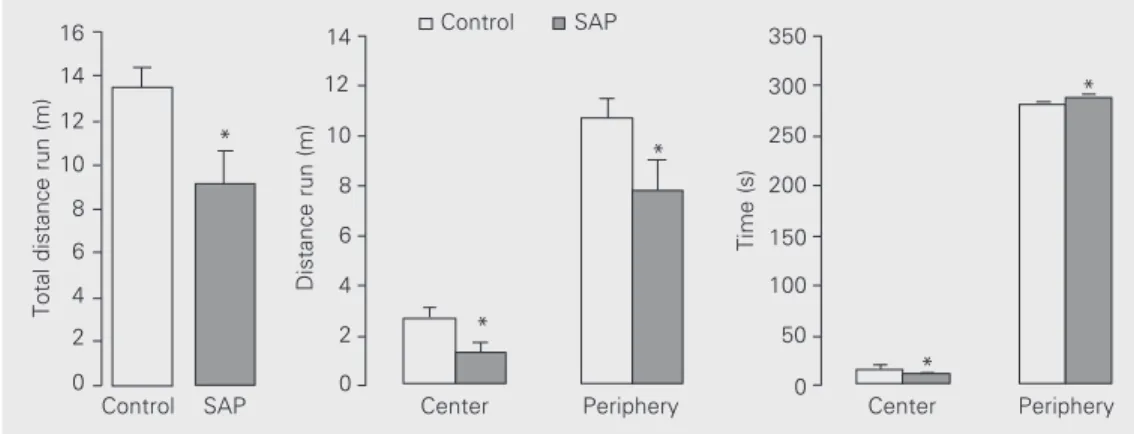
Behavioral comparisons of group means using the Mann-Whitney rank sum test to compare different groups (control x SAP) showed that intraseptal injection of SAP in-duced a significant decrease in the total dis-tance run in the open-field (Figure 1). Fur-ther analyses showed that this general reduc-tion was due to reducreduc-tions in the distance run in both central (T = 196.5, P = 0.003) and peripheral areas (T = 225.5, P = 0.03) (Fig-ure 1). On the other hand, SAP-treated rats
Total distance run (m)
16
14
12
10
8
6
4
2
0
Distance run (m)
14
12
10
8
6
4
2
0
Time (s)
350
300
250
200
150
100
50
0
Control SAP Center Periphery Center Periphery
*
*
*
*
*
Figure 1. Effect of intraseptal le-sion of the cholinergic compo-nent of the rat septohippocam-pal system. The figure reports total distance and distance run in the center and periphery (mean ± SEM) of an open-field by control and 192 IgG-saporin (SAP)-treated rats during a 5-min session. *P<0.05 compared to control (Mann-Whitney rank sum test).
spent significantly more time in the periph-eral area (T = 371.5, P = 0.04) and less time in the central area (T = 223.0, P = 0.03) when compared with the control animals (Figure 1). Finally, rearing and grooming did not differ between SAP-treated and control groups.
Additionally, analysis by the Wilcoxon signed rank test to compare different areas of the open-field (center x periphery) showed that control and SAP animals exhibited the standard profile of anxiety-related behaviors when placed in the open-field for the 5-min test session (10). They avoided entry into the central area (W = 300, P<0.001) and spent little time there (W = 300, P<0.001) (Figure 1). Finally, to compare behavioral effects along the session in a minute-by-minute fash-ion we used the Friedman rank sum test for repeated measures followed by all pairwise multiple comparison procedures (Student-Newman-Keuls method). The analysis re-vealed that the control group showed a sig-nificant and gradual decrease in total (c2 =
46.8, P<0.001), peripheral (c2 = 60.9,
P<0.001) and central (c2 = 12.6, P = 0.013)
distance run along the session; SAP-treated animals, on the other hand, did not exhibit such a gradual activity reduction along the session but instead tended to explore uni-formly, except in the central area (c2 = 19.3,
P<0.001) (Figure 2).
SAP-treated animals exhibited a shorter distance run (total: t = 181.0, P<0.001; pe-riphery: t = 184.5, P<0.001; center: t = 223.5, P = 0.028) compared to control rats in the first minute (Figure 2). In the second minute, SAP-treated animals exhibited a shorter dis-tance run (total: t = 200.0, P = 0.004; periph-ery: t = 210.5, P = 0.010; center: t = 235.5, P = 0.064). Finally, in the third minute SAP-treated animals explored the central area less than the control group (t = 202.0, P = 0.005). In general, the SAP-treated group exhib-ited less activity at the beginning of exposure to the open-field and less habituation of exploratory behavior over the 5-min test ses-sion than the control group.
Intraseptal injection of SAP produced specific behavioral effects associated with an extensive decrease in AChE activity in the hippocampus and, to a lesser extent, in the frontal cortex. AChE activity in the hip-pocampus has been shown to correspond to the innervation of hippocampal neurons by terminals originating in the cell bodies in the medial septum (17). Moreover, histochemi-cal studies have demonstrated that disrupt-ing septohippocampal connections by septal lesions (18) or fimbria-fornix transections decreases AChE in the hippocampus (17).
Previous reports have shown that the dose of SAP used in the present study (237.5 ng/ 0.5 µl) has no long-term effect on the levels of serotonin, norepinephrine, dopamine, or their metabolites in the hippocampus, fron-tal cortex, or striatum (14). In addition, this dose of SAP has been shown to produce a delay-dependent deficit in a spatial working memory task (14) without affecting attentional processes in a latent inhibition
Total distance run (m)
5 4 3 2 1 0
* *
1 2 3 4 5 1 2 3 4 5
Control SAP
* *
1 2 3 4 5 1 2 3 4 5
1 2 3 4 5 1 2 3 4 5
Periphery (m)
4
3
2
1
0
Center (m)
1.0
0.8
0.6
0.4
0.2
0.0
* *
Time (min) Figure 2. Effect of intraseptal
le-sion of the cholinergic compo-nent of the rat septohippocam-pal system. The figure reports total distance and distance run in the center and periphery (mean ± SEM) of an open-field by control and 192 IgG-saporin (SAP)-treated rats during each minute of a 5-min session. oP<0.05 compared to the
re-spective first minute (Dunnett’s test); *P<0.05 compared to con-trol (Mann-Whitney rank sum test).
paradigm (19).
The data presented here demonstrate that intraseptal SAP does not affect the distance run in the central area of the open-field, considered by some a measure of anxiety (8,9). In fact, SAP-treated rats exhibited a clear reduction in exploratory behavior in all areas of the open-field. As mentioned above, electrolytic lesions of the medial septum abolish the motivation to initiate exploration of an open-field voluntarily accessible from the home cage (5) and decrease the explora-tion of novel objects in an open-field (6). The data of the present experiment allow us to attribute the deficits in exploratory behav-ior to the selective destruction of the cholin-ergic component of the septohippocampal pathway and not to other neurotransmitter systems also present in this pathway.
Additionally, the decrease in exploratory behaviors is consistent with previous reports of the role of cholinergic processes in the modulation of locomotor activity and in the exploratory repertoire of rodents. For ex-ample, infusion of the muscarinic agonist carbachol into the septal area or into the hippocampus increases the frequency of be-haviors associated with exploration, such as rearing, ambulation, scanning, and move-ments of the vibrissae (20,21). These behav-ioral effects were reversed by a small dose of atropine, suggesting that muscarinic recep-tors in the hippocampus play a crucial role in
the initiation of locomotor activity. Litera-ture data about SAP lesions plus the results obtained in the present study suggest that the septohippocampal cholinergic system, to-gether with other cholinergic projections probably originating in the tegmental pedun-culopontine and laterodorsal nuclei (see Ref. 22), contribute to the modulation of behav-iors associated with exploration.
On the other hand, the persistence of exploration by SAP-treated animals (but not by controls) throughout the 5-min period of testing allows us to infer a deficit in spatial memory processes, as reported by us in the elevated plus-maze (11). Intraseptal injec-tion of SAP (2,14) has been shown to pro-duce deficits in other spatial memory tasks, indicating alterations in goal-directed ex-ploratory behavior. The results of the pres-ent study suggest that septohippocampal cho-linergic mechanisms contribute to at least two critical behavioral processes, one re-lated to the motivation to explore new envi-ronments and the other related to the acqui-sition and storage of spatial information (i.e., spatial memory).
Acknowledgments
The authors are indebted to Juan Carlos Martinez, Universidad de la Sabana, Colom-bia, for help with the behavioral experi-ments.
References
1. Dutar MH, Bassant MH, Senut MC & Lamour Y (1995). The septo-hippocampal pathway: structure and function of a central cholinergic system. Physiological Reviews, 75: 393-427.
2. Shen J, Barnes CA, Wenk GL & McNaughton BL (1996). Differential effects of selective immunotoxic lesions of medial septal cholin-ergic cells on spatial working and reference memory. Behavioral Neuroscience, 110: 1181-1186.
3. McNaughton N & Gray JA (2000). Anxiolytic action on the behaviour-al inhibition system implies multiple types of arousbehaviour-al contribute to anxiety. Journal of Affective Disorders, 61: 161-176.
4. Walsh TJ & Chrobak JJ (1991). Animal models of Alzheimer’s dis-ease. Role of hippocampal cholinergic system in working memory. In: Dachowsky L & Flaherty C (Editors), Current Topics in Animal
Learning, Brain, Emotion and Cognition. Lawrence Erlbaum, Hillsdale, NJ, USA, 347-379.
5. Kohler C & Srebro B (1980). Effects of lateral and medial septal lesions on exploratory behavior in the albino rat. Brain Research, 182: 423-440.
6. Myhrer T (1989). Exploratory behavior and reaction to novelty in rats: effects of medial and lateral septal lesions. Behavioral Neurosci-ence, 103: 1226-1233.
7. Lee EHY, Lin YP & Yin TH (1988). Effects of lateral and medial septal lesions on various activity and reactivity measures in rats. Physiolo-gy and Behavior, 42: 97-102.
and Biobehavioral Reviews, 3: 247-263.
9. Lee EHY, Tsai MJ & Chai CY (1986). Stress selectively influences central region activity of mice in an open-field. Physiology and Be-havior, 37: 659-662.
10. Lee EHY, Tang YP & Chai CY (1987). Stress and corticotrophin releasing factor potentiate center region activity of mice in an open-field. Psychopharmacology, 93: 320-323.
11. Lamprea MR, Cardenas FP, Silveira R, Morato S & Walsh TJ (2000). Dissociation of memory and anxiety in a repeated elevated plus maze paradigm: forebrain cholinergic mechanisms. Behavioural Brain Research, 117: 97-105.
12. Wiley RG & Lappi DA (1994). Suicide Transport and Immunolesion-ing. RG Landes Co., Austin, TX, USA.
13. Wiley RG & Lappi DA (1992). Neural lesioning with ribosome-inacti-vating proteins: Suicide transport and immunolesioning. Trends in Neurosciences, 15: 285-290.
14. Walsh TJ, Herzog CD, Gandhi C, Stackman RW & Wiley RG (1996). Injection of IgG 192-saporin into the medial septum produces cholin-ergic hypofunction and dose-dependent working memory deficits. Brain Research, 726: 69-79.
15. Paxinos G & Watson C (1997). The Rat Brain in Stereotaxic Coordi-nates. 3rd edn. Academic Press, San Diego, CA, USA.
16. Augustinsson KB, Eriksson H & Faigersson Y (1978). A new
ap-proach to determining cholinesterase activities in samples of whole blood. Clinica Chimica Acta, 89: 239-252.
17. Lewis PR, Shute CCD & Silver A (1967). Confirmation from choline acetylase analysis of a massive cholinergic innervation to rat hippo-campus. Journal of Physiology, 191: 215-224.
18. Mellgren SI & Srebro B (1973). Changes in acetylcholinesterase and distribution of degenerating fibers in the hippocampal region after septal lesions in the rat. Brain Research, 52: 19-36.
19. Dougherty KD, Salat D & Walsh TJ (1996). Intraseptal injections of the cholinergic immunotoxin 192-IgG saporin fails to disrupt latent inhibition in a conditioned test aversion paradigm. Brain Research, 736: 260-269.
20. Flicker C & Geyer MA (1982). Behavior during hippocampal microinfusions II: muscarinic locomotor activation. Brain Research, 257: 105-127.