Bi-dire ctio nal actio ns o f e stro ge n
o n the re nin-angio te nsin syste m
1The Hypertension and Vascular Disease Center,
Wake Forest University School of Medicine, Winston-Salem, NC, USA
2Eye Institute, Cleveland Clinic Foundation, Cleveland, O H, USA
K.B. Brosnihan1,
P.S. Senanayake2, P. Li1
and C.M. Ferrario1
Abstract
Estrogen stimulates the renin-angiotensin system by augmenting both tissue and circulating levels of angiotensinogen and renin. We show, however, that angiotensin converting enzyme (ACE) activity in the circulation and in tissues is reduced in two animal models of post-menopausal chronic hormone replacement. We observed a reduction of ACE activity in association with a significant increase in plasma angiotensin I (Ang I) and hyperreninemia in ovariectomized monkeys treated with Premarin (conjugated equine estrogen) replacement for 30 months. Plasma angiotensin II (Ang II) levels were not increased in monkeys treated with estrogen, suggesting that the decrease in ACE curtailed the formation of the peptide. The Ang II/Ang I ratio, an in vivo index of ACE activity, was significantly reduced by estrogen
treatment, further supporting the biochemical significance of estrogens inhibition of ACE. In ovariectomized transgenic hypertensive (mRen2)27 rats submitted to estrogen replacement treatment for 3 weeks, ACE activity in plasma and tissue (aorta and kidney) and circulating Ang II levels were reduced, whereas circulating levels of angiotensin-(1-7) (Ang-(1-7)) were increased. Ang-(1-7), the N-ter-minal fragment of Ang II, is a novel vasodilator and antihypertensive peptide. Thus, the net balance of these effects of estrogen on the renin-angiotensin vasoconstrictor/vasodilator system is to promote the anti-hypertensive effect.
Co rre spo nde nce
K.B. Brosnihan
Hypertension and Vascular Disease Center
Wake Forest University School of Medicine
Winston-Salem, NC 27157-1032 USA
Fax: + 1-336-716-2456 E-mail: bbrosnih@ wfubmc.edu
Presented at the XIII Annual Meeting of the Federação de Sociedades de Biologia Experimental, Caxambu, MG, Brasil, August 26-29, 1998.
Received January 8, 1999 Accepted January 26, 1999
Ke y wo rds
·Renin
·Angiotensin converting enzyme
·Angiotensin peptides
·Angiotensin receptors
·Bradykinin
·Nitric oxide
·Kinins
·Vasodilation
·Hormone replacement
Intro ductio n
Cardiovascular disease is the leading cause of female mortality, resulting in more deaths in women over 50 than all cancers (1). The incidence of cardiovascular disease in women rises steadily, approaching the inci-dence in men during the fifth to seventh decade of life (2). Estrogen protects women from cardiovascular disease by inhibiting atherosclerosis (3) and through effects on
ef-fect (1,15), suggesting that estrogen main-tains vascular health through additional pro-cesses. One potential mechanism is the ef-fect of estrogen on the renin-angiotensin system (RAS).
In this review we discuss the effects of estrogen on components of the RAS and suggest that estrogen shifts the generation of angiotensin peptides away from angiotensin II (Ang II) and towards the N-terminal heptapeptide, angiotensin-(1-7) (Ang-(1-7)). This fragment has little or none of the vaso-constrictor properties of Ang II (16,17), but instead releases prostaglandins (PGs) (18-22) and nitric oxide (NO) (23,24) and aug-ments the vasodilator action of bradykinin (BK) (25,26).
Estro ge n re gulatio n o f RAS co mpo ne nts
The RAS is the primary regulator of blood pressure and fluid and sodium balance. En-hanced activation of the RAS contributes to the evolution of hypertension (17-29), salt retention (30,31), and hyperaldosteronism (32-34). Angiotensinogen (Aogen), the pre-cursor of angiotensin peptides, is converted to angiotensin I (Ang I) by renin, an aspartyl protease. Angiotensin converting enzyme (ACE; EC 3.4.15.1) is the major enzyme responsible for the formation of the vaso-constrictor peptide Ang II from Ang I. This enzyme, also called kininase II, plays a piv-otal role both in the RAS and the kinin system. ACE is a metalloprotease that re-leases C-terminal dipeptides from substrates such as Ang I and BK (35) and degrades BK, a vasodilator, to its inactive metabolite. ACE inhibition was associated with a 5-50-fold increase in Ang-(1-7) and BK in tissues and the circulation (36-38). Ang-(1-7), a heptapeptide with novel vasodilator and an-tihypertensive properties (39-42), is gener-ated from either Ang I or Ang II by specific peptidases (43,44). In bovine, porcine, and human aortic and venous endothelial cells,
Ang I is primarily processed to Ang-(1-7) by neutral endopeptidase 24.11 (40-50%) and prolylendopeptidase (45). In vascular smooth muscle cells, Ang-(1-7) was the major prod-uct generated from Ang I, and its prodprod-uction was dependent upon metalloendopeptidase 24.15 (46). Further metabolism of Ang-(1-7) or Ang II by aminopeptidases and dipepti-dases leads to the formation of smaller frag-ments, Ang-(3-7) and Ang IV, which may also have a biological function (47,48). Im-portantly, Ang-(1-7) is both a substrate (49,50) and an inhibitor of ACE (23,50).
luteal phase when both estrogen and proges-terone are elevated (61). Thus, Aogen and renin are elevated in response to increased plasma estrogen due to normal cycling events or pharmacological hormone replacement.
If estrogen treatment activates the RAS, why do the reninemia and hyper-aogenemia states not result in significant increases in blood pressure? The effects of hormone replacement on blood pressure are conflicting, with findings of either no change or a decrease in blood pressure (12,62-64). In investigating this question, we found that estrogen shifts the pattern of angiotensin peptide formation in a tissue-specific man-ner, reducing production of Ang II, while augmenting the production of the N-termi-nal Ang II fragment, Ang-(1-7). This shift in the pattern of peptides arises in part due to estrogen decreasing the activity of ACE.
Chro nic ho rmo ne re place me nt in cyno mo lgus mo nke ys
In the first study we evaluated the effects of chronic hormone replacement in a model of post-menopausal hormone replacement, i.e., ovariectomized monkeys receiving premarin (conjugated equine estrogens; CEE) orally. At the time the study was initiated 29 feral adult female cynomolgus monkeys (Macaca fascicularis) ranging in age from 5 to 13 years had been fed a moderately genic diet for 22 months to induce athero-sclerosis. Bilateral ovariectomies were per-formed to initiate surgical menopause on all animals 4 months after the diet started. At the end of 22 months of continuous feeding of the atherosclerotic diet, all animals re-ceived a lipid-lowering diet and were ran-domly assigned to a replacement therapy protocol lasting 30 months as indicated: group 1 (placebo, N = 14) and group 2 (CEE, N = 15). Groups 2 received 7.2 µg/day of CEE (Premarin®
, Wyeth-Ayerst, Radnor, PA, USA) for the first eight months and 166 µg/ day of CEE for the remaining 22 months.
The latter dose of CEE was used to raise the levels of circulating 17 ß-estradiol to ap-proximately 150 pg/ml, a concentration equivalent to the therapeutic concentrations achieved in women (~0.625 mg/day) (65). Hormones were administered twice daily in the diet. Blood samples characterizing the levels of sex hormones were taken 4 h after administration of the oral drug (peak level) under ketamine hydrochloride sedation (10 to 15 mg/kg body weight, im) (Fort Dodge Laboratories, Inc., Fort Dodge, IA, USA) 5 months before necropsy.
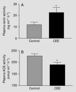
There was no significant effect of hor-mone replacement on mean arterial blood pressure or body weight. As expected, plasma 17-ß-estradiol levels were near the mini-mum detectable level of the assay in un-treated control animals (2.4 ± 1.1 pg/ml) but rose significantly (148.4 ± 13.4 pg/ml, P<0.05) in CEE-treated animals. Figure 1 shows that long-term replacement therapy with CEE produced significant increases in PRA. These changes were accompanied by a significant reduction in plasma ACE activity in CEE-treated animals. Replacement of es-trogen resulted in a significant, three-fold increase in plasma Ang I in animals given CEE (Figure 2A). On the other hand, there
P
la
s
m
a
r
e
n
in
a
c
ti
v
it
y
(n
g
m
l
-1 h -1)
30
20
10
0
Control CEE
Control CEE 250
200
150
100
P
la
s
m
a
A
C
E
a
c
ti
v
it
y
(n
m
o
l
m
l
-1 h -1)
*
*
Figure 1 - Effects of estrogen on plasma renin activity (A) and an-giotensin converting enzyme ac-tivity (B). Values are reported as mean ± SEM . * P<0.05 vs con-trol (Student t-test). All cynomol-gus monkeys w ere ovariecto-mized and left either untreated (control, N = 14) or treated w ith conjugated equine estrogens (CEE; N = 15). (Reproduced from Ref. 72 w ith permission).
A
was no significant effect of CEE replace-ment on the circulating levels of Ang II or Ang-(1-7) (Figure 2B and C). The Ang II/ Ang I ratio (1.22 ± 0.56 vs 0.38 ± 0.08, P<0.05; control vs CEE), an in vivo estimate of ACE activity (66), was significantly re-duced in hormone-treated animals. Thus, in cynomolgus monkeys with chronic experi-mental atherosclerosis, replacement therapy with estrogen for 30 months in addition to a lipid-lowering diet resulted in significant and reciprocal changes in the activity of the two enzymes that account for the generation of angiotensin peptides. In our study estrogen replacement therapy augmented renin activ-ity and reduced ACE activactiv-ity. The increase in PRA was sufficient to be reflected in
higher plasma Ang I concentrations in the CEE-treated group. By curtailing the forma-tion of the vasoconstrictor end-product Ang II, the effect of estrogen on ACE activity prevented an otherwise expected increase in the production of Ang II. These experiments provided new information on the potential mechanisms that may contribute to the de-scribed cardio-protective action of estrogen replacement therapy on post-menopausal women.
Estro ge n re place me nt in transge nic hype rte nsive (mRe n-2)27 rats
In the next study, we evaluated hormone replacement in hypertensive and normoten-sive rats. Fifty-three female transgenic nega-tive Tg(-) and heterozygous hypertensive Tg(+) rats (body weight: 220~250 g) from the Hypertension Center Transgenic Rat Colony of Wake Forest University School of Medicine underwent bilateral ovariectomy at age 12 weeks under general anesthesia with ketamine (30 mg/kg, im) and xylazine (5 mg/kg, im). Pellets containing either 17 ß-estradiol (E2) (1.5 mg/rat, for 3-week
re-lease; Innovative Research of America, To-ledo, OH, USA) or vehicle (VEH) were im-planted into the subcutaneous tissue.
Chronic 17 ß-estradiol treatment pro-duced a small but statistically significant decrease in the mean blood pressure of both transgenic hypertensive (159 ± 4 vs 145 ± 5 mmHg, P<0.05) and normotensive (119 ± 4
vs 108 ± 2 mmHg, P<0.05) rats. There was no change in heart rate with estrogen treat-ment. Plasma 17 ß-estradiol concentration averaged 190 ± 20 pg/ml in E2-treated rats
and <15 pg/ml in VEH-treated rats.
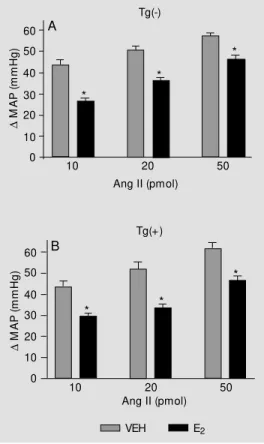
The magnitude of the pressor response produced by the injection of three doses of Ang II was similar in VEH-treated Tg(+) and Tg(-) rats. In contrast, chronic 17 ß-estradiol treatment significantly attenuated the pres-sor responses to intravenous injection of Ang II in both strains at all doses tested
P
la
s
m
a
A
n
g
l
(
p
g
/m
l)
300
200
100
0
Control CEE
Control CEE 75
50
25
0
*
P
la
s
m
a
A
n
g
l
I
(p
g
/m
l)
20
10
0
P
la
s
m
a
A
n
g
-(
1
-7
)
(p
g
/m
l)
Control CEE
A
B
C
(Figure 3A and B). Intravenous injections of Ang-(1-7) in normotensive and transgenic rats elicited a biphasic response consisting of a rapid (55.6 s) pressor component fol-lowed by a longer lasting (138.9 s) depressor component. Estrogen replacement therapy had a small but significant effect on the pressor but not the depressor component of the response to intravenous injections of Ang-(1-7) in normotensive rats (Figure 4A and C). In Tg(+) rats treated with estrogen, the blunting of the pressor component of the Ang-(1-7) response was comparable to that obtained in Tg(-) rats (Figure 4A and B). In hypertensive rats, estrogen potentiated the magnitude of the fall in blood pressure pro-duced by Ang-(1-7) (Figure 4D).
Replacement with estrogen resulted in a nearly 2-fold reduction in the circulating levels of Ang II in Tg(+) rats while it had no effect on plasma Ang II in Tg(-) rats (Figure 5A). In contrast, estrogen replacement sig-nificantly increased the levels of plasma Ang-(1-7) in Tg(+) animals (Figure 5B). In accor-dance with the reduction in plasma Ang II levels in Tg(+) rats, estrogen significantly reduced plasma ACE activity in Tg(+) ani-mals (Figure 6A). A similar reduction in plasma ACE was observed in Tg(-) rats. There was no difference in plasma, kidney, or aorta ACE activity levels between Tg(-) and Tg(+) animals on similar treatment (Fig-ure 6A-C), whereas chronic estrogen replace-ment therapy significantly decreased both kidney and aorta ACE activity in Tg(+) but not in Tg(-) rats.
In summary, this study combined a mono-genetic model of renin-dependent hyperten-sion with a surgically induced postmeno-pausal model. Hormone replacement in this model attenuates hypertension in associa-tion with reduced generaassocia-tion of Ang II and increased production of Ang-(1-7). In spite of the overall impression in the literature that estrogen activates the RAS (51,52,67) by increasing the levels of angiotensinogen and renin, estrogen acts downstream in relation
Figure 4 - Effects of 3-w eek estrogen (E2) replacement on Ang-(1-7) pressor/depressor responses of conscious, resting Tg(-) (A, C) and Tg(+) rats (B, D). E2 attenuates the Ang-(1-7)-induced pressor responses of both Tg(-) (A) and Tg(+) (B) animals, but augments the depressor responses of Tg(+) rats (D). Values are reported as mean ± SEM . * P<0.05, VEH vs E2 (ANOVA).VEH = Vehicle. (Reproduced from Ref. 62 w ith permission).
60
50
40
30
D
M
A
P
(
m
m
H
g
)
20
10
0
10 20 50
Tg(-)
*
*
* A
Figure 3 - Effects of 3-w eek es-trogen (E2) replacement on Ang II pressor responses of con-scious, resting Tg(-) (A) and Tg(+) (B) rats. E2 attenuates the Ang II-induced pressor responses. Values are reported as mean ± SEM . * P< 0.05, VEH vs E2 (ANOVA). Each group included 6~8 rats. VEH, Vehicle. (Repro-duced from Ref. 62 w ith permis-sion).
60
50
40
30
D
M
A
P
(
m
m
H
g
)
20
10
0
Tg(+)
*
* *
Ang II (pmol)
10 20 50
B
VEH E2 Ang II (pmol)
60
40
D
M
A
P
(
m
m
H
g
)
20
0
0
-2
D
M
A
P
(
m
m
H
g
)
-4
-6
-8
-10
100 300 600 100 300 600
60
40
D
M
A
P
(
m
m
H
g
)
20
0
0
-2
D
M
A
P
(
m
m
H
g
)
-8
-10 -4
-6
Tg(-) Tg(+)
Ang-(1-7) (nmol)
* *
*
* *
*
* *
*
A B
C D
Ang-(1-7) (nmol)
to these two proteins by reducing ACE activ-ity and shifting the profile of the circulating angiotensin peptides. Thus, estrogen acts as a fulcrum reducing the magnitude of the response to and levels of Ang II, while in-creasing the formation and vasodilator ef-fect of Ang-(1-7). These studies provide new information on the potential mechanisms that may contribute to the therapeutic action of estrogen replacement therapy in postmeno-pausal women who are at an increased risk of cardiovascular morbidity.
Estro us cycle influe nce o n angio te nsin pe ptide s
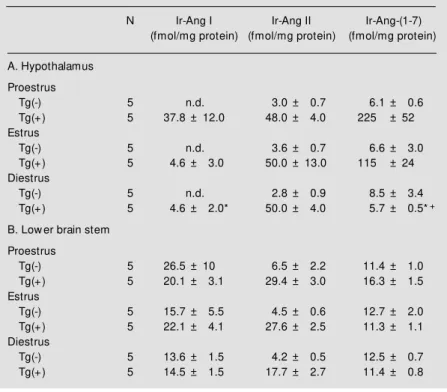
A third study was conducted to evaluate if local regional angiotensin peptide levels are modulated by the estrous cycle. Ten-week-old normotensive (N = 15) and hyper-tensive (mRen2)27 (N = 15) rats were sacri-ficed by decapitation without prior anesthe-sia. Vaginal smears were obtained from each rat to determine the stage of the estrous cycle at sacrifice, as described by Long and Evans (68). Immunoreactive (Ir) peptide levels were measured using three different radioimmu-noassays, as previously described (69). Table 1A shows the influence of the estrous cycle on angiotensin peptides in the hypothala-mus. There was no influence of the estrous cycle on hypothalamic peptide levels of nor-motensive rats. Ir-Ang I was significantly elevated during proestrus as compared to diestrus in hypertensive rats. Ir-Ang II levels were unchanged with the estrous cycle. Ir-Ang-(1-7) levels were significantly elevated during both proestrus and estrus as com-pared to diestrus. Table 1B illustrates that the peptide levels in the lower brain stem of normotensive or hypertensive rats did not change with the estrous cycle. In summary, the estrous cycle exerts an influence on hy-pothalamic angiotensin peptides, enhancing the expression of Ang I and Ang-(1-7) in hypertensive rats.
Gender differences in cardiovascular
dis-P la s m a A n g I I (p g /m l) 40 30 20 0 A Tg(-) 10 Tg(+) 200 150 100 0 B 50 P la s m a A n g -( 1 -7 ) (p g /m l) Tg(-) Tg(+) # * # *
Figure 5 - A, Effects of estrogen replacement (E2) on plasma lev-els of Ang II. Estrogen reduces the circulating levels of plasma Ang II in Tg(+) animals. B, Ef-fects of estrogen replacement (E2) on plasma levels of Ang-(1-7). Estrogen increases the circu-lating levels of plasma Ang-(1-7) in Tg(+) animals. Values are re-port ed as m ean ± SEM . * P< 0.05, VEH vs E2 (Tukey-Kramer test); #P<0.01 Tg(+) vs Tg(-) (Tukey-Kramer test). Each group included 6~8 rats. VEH = Vehicle. (Reproduced from Ref. 62 w ith permission).
150 100 50 0 P la s m a A C E a c ti v it y (n m o l m in
-1 m
l -1) Tg(-) Tg(+) 1.5 1.0 0.5 0.0 K id n e y A C E a c ti v it y (n m o l m g
-1 m
l -1) 7.5 5.0 2.5 0.0 A o rt a A C E a c ti v it y (n m o l m g
-1 m
l -1) Tg(-) Tg(+) Tg(-) Tg(+) A B C * * *
Figure 6 - Effect of estrogen re-placement (E2) on plasma (A), kidney (B) and thoracic aorta (C) levels of angiotensin converting enzyme. Estrogen reduces the plasma angiotensin converting enzym e activity in Tg(+ ) and Tg(-) animals, w hereas it only re-duces the tissue levels of kidney and thoracic aorta in Tg(+) rats. Values are reported as mean ± SEM . * P< 0.05, VEH vs E2 (Tukey-Kramer test). Each group included 6~8 rats. VEH = Ve-hicle. (Reproduced from Ref. 62 w ith permission).
*
VEH E2
ease incidence have been established (70). After menopause, the prevalence of hyperten-sion and coronary heart disease increases mark-edly in women (59,70). Hormone replacement with estrogen provides cardioprotective ef-fects. However, estrogen also increases the production of Aogen and renin, two compo-nents of the RAS. An elevation of Aogen with unchanged or increased levels of renin would appear to favor the development of hyperten-sion and increase cardiovascular risk. Yet women taking estrogen replacement have ei-ther unchanged or slightly lower blood pres-sure (8,71). The literature reviewed above and our studies offer a new hypothesis for the effects of estrogen on the RAS and its potential relevance to hypertension and local vasoreac-tivity. Although renin and Aogen are increased, we show that ACE activity is reduced by estrogen. The reduction in ACE activity acts to curb the activated RAS by shifting the amount of the peptides which are formed, i.e., increas-ing Ang I, reducincreas-ing the production of Ang II and enhancing formation of Ang-(1-7). Our findings that estrogen reduces ACE activity require a re-evaluation of the long held tenet that estrogens activation of the RAS increases circulating Ang II. Estrogens reduction of ACE activity leads to increased levels of Ang-(1-7) due to the diversion of the processing pathway from Ang II to increased Ang I. Thus, we schematically propose in Figure 7 that estrogen acts as a fulcrum reducing the magni-tude of the response to and levels of Ang II, while increasing the formation and vasodila-tor effect of Ang-(1-7). In addition, new stud-ies carried out by Dr. Chappell (49) of our group showed that ACE metabolizes Ang-(1-7). By reducing ACE activity, estrogen would decrease the breakdown of Ang-(1-7) and add further to the increased levels of Ang-(1-7), potentially resulting in an enhancement of vasodilation. Because ACE reduction also leads to less degradation of bradykinin, another as-pect of estrogens action would be to enhance
Figure 7 - Schematic diagram demonstrating the effects of estrogen on the renin-angio-t ensin sysrenin-angio-t em . ACE, Angio-tensin converting enyme; BK, bradykinin.
Table 1 - Influence of estrous cycle on immunoreactive (Ir) angiotensin peptides in hypothalamus (A) and low er brain stem (B).
Values are reported as mean ± SEM . * P<0.05 vs proestrus; +P<0.05 vs estrus. n.d., Not detectable.
N Ir-Ang I Ir-Ang II Ir-Ang-(1-7) (fmol/mg protein) (fmol/mg protein) (fmol/mg protein)
A. Hypothalamus
Proestrus
Tg(-) 5 n.d. 3.0 ± 0.7 6.1 ± 0.6 Tg(+) 5 37.8 ± 12.0 48.0 ± 4.0 225 ± 52 Estrus
Tg(-) 5 n.d. 3.6 ± 0.7 6.6 ± 3.0 Tg(+) 5 4.6 ± 3.0 50.0 ± 13.0 115 ± 24 Diestrus
Tg(-) 5 n.d. 2.8 ± 0.9 8.5 ± 3.4 Tg(+) 5 4.6 ± 2.0* 50.0 ± 4.0 5.7 ± 0.5*+
B. Low er brain stem
Proestrus
Tg(-) 5 26.5 ± 10 6.5 ± 2.2 11.4 ± 1.0 Tg(+) 5 20.1 ± 3.1 29.4 ± 3.0 16.3 ± 1.5 Estrus
Tg(-) 5 15.7 ± 5.5 4.5 ± 0.6 12.7 ± 2.0 Tg(+) 5 22.1 ± 4.1 27.6 ± 2.5 11.3 ± 1.1 Diestrus
Tg(-) 5 13.6 ± 1.5 4.2 ± 0.5 12.5 ± 0.7 Tg(+) 5 14.5 ± 1.5 17.7 ± 2.7 11.4 ± 0.8
vasodilation by both Ang-(1-7) and BK. Our studies enhance the understanding of the role of estrogen in the regulation of the RAS and may provide a new rationale for the use of estrogen to prevent cardiovascular disease in postmenopausal women who are at increased cardiovascular risk.
Ang-(1-5)
Ang-(1-7)
Ang I
Ang II
Vasoconstriction
Vasodilation BK metabolite
Vasodilation
ESTROGEN
(-)
Bradykinin
Re fe re nce s
1. Wentz AC (1994). Women’s health is-sues. In: Wentz AC (Editor), Advances in Internal M edicine. M osby-Year Book, Inc. St. Louis, M O, 1-30.
2. Castelli WP (1988). Cardiovascular dis-ease in w omen. American Journal of Ob-stetrics and Gynecology, 158: 1553-1560. 3. Pennisi E (1997). Differing roles found for estrogen’s tw o receptors. Science, 277: 1439.
4. Ross RK, M ack TM , Paganini-Hill A, Arthur M & Henderson BE (1981). M enopausal oestrogen therapy and protection from death from ischaemic heart disease. Lan-cet, 1 (8225): 858-860.
5. M aynard C & Weaver WD (1992). Treat-ment of w omen w ith acute M I: New find-ings from the M ITI registry. Journal of M yocardial Ischemia, 4: 27-37.
6. Keresztes PA & Dan AJ (1992). Estrogen and cardiovascular disease. Cardiovascu-lar Nursing, 28: 1-6.
7. Stamper M J, Colditz GA, Willett WC, M anson JAE, Rosner B, Speizer FE & Hennekens CH (1991). Postmenopausal estrogen therapy and cardiovascular dis-ease: ten-year follow -up from the nurses’ health study. New England Journal of M edicine, 325: 756-762.
8. Stevenson JC, Crook D, Godsland IF, Collins P & Whitehead M I (1994). Hor-mone replacement therapy and the car-diovascular system. Drugs, 47: 35-41. 9. Williams JK, Honore EK, Washburn SA &
Clarkson TB (1994). Effects of hormone replacement therapy on reactivity of ath-erosclerotic coronary arteries in cynomol-gus monkeys. Journal of the American College of Cardiology, 24: 1757-1761. 10. Gisclard V, M iller VM & Vanhoutte PM
(1988). Effect of 17-ß estradiol on endo-thelium-dependent responses in the rab-bit. Journal of Pharmacology and Experi-mental Therapeutics, 244: 19-22. 11. Kauser K & Rubanyi GM (1994). Gender
difference in bioassayable endothelium-derived nitric oxide from isolated rat aor-tae. American Journal of Physiology, 267: H2311-H2317.
12. Li P, Ferrario CM , Ganten D & Brosnihan KB (1997). Chronic estrogen treatment in female transgenic (mRen2)27 hyperten-sive rats augments endothelium-derived nitric oxide release. American Journal of Hypertension, 10: 662-670.
13. Barrett-Connor E & Bush TL (1991). Estro-gen and coronary heart disease in w omen. Journal of the American M edical Associa-tion, 265: 1861-1867.
14. Kolata G (1983). New puzzles over estro-gen and heart disease. Science, 220: 1137-1138.
15. PEPI Trial Working Group (1995). Effects of estrogen or estrogen/progestin regi-mens on heart disease risk factors in post menopausal w oman. Journal of the Ameri-can M edical Association, 273: 199-208. 16. Peach M J & Levens NR (1980). M olecular
Approaches to the Study of Angiotensin Receptors. Plenum Press, New York, 171-194.
17. Bumpus FM & Smeby RR (1962). Impor-tance of amino acid side groups for bio-logic activity of angiotensin II. Circulation, 25: 183-185.
18. Jaisw al N, Diz DI, Tallant EA, Khosla M C & Ferrario CM (1991). The non-peptide angiotensin II antagonist DuP 753 is a potent stimulus for prostacyclin release. American Journal of Hypertension, 4: 228-233.
19. Jaisw al N, Tallant EA, Diz DI, Khosla M C & Ferrario CM (1991). Subtype 2 angio-tensin receptors mediate prostaglandin synthesis in human astrocytes. Hyperten-sion, 17: 1115-1120.
20. Jaisw al N, Diz DI, Tallant EA, Khosla M C & Ferrario CM (1991). Characterization of angiotensin receptors mediating prosta-glandin synthesis in C6 gliom a cells. American Journal of Physiology, 260: R1000-R1006.
21. Tallant EA, Jaisw al N, Diz DI & Ferrario CM (1991). Human astrocytes contain tw o distinct angiotensin receptor subtypes. Hypertension, 18: 32-39.
22. Tallant EA, Diz DI, Khosla M C & Ferrario CM (1991). Identification and regulation of angiotensin II receptor subtypes on NG108-15 cells. Hypertension, 17: 1135-1143.
23. Li P, Chappell M C, Ferrario CM & Brosnihan KB (1997). Angiotensin-(1-7) augments bradykinin-induced vasodilation by competing w ith ACE and releasing ni-tric oxide. Hypertension, 29: 394-400. 24. Osei SY, Ahima RS, M inkes RK, Weaver
JP, Khosla M C & Kadow itz PJ (1993). Dif-ferential responses to angiotensin-(1-7) in the feline mesenteric and hindquarters vascular beds. European Journal of Phar-macology, 234: 35-42.
25. Porsti I, Bara AT, Busse R & Hecker M (1994). Release of nitric oxide by angio-tensin-(1-7) from porcine coronary endo-thelium: implications for a novel angio-tensin receptor. BritishJournal of Phar-macology, 111: 652-654.
26. Brosnihan KB, Li P & Ferrario CM (1996). Angiotensin-(1-7) dilates canine coronary arteries through kinins and nitric oxide. Hypertension, 27: 523-528.
27. M acGregor GA, M arkandu ND, Roulston JE, Jones JC & M orton JJ (1981). M ainte-nance of blood pressure by the renin-an-giotensin system in normal man. Nature, 291: 329-331.
28. Laragh JH (1992). The renin system and four lines of hypertension research: neph-ron heterogeneity, the calcium connec-tion, the prorenin vasodilator limb, and plasma renin and heart attack. Hyperten-sion, 20: 267-279.
29. M ullins JJ, Peters J & Ganten D (1990). Fulminant hypertension in transgenic rats harbouring the mouse Ren-2 gene. Na-ture, 344: 541-544.
30. Hall JE & Brands M W (1992). The renin-angiotensin-aldosterone systems - Renal mechanisms and circulatory homeostasis. In: Seldin DW & Giebisch G (Editors), The Renin-Angiotensin-Aldosterone Systems. 2nd edn. Raven Press Ltd., New York, 1455-1504.
31. Hall JE, M izelle HL, Brands M W & Hildebrandt DA (1992). Pressure natriure-sis and angiotensin II in reduced kidney mass, salt-induced hypertension. Ameri-can Journal of Physiology, 262: R61-R71. 32. Bravo EL, Tarazi RC & Dustan HP (1977). M ultifactorial analysis of chronic hyper-tension induced by electrolyte-active ste-roids in trained unanesthetized dogs. Cir-culation Research, 40 (Suppl I): 140-145. 33. Gordon RD, Klem m SA, Tunny TJ &
Stow asser M (1992). Primary aldoster-onism: Hypertension w ith a genetic ba-sis. Lancet, 340: 159-161.
34. Genest J, Now aczynski W, Kuchel O, Boucher R & Rojo-Ortega JM (1977). The role of the adrenal cortex in human essen-tial hypertension. M ayo Clinic Proceed-ings, 52: 291-307.
35. Welches WR, Brosnihan KB & Ferrario CM (1993). A comparison of the proper-ties, and enzymatic activity of three angio-tensin processing enzymes: angioangio-tensin converting enzyme, prolyl endopeptidase and neutral endopeptidase 24.11. Life Sci-ences, 52: 1461-1480.
36. Kohara K, Brosnihan KB & Ferrario CM (1993). Angiotensin-(1-7) in the spontane-ously hypertensive rat. Peptides, 14: 883-891.
vascular endothelium. Hypertension, 19 (Suppl II): II-56-II-61.
38. Campbell DJ, Kladis A & Duncan AM (1994). Effects of converting enzyme in-hibitors on angiotensin and bradykinin peptides. Hypertension, 23: 439-449. 39. Iyer SN, Chappell M C, Averill DB, Diz DI &
Ferrario CM (1998). Vasodepressor ac-tions of angiotensin-(1-7) unmasked dur-ing combined treatment w ith lisinopril and losartan. Hypertension, 31: 699-705. 40. Ferrario CM , Chappell M C, Tallant EA,
Brosnihan KB & Diz DI (1997). Counter-regulatory actions of angiotensin-(1-7). Hypertension, 30: 535-541.
41. Benter I, Ferrario CM , M orris M & Diz DI (1995). Antihypertensive actions of angio-tensin-(1-7) in spontaneously hyperten-sive rats. American Journal of Physiology, 269: H313-H319.
42. Ferrario CM , Brosnihan KB, Tallant EA & Diz DK (1995). Angiotensin-(1-7). A novel hormone of the RAS. American Journal of Hypertension, 8: 28 (Abstract).
43. Brosnihan KB, Santos RAS, Block CH, Schiavone M T, Welches WR, Chappell M C, Khosla M C, Greene LJ & Ferrario CM (1988). Biotransformation of angiotensins in the central nervous system. Therapeu-tic Research, 9: 48-59.
44. Ferrario CM & Chappell M C (1994). A new myocardial conversion of angiotensin I. Current Opinion in Cardiology, 9: 520-526. 45. Yamamoto K, Chappell M C, Brosnihan KB & Ferrario CM (1992). In vivo metabolism of angiotensin I by neutral endopeptidase (EC 3.4.24.11) in spontaneously hyperten-sive rats. Hypertension, 19: 692-696. 46. Chappell M C, Tallant EA, Brosnihan KB &
Ferrario CM (1994). Conversion of angio-tensin I to angioangio-tensin-(1-7) by thimet oli-gopeptidase (EC 3.4.24.15) in vascular smooth muscle cells. Journal of Vascular M edicine and Biology, 5: 129-137. 47. Sardinia M F, Hanesw orth JM , Krebs LT &
Harding JW (1993). AT4 receptor binding characteristics: D-amino acid- and glycine-substituted peptides. Peptides, 14: 949-954.
48. Hanesw orth JM , Sardinia M F, Krebs LT, Hall KL & Harding JW (1993). Elucidation of a specific binding site for angiotensin II (3-8), angiotensin IV, in mammalian heart membranes. Journal of Pharmacology and Experimental Therapeutics, 266: 1036-1042.
49. Chappell M C, Pirro NT, Sykes A & Ferrario CM (1998). M etabolism of angiotensin-(1-7) by angiotensin converting enzyme. Hy-pertension, 31: 362-367.
50. Deddish PA, Jackman HL, Wang H-Z, Skidgel RA & Erdos EG (1997). An N-do-main specific substrate and C-doN-do-main spe-cific inhibitor of angiotensin converting en-zyme: Angiotensin1-7 and Keto-ACE.
Hy-pertension, 30: 494 (Abstract).
51. Nasjletti A & M asson GM C (1972). Stud-ies on angiotensinogen formation in a liver perfusion system. Circulation Research, 30 (Suppl II): 187-202.
52. Tew ksbury DA (1990). Angiotensinogen -biochemistry and molecular biology. In: Laragh JH & Brenner BM (Editors), Hyper-tension: Pathophysiology, Diagnosis and M anagement. Raven Press, New York, 1197-1216.
53. Chen Y, Naftilan AJ & Oparil S (1992). Androgen-dependent angiotensinogen and renin messenger RNA expression in hypertensive rats. Hypertension, 19: 456-463.
54. Rubattu S, Quim by FW & Sealey JE (1991). Tissue renin and prorenin increase in female cats during the reproductive cycle w ithout commensurate changes in plasma, amniotic or ovarian follicular fluid. Journal ofHypertension, 9: 525-535. 55. Glorioso N, At las SA, Laragh JH,
Jew elew icz R & Sealey JE (1986). Prorenin in high concentrations in human ovarian follicular fluid. Science, 233: 1422-1424.
56. How ard RB, Pucell AG, Bumpus FM & Husain A (1988). Rat ovarian renin: Char-acterization and changes during the es-trous cycle. Endocrinology, 123: 2331-2340.
57. Alhenc-Gelas F, Tache A, Saint-Andre JP, M illiez J, Sureau C, Corvol P & M enard J (1986). The renin-angiotensin system in pregnancy and parturition. In: Year Book M edical Publishers, Inc., Chicago, IL, 25-33.
58. Jespersen CM , Arnung K, Hagen C, Hilden T, Nielsen F, Nielsen M D & Giese J (1983). Effects of natural oestrogen therapy on blood pressure and renin-an-giotensin system in normotensive and hypertensive menopausal w omen. Jour-nal of Hypertension, 1: 361-364. 59. Oelkers WKH (1996). Effects of estrogens
and progestogens on the renin-aldoste-rone system and blood pressure. Ste-roids, 61: 166-171.
60. Cain M D, Walters WA & Catt KJ (1971). Effects of oral contraceptive therapy on the renin-angiotensin system. Journal of Clinical Endocrinology, 33: 671-676. 61. Sealey JE, Cholst I, Glorioso N, Troffa C,
Weintraub ID, James G & Laragh JH
(1987). Sequential changes in plasma luteinizing hormone and plasma prorenin during the menstrual cycle. Journal of Clinical Endocrinology and M etabolism, 63: 1-1.
62. Brosnihan KB, Li P, Ganten D & Ferrario CM (1997). Estrogen protects transgenic hypertensive rats by shifting the vasocon-strictor-vasodilator balance of RAS. Ameri-can Journal of Physiology, 273: R1908-R1915.
63. Dunne FP, Barry DG, Ferriss JB, Grealy G & M urphy D (1991). Changes in blood pressure during the normal menstrual cycle. Clinical Science, 81: 515-518. 64. Pfeffer RI, Kurosaki TT & Charlton SK
(1979). Estrogen use and blood pressure in later life. American Journal of Epidemi-ology, 110: 469-478.
65. Williams JK, Anthony M S, Honore EK, Herrington DM , M organ TM , Register TC & Clarkson TB (1995). Regression of ath-erosclerosis in female monkeys. Arterio-sclerosis, Thrombosis, and Vascular Biol-ogy, 15: 827-836.
66. Juillerat L, Nussberger J, M enard J, M ooser V, Christen Y, Waeber B, Graf P & Brunner HR (1990). Determinants of an-giotensin II generation during converting enzyme inhibition. Hypertension, 16: 564-572.
67. Kaplan NM (1995). The treatment of hy-pertension in w omen. Archives of Inter-nal M edicine, 155: 563-567.
68. Long D & Evans R (1922). M ammalian reproduction. M emoir of the University of California, 6: 1-148.
69. Senanayake PD, M origuchi A, Kumagai H, Ganten D, Ferrario CM & Brosnihan KB (1994). Increased expression of angio-tensin peptides in the brain of transgenic hypertensive rats. Peptides, 15: 919-926. 70. Wenger NK (1995). Hypertension and ot her cardiovascular risk f act ors in w omen. American Journal of Hyperten-sion, 8: 94S-99S.
71. Cacciabaudo JM & Sealey JE (1994). Hor-mone replacement therapy and hyperten-sion: Relationship to the renin-angiotensin syst em . In: Saf ar M , St im pel M & Zanchetti A (Editors), Hypertension in Post m enopausal W om en. Springer-Verlag, Berlin, Heidelberg, 53-63. 72. Brosnihan KB, Weddle D, Anthony M S,