Motivation and Spatial Cognition in Young Male Rats
Katrin Meyer1,2, Volker Korz2,3*
1Leibniz Institute for Neurobiology, Magdeburg, Germany, 2Institute for Biology, Otto-von-Guericke University, Magdeburg, Germany, 3Center for Behavioral Neuroscience, Magdeburg, Germany
Abstract
Estrogenic functions in regulating behavioral states such as motivation, mood, anxiety, and cognition are relatively well documented in female humans and animals. In males, however, although the entire enzymatic machinery for producing estradiol and the corresponding receptors are present, estrogenic functions have been largely neglected. Therefore, and as a follow-up study to previous research, we sub-chronically applied a specific estrogen receptora(ERa) antagonist in young male rats before and during a spatial learning task (holeboard). The male rats showed a dose-dependent increase in motivational, but not cognitive, behavior. The expression of hippocampal steroid receptor genes, such as glucocorticoid (GR), mineralocorticoid (MR), androgen (AR), and the estrogen receptor ERabut not ERbwas dose-dependently reduced. The expression of the aromatase but not the brain-derived neurotrophic factor (BDNF) encoding gene was also suppressed. Reduced gene expression and increased behavioral performance converged at an antagonist concentration of 7.4mmol.
The hippocampal and blood serum hormone levels (corticosterone, testosterone, and 17b-estradiol) did not differ between the experimental groups and controls. We conclude that steroid receptors (and BDNF) act in a concerted, network-like manner to affect behavior and mutual gene expression. Therefore, the isolated view on single receptor types is probably insufficient to explain steroid effects on behavior. The steroid network may keep motivation in homeostasis by supporting and constraining the behavioral expression of motivation.
Citation:Meyer K, Korz V (2013) Estrogen ReceptoraFunctions in the Regulation of Motivation and Spatial Cognition in Young Male Rats. PLoS ONE 8(11): e79303. doi:10.1371/journal.pone.0079303
Editor:Manabu Sakakibara, Tokai University, Japan
ReceivedFebruary 15, 2013;AcceptedSeptember 22, 2013;PublishedNovember 13, 2013
Copyright:ß2013 Meyer, Korz. This is an open-access article distributed under the terms of the Creative Commons Attribution License, which permits unrestricted use, distribution, and reproduction in any medium, provided the original author and source are credited.
Funding:This work was funded by the federal state of Saxony-Anhalt and the ‘‘European Regional Development Fund’’ (ERDF 2007–2013; Center for Behavioral Brain Studies; FKZ: 1211080005)(http://www.med.uni-magdeburg.de/neuromd). The funders had no role in study design, data collection and analysis, decision to publish, or preparation of the manuscript.
Competing Interests:The authors have declared that no competing interests exist. * E-mail: [email protected]
Introduction
In recent years, estrogen receptors have increasingly been identified as involved in modulating motivation and cognition in female human development, postmenopausal mood disorders, and corresponding animal models [1], [2], [3]. The effects in male subjects, however, have been largely neglected, although the entire enzymatic machinery for locally producing estrogens as well as both estrogen receptors (ERaand ERb) are present in male brains. Moreover, there is evidence that cognitive deficits can be rescued by estrogens [4]. Most studies focused on sexual and aggressive behavior [5], [6], [7]. The large body of evidence of estrogenic effects on neuronal plasticity, such as long-term potentiation, spine plasticity, and neurogenesis [8], [9], [10], [11], is contrasted by only a few studies on the effects of more general states such as motivation and mood and their outcome in behavioral perfor-mance. Aggression and modulations of the stress axis activity have been reported to be affected by estrogenic mechanisms in male mice and rats [12], [13], [14]. Aggression, therefore, may be stimulated by ERaand suppressed by ERbactivation in male rats [15]. Estrogenic effects in spatial learning have also been reported in both sexes [16], [17], [18], [19], [20], [21].
Hippocampal synthesis of estradiol in male rats is realized by the enzyme aromatase that converts testosterone into estradiol. Aromatase as well as synaptic and nuclear ERa have been
identified in all subregions of the hippocampus and in the dentate gyrus. Estradiol can induce rapid upregulation of spine number and fast modulation of hippocampal synaptic plasticity [22]. Accordingly, rapid alternation and control of various behaviors in males, including learning, are controlled by brain-derived estrogens [23].
target genes including those of other steroid receptors. Thus, we measured the expression of hippocampal corticosterone binding receptors, the MR and GR genes, as well as the AR and both estradiol binding estrogen receptors. In addition to these slow genomic functions, membrane-bound steroid receptors can mediate fast non-genomic functions by activating different intracellular pathways [28]. Membrane receptors that are at least closely related to ERaand ERbdue to their ability to be activated by ERa- or ERb-specific agonists have been identified [29]. Membrane-bound estrogen receptors are also involved in hippo-campal-dependent object memory consolidation [30].
Therefore, while the MPP binds to cytosolic ER, we cannot rule out that membrane-bound receptors are blocked as well. The blood serum and hippocampal stress and sex hormone concen-trations were also measured. In addition, aromatase and BDNF gene expression was measured. The latter was considered because rat hippocampal BDNF has been reported to interact with estrogens such that the administration of estradiol enhances hippocampal BDNF-mRNA [31], [32], by activation of extranu-clear ER [33], whereas corticosterone [34] reduces BDNF-mRNA, and thus have opposite effects on BDNF expression and possibly on BDNF effects on learning and memory.
Based on our previous data, we hypothesized reducing the motivation of males treated with an ERaantagonist, whereas the spatial cognitive aspect should be unaffected. The latter hypothesis was confirmed, whereas MPP treatment resulted in increased motivation. Possible reasons for these results are discussed, and advanced hypotheses are outlined.
Methods
Ethics Statement
All experiments were performed in accordance with the European Communities Council Directive of 24th Nov. 1986 (86/609/EEC) and the German guidelines for the care and use of animals in laboratory research. The experimental protocols were approved by the ethics committee of Saxony-Anhalt. All efforts were made to reduce the number of rats used in this study and their suffering.
Animal Keeping
Wistar rats from the institute’s breeding colony were weaned at post-natal day 21 and then housed in groups of 5 males in standard cages (59 cm638 cm625 cm). They were maintained under a 12 h: 12 h light regimen with lights on at 6:00 a.m.; the ground was covered with commercial bedding material (wood spans, ssniff, Soest, Germany) and food pellets (ssniff,/M-H), and tap water was givenad libitum. After the implantation of a cannula when the rats were 7 weeks old, they were transferred to individual cages (40 cm625 cm618 cm).
Surgery
Seven-week-old rats were anesthetized with Nembutal (40 mg/ kg, intraperitoneal.). An intra-cerebroventricular (i.c.v.) cannula was stereotactically implanted in the lateral ventricle of the right hemisphere (coordinates: AP –0.8; L 1.5 from the bregma). The animals were allowed to recover from surgery for at least 3 days. After recovery, the animals were mildly food deprived for 4 days (receiving 5–6 g food per day, thus reaching about 80% of the initial body weight); food deprivation persisted during the training procedure. Body weights between groups were not statistically different at all time points:day23 (three days before habituation; F5,80= 1.44, p = 0.219), day 0 (habituation; F5,80= 1.27,
p = 0.287), day1(first training day; F5,80= 1.87, p = 0.109),and
day 2 (second training day; F5,80= 1.52, p = 0.194).
Behavioral Tests
Holeboard. The holeboard apparatus consisted of a black board (1 m61 m) with 16 regularly arranged holes, 7.5 cm in diameter and 7 cm deep. The board was surrounded with Plexiglas walls 50 cm high. The walls were covered with white paper on the outside and marked with a different black cue at each side. Four out of 16 holes were baited in a fixed pattern with standard food pellets (dustless precision pellets, 45 mg, BioServ).
Path trajectories were recorded with the tracking system BiObserve Viewer software (Version 3.0.0.92), and behavioral parameters were measured. The head-dips of the animals were registered by photobeams at the middle of each hole. The photobeam signals were detected and counted by the holeboard-plugIn of the BiObserve Viewer software.
Thus, the time to find all pellets (the latency for rats that did not find all four pellets was scored as 120 s), the average velocity (given as the average speed in cm/s during a given trial), the number of pellets found, the hole dips/s, the total hole dips, the mean distance to the wall (the distance from the animal’s body to the nearest wall, given as the mean over all time points of a trial), the working memory errors (a rat revisits a hole that it already took bait from during a specific trial), and the reference memory errors (visiting an unbaited hole) were recorded. We calculated an index of performance for both types of memory. This calculation helps to divide the animals that made no errors, because they did not move at all on the holeboard from those that made no errors because they performed the task correctly. The index was calculated as total visits/(error+total visits). Thus, an index of 0 indicates no hole visit at all (0 errors because of 0 attempts were set to 0), 0.5 the number of hole visits equals the number of errors, and 1 indicates no error (four dips coincides with four pellets found).
Animals stayed in their home cages in the testing room during the entire experiment. Animals were transferred from the cage to the test arena for each training trial. A trial was automatically stopped after 2 min or when the animal had found all 4 pellets. All experimental animals were familiarized with the test set-up 1 day before training. Then they received spatial training on a fixed pattern of baited holes over 10 trials (day 1: five trials, day 2: four trials, and the retention trial on day 3), with a 15 min inter-trial interval [35]. Training started at 9:00 am, and the retention trial (day 3) started 24 h after the last trial of day 2 (10:00 am). The board was cleaned after each trial with 20% ethanol.
Open-field test and elevated plus maze test. Animals were tested for 5 min in each test twice: open field at 9:00 a.m. followed by the elevated plus test 1 h later. This procedure was repeated 24 h later. Animals stayed in the test room overnight. For these tests, we used non-food-deprived animals. The arenas were cleaned after each trial with 20% ethanol.
Open-field test. The holeboard served also as an open-field arena, with the exception that the floor with the holes was covered with a black plastic plate. The arena was, via the tracking system, divided into different zones: four corner zones (25 cm625 cm),
four wall zones (25 cm650 cm), and one center zone (50 cm650 cm). The percentage of time spent in each zone, the mean velocity, and the track length were measured.
Elevated plus maze. The elevated plus maze consisted of two closed and two open arms of black plastic at a height of 80 cm above floor. The arms were 10 cm wide and 50 cm long. The closed arms were equipped with black walls (30 cm high). From a center arena (10 cm610 cm), the animals started to explore the
velocity, and the track length were measured via the BiObserve Viewer software.
Pharmacology
The ERaantagonist MPP binds to extranuclear receptors with very high specificity [36] and has been proved to antagonize estradiol-induced gene transactivation and -repression with no effect on these processes mediated through ERb [37]. MPP, dissolved in water, was administered in four concentrations, 1.5mmol (n = 18), 3.7mmol (n = 13), 7.4mmol (n = 18), and 12.6mmol (n = 12), and applied over 7 consecutive days (starting
5 days before the experiments and ending at training day 2) via a Hamilton syringe (5ml volume over 5 min). A flexible tube allowed the animals to move freely during administration. Two food-deprived, vehicle-treated groups, one trained (n = 13) and one untrained (n = 7), served as time-matched controls. The untrained control group remained in the testing room throughout the experiments. All animals in the various groups were killed euthanized at the same time point (15 min after the retention trial, i.e., between 10:15 and 10:30 a.m.).
Similar as in previous studies, we investigated the right hippocampus (genes, hormones) as well as the hormone levels in blood serum.
The rats used for the open-field and elevated plus maze tests were treated with 7.4mmol MPP i.c.v. (the most effective dose in the holeboard task) or vehicle. In accordance with the holeboard procedure, daily injections started 5 days before the first test day and continued during the two test days.
Tissue Sampling and Hormone Assaying
Animals were decapitated 15 min after the last trial, and trunk blood was collected in vials containing clot activator (REF 41.1500.005, Sarstedt; Germany). Tissue from the right hippo-campus was rapidly dissected and frozen (220uC) until measure-ments. Samples were homogenized (Biovortexer No. 1083; BioSpec products), diluted (Sample diluent; IBL Hamburg; REF KLZZ731) to reach a final volume of 25ml/mg tissue weight, and centrifuged (10 min, 10000 rpm). The supernatant was stored at
220uC. For the hormone assays, we used the enzyme-linked immunosorbent assay (ELISA). Briefly, samples were thawed and diluted (brain samples 1:3, 1:2, and 1:1, serum samples 1:10, 1:2, and 1:5 for the testosterone, 17b-estradiol, and corticosterone assays, respectively). Samples and standards were applied in duplicate. OD values were measured at 450 nm in a micro-plate reader (Thermo Scientific MultiSkan FC ELISA Reader) and calculated via a standard four-parameter logistics plot. For the testosterone assay (Testosterone Saliva ELISA by IBL Hamburg; Germany), the limit of detection (LOD) was 2.0 pg/ml, and the intra-assay and inter-assay coefficients of variation were 8.2% and 5.5%, respectively. The estradiol assay (17beta-Estradiol Saliva ELISA by IBL Hamburg) had an LOD of 0.4 pg/ml. The intra-assay and inter-intra-assay coefficients of variation were a maximum of 9.9% and 11.1%, respectively. For the corticosterone kit (Corticosterone ELISA by IBL Hamburg), the LOD was 1.631 nmol/L, and the intra-assay and inter-assay coefficients of variations were 2.77% and 6.14%, respectively. Randomly chosen subsets of the animals used in the behavioral experiments were analyzed.
Quantitative Real-time RT-polymerase Chain Reaction
Tissue from the right hippocampus, added with an mRNA stabilizing agent (RNA later, Qiagen, Hilden; Germany), was stored at280uC. For the analysis, mRNA was isolated (RNeasy Plus MiniKit, Qiagen) and transcribed to cDNA (high-capacity
cDNA reverse Transcription Kit from Applied Biosystems (Carlsbad, CA, USA, now Life Technologies).
Gene expression analysis was performed with a StepOnePlus machine from Applied Biosystems. TaqMan Gene Expression assays with the classification ‘‘m’’ were used exclusively, whose primers (Life Technologies) span an exon junction and detect only genomic DNA, except PGK, where no assay with the classification ‘‘m’’ was available (AR/AR: NM_012502.1 (Rn00560747_m1); ERa/ESR1: NM_012689.1 (Rn01430443_m1); ERb/ESR2: NM_012754.1 (Rn00562610_m1); GR/NR3C1: NM_012576.2 (Rn01405584_m1); MR/NR3C2: NM_037263.1 (Rn00565562_ m1); BDNF/BDNF: BC_087634.1 (Rn01484928_m1); Aromatase/
Cyp19: NM_017085.2 (Rn01422547_m1); PGK/PGK: NM_ 053291.3 (Rn00821429_g1); HPRT/HPRT: NM_012583.2 (Rn01527840_m1);TaqMan Universal PCR Master Mix). Assays were performed in triplicate. As endogenous control, we used rat GAPDH (Vic/MGB Probe), while HPRT and PGK served as reference genes. All reagents were from Applied Biosystems. Differences in relative gene expression were calculated with the comparative cycle threshold method [38]. The relative expression levels of estrogen receptor genes ERaand ERb, AR, MR, and GR as well as the BDNF and aromatase encoding genes were measured. Subsets of the animals used in the behavioral experiments were analyzed because of material limitations (serum) or were chosen randomly (brain samples).
Statistical Analyses
All statistical analyses were conducted with SPSS (V. 20). The distribution of all data was tested with the Shapiro-Wilk-test. Differences in gene expression data and hormone concentrations were analyzed with the univariate general linear model (GLM) with the treatment as a factor followed (if significant) by post-hoc tests (Tukey). Data that were not normally distributed (reference and working memory errors) were analyzed with the Kruskal-Wallis test for k-group comparisons followed (if significant) by the Mann-Whitney-U-test for pairwise comparisons. Behavioral data over the trials were tested with a linear mixed model with trials, learning phases (days), treatment, and phase6treatment interac-tion as fixed factors and trials and learning phases as repeated measure variables. The linear mixed model, in contrast to the general linear model, compares the phases with different numbers of trials and handles missing values. Behavior in the open-field and elevated plus maze tests was analyzed with the GLM for repeated measures. All tests were two-tailed, and the level of significance was set at p#0.05.
Results
Behavior
revealed significantly faster times to find all pellets, higher velocity, more pellets found, more total hole dips and hole dips/s and a larger wall distance for the group treated with 7.4mmol MPP
(Video S1), faster times to find all pellets and higher velocity for the animals treated with 3.7mmol MPP, and larger wall distances of rats treated with 12.6mmol MPP compared to vehicle-treated rats (Video S2).
In addition to these behavioral parameters, we analyzed the working and reference memories. For investigating and comparing different memory states, we calculated the error indices for the end of the acquisition (trial 9) and for retention trial 10 (Figure 2). We found a significant overall difference in the reference memory (chi2= 9.75, df = 4, p = 0.045) but not working memory (chi2= 1.19, df = 4, p = 0.880) indices in trial 9. However, single group comparisons revealed no significant differences in reference
Figure 1. Treatment with MPP at a 3.7 and 7.4mmol concentration decreased the time to find all pellets and increased the average velocity when compared to vehicle controls.Only animals treated with 7.4mmol MPP found significantly more pellets within 2 min, showed increased numbers of hole dips/s and total hole dips as compared to vehicle treated rats and larger mean wall distances were shown in rats treated with 7.4 and 12.6mmol MPP when compared to controls. Given are the arithmetic means and the s.e.m.
memory indices between any groups (p.0.1, each). In trial 10, no significant differences in the reference memory (chi2= 8.38, df = 4, p = 0.079) or working memory (chi2= 7.85, df = 4, p = 0.097) indices were identified.
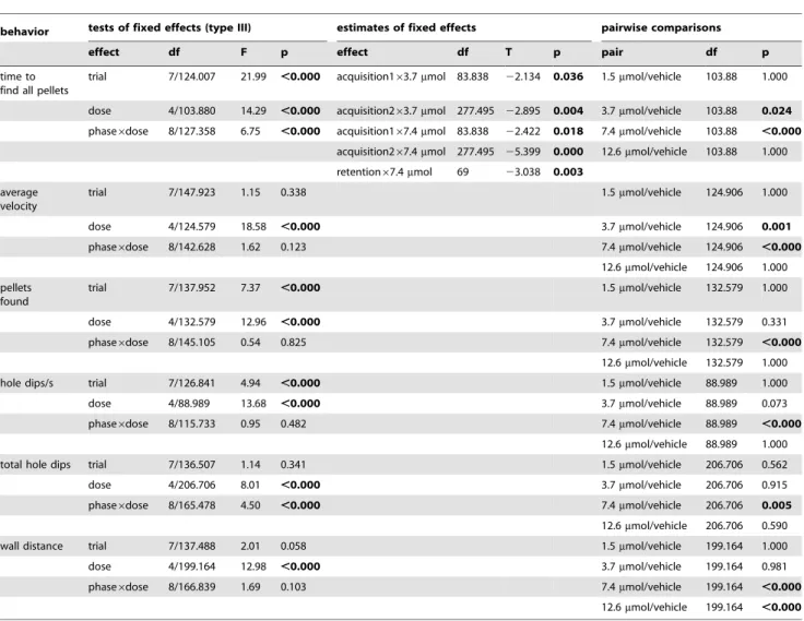
Hormones
We found significant differences in the hippocampal corticoste-rone (F5,48= 3.268, p = 0.014) levels, but not in the serum
concentrations (F5,43= 1.125, p = 0.364). The statistical analyses
of the testosterone and estradiol concentrations (Figure 3) revealed no differences in the hippocampal (F5,47= 1.343, p = 0.265, and
F5,47= 1.665, p = 0.164, respectively) and serum samples
(F5,45= 1.017, p = 0.420, and F5,38= 1.193, p = 0.334,
respective-ly).
The post hoc tests revealed higher hippocampal corticosterone concentrations in the animals treated with 3.7mmol MPP compared to the rats treated with 1.5mmol (p = 0.044) and 12.6mmol (p = 0.026) MPP.
Genes
The gene expression data are given in Figure 4. The GLM provides significant differences for all genes (ERa: F5,54= 6.509,
p,0.001; ERb: F5,54= 2.852, p = 0.024; AR: F5,54= 7.567,
p,0.001; MR: F5,54= 28.802, p,0.001; GR: F5,54= 11.714,
p,0.001; BDNF: F5,54= 11.805, p,0.001) except the aromatase
geneCyp19(F5,54= 1.642, p = 1.670).
The single group comparisons revealed that animals treated with 7.4mmol MPP showed the highest suppression of relative gene expression of ERa compared with animals treated with 1.5mmol (p = 0.008), 3.7mmol (p = 0.017), and vehicle (p = 0.004)
and untrained vehicle-treated control rats (p = 0.002). Further-more, animals administered 12.6mmol MPP had a significantly
lower ERa-mRNA level than the vehicle-treated trained (p = 0.036) and untrained (p = 0.014) controls. The relative gene expression of ERb, however, differed significantly only in the 7.4mmol MPP-treated rats compared to the untrained
vehicle-treated rats (p = 0.043).
The expression pattern of the androgen receptor was similar to that of ERa (7.4mmol/1.5mmol: p = 0.011; 7.4mmol/3.7mmol:
Table 1.Statistical analysis of differences in behavior.
behavior tests of fixed effects (type III) estimates of fixed effects pairwise comparisons
effect df F p effect df T p pair df p
time to find all pellets
trial 7/124.007 21.99 ,0.000 acquisition163.7mmol 83.838 22.134 0.036 1.5mmol/vehicle 103.88 1.000
dose 4/103.880 14.29 ,0.000 acquisition263.7mmol 277.495 22.895 0.004 3.7mmol/vehicle 103.88 0.024 phase6dose 8/127.358 6.75 ,0.000 acquisition167.4mmol 83.838 22.422 0.018 7.4mmol/vehicle 103.88 ,0.000
acquisition267.4mmol 277.495 25.399 0.000 12.6mmol/vehicle 103.88 1.000
retention67.4mmol 69 23.038 0.003 average
velocity
trial 7/147.923 1.15 0.338 1.5mmol/vehicle 124.906 1.000
dose 4/124.579 18.58 ,0.000 3.7mmol/vehicle 124.906 0.001
phase6dose 8/142.628 1.62 0.123 7.4mmol/vehicle 124.906 ,0.000
12.6mmol/vehicle 124.906 1.000
pellets found
trial 7/137.952 7.37 ,0.000 1.5mmol/vehicle 132.579 1.000
dose 4/132.579 12.96 ,0.000 3.7mmol/vehicle 132.579 0.331
phase6dose 8/145.105 0.54 0.825 7.4mmol/vehicle 132.579 ,0.000
12.6mmol/vehicle 132.579 1.000
hole dips/s trial 7/126.841 4.94 ,0.000 1.5mmol/vehicle 88.989 1.000
dose 4/88.989 13.68 ,0.000 3.7mmol/vehicle 88.989 0.073
phase6dose 8/115.733 0.95 0.482 7.4mmol/vehicle 88.989 ,0.000
12.6mmol/vehicle 88.989 1.000
total hole dips trial 7/136.507 1.14 0.341 1.5mmol/vehicle 206.706 0.562
dose 4/206.706 8.01 ,0.000 3.7mmol/vehicle 206.706 0.915
phase6dose 8/165.478 4.50 ,0.000 7.4mmol/vehicle 206.706 0.005
12.6mmol/vehicle 206.706 0.590
wall distance trial 7/137.488 2.01 0.058 1.5mmol/vehicle 199.164 1.000
dose 4/199.164 12.98 ,0.000 3.7mmol/vehicle 199.164 0.981
phase6dose 8/166.839 1.69 0.103 7.4mmol/vehicle 199.164 ,0.000
12.6mmol/vehicle 199.164 ,0.000
p = 0.042; 7.4mmol/vehicle: p,0.001; 7.4mmol/untrained vehi-cle: p = 0.013; 12.6mmol/vehicle: p = 0.001; 12.6mmol/untrained vehicle: p = 0.015). In addition, the 12.6mmol MPP group showed a difference in AR expression compared with the 1.5mmol MPP-treated animals (p = 0.017).
The mineralocorticoid receptor also had the lowest gene expression in animals treated with 7.4mmol MPP compared to all other groups (7.4mmol/1.5mmol: p,0.001; 7.4mmol/ 3.7mmol: p,0.001; 7.4mmol/12.6mmol: p,0.001; 7.4mmol/ vehicle: p,0.001; 7.4mmol/untrained vehicle: p,0.001). More-over, the 3.7mmol MPP group differed significantly from the 1.5mmol MPP group (p = 0.030), as well as from the vehicle-treated trained (p = 0.005) and untrained (p = 0.028) control groups.
For the BDNF and glucocorticoid receptor, we observed decreased gene expression in animals treated with 7.4mmol compared to the 1.5mmol (both: p,0.001), 3.7mmol (BDNF: p,0.001; GR: p = 0.005), 12.6mmol (both: p,0.001), and vehicle-treated trained (both: p,0.001) and untrained (both: p,0.001) groups.
Dose Effects
To compare dose effects on behavior and gene expression, we calculated a dose-effect curve (Figure 5) at the individual level. We followed the trend indicated by the between-group statistical effects and calculated the curve as the percentage of treated individuals that showed measurements higher (behavior) or lower (gene expression) than the mean of the control animals. The
Figure 2. Treatment with MPP at any concentration had no statistically significant effect on the reference and working memory indices at the end of the second acquisition phase (trial 9) and during retention (trial 10).However, animals treated with 7.4mmol MPP showed the highest indices of both types of memory. Given are the arithmetic means and the s.e.m.
doi:10.1371/journal.pone.0079303.g002
Figure 3. Treatment with MPP at any concentration had a statistically significant effect on the hippocampal concentra-tions of corticosterone only between the group treated with 3.7mmol compared to the groups treated with 1.5mmol and 7.4mmol MPP. Serum concentrations were not different, and no difference between groups was found for testosterone and 17b -estradiol. Horizontal lines with asterisks indicate statistically significant differences. Numbers above columns indicate sample sizes. Longer vertical lines indicate the groups against which the other groups were tested. Given are the arithmetic means and the s.e.m.
percentage measures were plotted against the logarithm of the dosages. It became immediately apparent that all effects (except total hole dips) were highest at the 7.4mmol dosage. Thus, only
when almost all individuals showed suppression in the expression of all genes was a behavioral effect observed. In addition, ERa,
ERb, and AR gene expression was the most sensitive to the treatment. Effects of more than 50% were observed at the lowest concentration and reached a plateau at 3.7mmol MPP. Ninety to
hundred percent of the individuals responded at 3.7mmol MPP in MR and GR expression and maintained the effects at 7.4mmol, whereas the aromatase and BDNF gene expression was less sensitive but also reached 100% responders at 7.4mmol MPP. Interestingly, with the decay in responders at 12.6mmol in behavior, the expression of MR, GR, and BDNF increased, whereas ERa, ERb, and AR expression remained at the 100% level.
Figure 4. Treatment with MPP had an effect on hippocampal relative gene expression of ERa, MR, GR, AR, and BDNF, but not ERb and aromatase (Cyp 19) encoding genes in a dose-dependent manner with statistically significant lower expres-sion of only the group treated with 7.4mmol MPP compared to the trained vehicle-treated animals.Horizontal lines with asterisks indicate statistically significant differences. Numbers in brackets indicate sample sizes. Longer vertical lines indicate the groups against which the other groups were tested. Given are the arithmetic means and the s.e.m.
doi:10.1371/journal.pone.0079303.g004
Figure 5. Dose-response relation of behavior (upper panel; time: time to find all pellets, pellets: pellets found) and gene expression for MPP treatment.Effect is given as the percentage of treated animals showing a measurement above (behavior, except time to find all pellets) or below (gene expression, time to find all pellets) the mean for the controls during retention. Corresponding highest effects at dose 7.4mmol MPP.
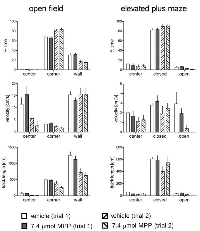
Open-field and Elevated Plus Maze Tests
To test whether the effects observed in the holeboard merely resulted from unspecific effects on locomotor activity or differences in anxiety induced by the pharmacological treatment, separate groups of animals underwent open-field and elevated plus maze tests (Figure 6). These animals were treated with 7.4mmol MPP (the most effective dose in the holeboard experiments, n = 12, one animal with missing values at day 1) or vehicle (n = 12; due to technical reasons, we lost the data for one animal in the elevated plus maze test at day 2). We found no statistically significant interaction between treatment and trial and no treatment effects in the open field (time spent in the center F1,22= 0.06, p = 0.815;
F1,22= 0.01, p = 0.932; time spent in corners F1,22= 0.54,
p = 0.471; F1,22= 0.01, p = 0.911; time spent in wall zones
F1,22= 0.71, p = 0.407; F1,22= 0.01, p = 0.941; velocity in the
center F1,22= 2.58, p = 0.122; F1,22= 0.02, p = 0.894; velocity in
the corners F1,22= 1.37, p = 0.254; F1,22= 0.39, p = 0.54; velocity
in wall zones F1,22= 0.51, p = 0.483; F1,22= 0.261, p = 0.615; track
length in the center F1,22= 0.00, p = 0.995; F1,22= 0.12, p = 0.734;
track length in the corners F1,22= 2.10, p = 0.161; F1,22= 1.81,
p = 0.192; track length in the wall zones F1,22= 0.04, p = 0.839;
F1,22= 0.66, p = 0.425). Similarly, no treatment trial interaction
and no treatment effect were determined in the elevated plus maze test (time spent in the center F1,20= 0.54, p = 0.473; F1,20= 0.00,
p = 0.956; time spent in closed arms F1,20= 0.00, p = 0.970;
F1,20= 0.05, p = 0.82; time spent in open arms F1,20= 2.23,
p = 0.15; F1,20= 0.02, p = 0.904; velocity in the center
F1,20= 0.18, p = 0.675; F1,20= 0.00, p = 0.987; velocity in closed
arms F1,20= 0.01, p = 0.907; F1,20= 0.79, p = 0.383; velocity in
open arms F1,20= 0.13, p = 0.720; F1,20= 0.78, p = 0.387; track
length in the center F1,20= 2.50, p = 0.129; F1,20= 0.54, p = 0.472;
track length in closed arms F1,20= 1.18, p = 0.291; F1,20= 0.61,
p = 0.443; track length in open arms F1,20= 2.22, p = 0.152;
F1,20= 0.07, p = 0.789). Discussion
We found dose-dependent changes (up to 7.4mmol MPP) in
behavioral performance with most significant effects in animals treated with 7.4mmol MPP. These animals needed less time to find all pellets, moved faster, had more hole dips, and found more pellets than the control animals. In contrast to our hypothesis, the MPP-treated animals performed better regarding motivation (velocity, hole dips/s), whereas, in line with our hypothesis, the cognitive component of the task was not affected by the treatment. Treatment effects on the general locomotor activity and changes in anxiety, which could have contributed to the observed differences, can be ruled out by our open-field and plus maze tests. Regarding exploration and anxiety, the MPP-treated rats did not behave differently from the controls.
The effects of the MPP treatment are particularly surprising, because we found previously that the upregulation of hippocampal ERa gene expression is a learning-related holeboard effect in males at the age tested here. We also found, although not significant, upregulation of ERagene expression in vehicle-treated males compared to untrained animals. However, the MPP-treated animals showed significantly reduced levels of gene expression not only for ERabut also for MR, GR, AR as well as the BDNF gene, whereas ERb and aromatase gene expression was unaffected in the trained animals. These results support two conclusions: i. ‘‘Specific’’ steroid receptor functions can be revealed only against the steroid network background, characterized by a stronger relation of ERaactivity to its own gene expression and to that of glucocorticoid and testosterone binding androgen receptors than
to ERb, and an effect on the BDNF but not on the aromatase encoding gene. ii. The training-induced increased expression of the ERa encoding gene does not positively correlate with the receptor function, because the receptor blockade results in increased rather than impaired performance of motivation-indicating behavior. Activation of extranuclear ER induces hippocampal BDNF signaling, thus mediating its effects on neuroprotection and plasticity [33], [39]. Because of these known interactions, we included the BDNF gene in the present study, and interestingly, we found the MPP treatment affected BDNF gene expression. Hippocampal BDNF and the related receptors are involved in learning and memory [40], [41]; thus, an ER-BDNF interaction may partly play a role in mediating behavior in the present study.
Thus, at least parts of the observed system of steroid receptors, and there are many others [42], react in a coordinated and dose-dependent manner to the MPP treatment, which suggests that compensating mechanisms exist within the network to keep the system in homeostasis and to provide a mechanism for reacting quickly to external signals. The apparent discrepancy between the previously observed positive correlation of ERa-mRNA with motivational behavior and the failure to reduce motivation by ERablockade on the protein level in the present study, may be explained by the translation of preexisting, training-induced, silent ERa-mRNA that could be quickly and locally translated into functional receptors [43], [44] by extracellular signaling. MR, that has been identified as the crucial acute stress-related receptor in our holeboard paradigm [45] may be involved in the transduction of external signals. These mechanisms could provide effects upon behavior independently from rapid changes in the availability of locally produced estrogens [23]. Future studies should prove this with in situ-hybridization and optical methods for identifying the hippocampal steroid receptor-mRNA distribution and trafficking in trained and untrained animals. Membrane- and cytosol-specific westernblots can detect training-induced changes in site-specific functional steroid receptors within the hippocampus. The behav-ioral effect that motivation increased with the ERa-blockade is likely a result of the MPP treatment that reduces ligand binding with functional receptors, which occupation with a natural ligand otherwise leads to a decrease in motivation. Thus, ERaactivation does not promote motivational behavior but may constrain over-motivated, high-risk behavior preserving the animal from life-threatening situations. This may be adaptive especially in young post-pubertal rats that ontogenetically are in the phase of migrating from their social groups and exploring new environ-ments. Importantly, we did not find upregulation of ERa gene expression in older individuals in the same situation [24].
The network characterization is further supported by the fact that we found significant effects in behavior only when the expression of all genes (except the aromatase and ERbencoding genes) was affected, although there were differences in the sensitivity to MPP treatment between genes, as suggested by the dose response curve. Second, changes in hormone concentrations, due to the treatment, were absent. Therefore, compensatory modulations of receptor expression resulting from altered hormonal states cannot explain the results. Last, the effects of MPP on gene expression are dose dependent (up to 7.4mmol MPP), thus indicating not unspecific treatment-induced downreg-ulation of gene expression. Further, not all genes were affected.
results in animals treated with 3.7 and 7.4mmol MPP (especially
for the learning phase6dose interactions) along with the
differ-ences in the effects on gene expression between the two dosages suggest complex effects of structural changes in the receptor network on motivation and behavior and may indicate a structural change in the receptor network over training. Training phase– specific changes in correlations of hippocampal steroid hormone concentrations with behavior has been described for the same learning procedure [46]. In that study, hippocampal testosterone
concentrations switched from a negative correlation with reference memory errors at acquisition phase 1 to no correlation during acquisition 2 and back to a negative correlation during retention. Corticosterone concentrations in the prefrontal cortex negatively correlated with reference memory errors during acquisition and positively during retention. This may be related to brain region– specific changes in steroid functions in behavioral regulation during training or reflect network adjustments over different brain regions.
Figure 6. Behavioral results for the open-field (left panel) and the elevated plus maze (right panel) tests of animals treated with 7.4mmol MPP or vehicle.Closed: closed arms; open: open arms. No group6trial interaction and no group differences were detected for any behavioral parameter. Given are the arithmetic means and the s.e.m.
Surprisingly, the dose-dependent effects of MPP on behavioral performance disappeared when the animals were treated with a higher dose than 7.4mmol MPP. Animals treated with 12.6mmol
MPP showed only a larger wall distance when compared to the vehicle-treated group. Notably, this group also showed similar MR and GR expression as the vehicle-treated animals and significantly higher expression of these receptor genes compared to the group treated with 7.4mmol MPP. Thus, higher concentrations of MPP may result in unspecificity of the receptor blockade with a subsequent rescue of MR and GR expression and behavior.
Due to the intra-cerebroventricular administration of MPP, resulting in brain-wide distribution, other brain regions such as the amygdala and hypothalamus, which contain estrogen receptors [47], [48], [49], are probably involved in the observed behavioral effects. Although no effects of ERa knockdown in the medial amygdala on sexual or aggressive behavior in 16-week-old male mice were observed, the same procedure in the ventromedial nucleus of the hypothalamus reduced both types of behavior [7]. The effects, however, may be age dependent [50], because different interactions and contributions of ERa and ERb in
maintaining hippocampal-dependent memory during aging in female mice have been reported [51]. Future studies, including local drug administration, should reveal the specific contributions of these brain regions to the motivational states and behavioral performance in young rats and the possible rules underlying the experience-dependent structural changes in the steroid network.
Supporting Information
Video S1 Representative example of the behavioral perfor-mance of a rat treated with 7.4mmol MPP (trial 10).
(MPG)
Video S2 Representative example of the behavioral perfor-mance of a rat treated with vehicle (trial 10).
(MPG)
Author Contributions
Conceived and designed the experiments: VK. Performed the experiments: KM. Analyzed the data: KM VK. Wrote the paper: VK KM.
References
1. Perlman WR, Tomaskovic-Crook E, Montague DM, Webster MJ, Rubinow DR et al. (2005) Alteration in estrogen receptor alpha mRNA levels in frontal cortex and hippocampus of patients with major mental illness. Biol Psychiatry 58: 812– 824.
2. Hajszan T, Milner TA, Leranth C (2007) Sex steroids and the dentate gyrus. Prog Brain Res 163: 399–415.
3. Gillies GE, McArthur S (2010) Estrogen actions in the brain and the basis for differential action in men and women: a case for sex-specific medicines. Pharmacol Rev 62: 155–198.
4. Lagunas N, Calmarza-Font I, Grassi D, Garcia-Segura LM (2011) Estrogen receptor ligands counteract cognitive deficits caused by androgen deprivation in male rats. Horm Behav 59: 581–584.
5. McEwen BS (1988) Genomic regulation of sexual behavior. J Steroid Biochem 30: 179–183.
6. Paisley JC, Huddleston GG, Carruth LL, Petrulis A, Grober MS et al. (2012) Sexual responses of the male rat medial preoptic area and medial amygdala to estrogen I: site specific suppression of estrogen receptor alpha. Horm Behav 62: 50–57.
7. Sano K, Tsuda MC, Musatov S, Sakamoto T, Ogawa S (2013) Differential effects of site-specific knockdown of estrogen receptorain the medial amygdala, medial pre-optic area, and ventromedial nucleus of the hypothalamus on sexual and aggressive behavior of male mice. Eur J Neurosci doi: 10.1111/ejn.12131. 8. Fester L, Ribeiro-Gouveia V, Prange-Kiel J, von Schassen C, Bo¨ttner M et al. (2006) Proliferation and apoptosis of hippocampal granule cells require local oestrogen synthesis. J Neurochem 97: 1136–1144.
9. Bowers JM, Waddell J, McCarthy MM (2010) A developmental sex difference in hippocampal neurogenesis is mediated by endogenous oestradiol. Biol Sex Differ 1: 8.
10. Gonza´lez-Burgos I, Rivera-Cervantes MC, Vela´zquez-Zamora DA, Feria-Velasco A, Garcia-Segura LM (2012) Selective estrogen receptor modulators regulate dendritic spine plasticity in the hippocampus of male rats. Neural Plast 2012: 309494, doi: 10.1155/2012/309494.
11. Fester L, Prange-Kiel J, Jarry H, Rune GM (2011) Estrogen synthesis in the hippocampus. Cell Tissue Res 345: 285–294.
12. Kuiper GG, Lemmen JG, Carlsson B, Corton JC, Safe SH et al. (1998) Interaction of estrogenic chemicals and phytoestrogens with estrogen receptor beta. Endocrinology 139: 4252–4263.
13. Scordalakes EM, Rissman EF (2003) Aggression in male mice lacking functional estrogen receptor alpha. Behav Neurosci 117: 38–45.
14. Nomura M, Andersson S, Korach KS, Gustafsson JA, Pfaff DW et al. (2006) Estrogen receptor-beta gene disruption potentiates estrogen-inducible aggression but not sexual behavior in male mice. Eur J Neurosci 23: 1860–1868. 15. Tetel MJ, Pfaff DW (2010) Contributions of estrogen receptor-aand estrogen
receptor-ß to the regulation of behavior. Biochim Biophys Acta 1800: 1084– 1089.
16. Frye CA, Duffy CK, Walf AA (2007) Estrogens and progestins enhance spatial learning of intact and ovariectomized rats in the object placement task. Neurobiol Learn Mem 88: 208–216.
17. Liu F, Day M, Muniz LC, Bitran D, Arias R et al. (2008) Activation of estrogen receptor-beta regulates hippocampal synaptic plasticity and improves memory. Nat Neurosci 11: 334–343.
18. Rissman EF (2008) Roles of oestrogen receptors a and b in behavioral neuroendocrinology: Beyond Yin/Yang. J Neuroendocrinol 20: 873–879.
19. Neese SL, Korol DL, Katzenellenbogen JA, Schantz SL (2010) Impact of estrogen receptor alpha and beta agonists on delayed alternation in middle-aged rats. Horm Behav 58: 878–890.
20. Galea LA, Uban KA, Epp JR, Brummelte S, Barha CK et al. (2008) Endocrine regulation of cognition and neuroplasticity: our pursuit to unveil the complex interaction between hormones, the brain, and behaviour. Can J Exp Psychol 62: 247–260.
21. Barha CK, Galea LA (2010) Influence of different estrogens on neuroplasticity and cognition in the hippocampus. Biochim Biophys Acta 1800: 1056–1067. 22. Mukai H, Kimoto T, Hojo Y, Kawato S, Murakami G et al. (2010) Modulation
of synaptic plasticity by brain estrogen in the hippocampus. Biochim Biophys Acta 1800: 1030–1044.
23. Cornil CA, Ball GF, Balthazart J (2012) Rapid control of male typical behaviors by brain-derived estrogens. Front Neuroendocrinol 33: 425–46.
24. Meyer K, Korz V (2013) Age dependent differences in the regulation of hippocampal steroid hormones and receptor genes: Relations to motivation and cognition in male rats. Horm Behav 63: 376–384.
25. Chen S, Wang J, Yu G, Liu W, Pearce D (1997) Androgen and glucocorticoid receptor heterodimer formation. A possible mechanism for mutual inhibition of transcriptional activity. J Biol Chem 272: 14087–14092.
26. Cvoro A, Yuan C, Paruthiyil S, Miller OH, Yamamoto KR et al. (2011) Cross Talk between Glucocorticoid and Estrogen Receptors Occurs at a Subset of Proinflammatory Genes. J Immunol 186: 4354–4360.
27. Savatier J, Jalaguier S, Ferguson ML, Cavaille`s V, Royer CA (2010) Estrogen receptor interactions and dynamics monitored in live cells by fluorescence cross-correlation spectroscopy. Biochemistry 49: 772–781.
28. McEwen BS (1992) Steroid hormones: effect on brain development and function. Horm Res 37 Suppl 3: 1–10.
29. Wu TW, Chen S, Brinton RD (2011) Membrane estrogen receptors mediate calcium signaling and MAP kinase activation in individual hippocampal neurons. Brain Res 1379: 34–43.
30. Fernandez SM, Lewis MC, Pechenino AS, Harburger LL, Orr PT et al. (2008) Estradiol-induced enhancement of object memory consolidation involves hippocampal extracellular signal-regulated kinase activation and membrane-bound estrogen receptors. J Neurosci 28: 8660–8667.
31. Solum DT, Handa RJ (2002) Estrogen regulates the development of brain-derived neurotrophic factor mRNA and protein in the rat hippocampus. J Neurosci 22: 2650–2659.
32. Spencer-Segal JL, Tsuda MC, Mattei L, Waters EM, Romeo RD et al. (2012) Estradiol acts via estrogen receptors alpha and beta on pathways important for synaptic plasticity in the mouse hippocampal formation. Neuroscience 202: 131– 146.
33. Yang LC, Zhang QG, Zhou CF, Yang F, Zhang YD et al. (2010) Extranuclear estrogen receptors mediate the neuroprotective effects of estrogen in the rat hippocampus. PLoS One 5: e9851.
34. Schaaf MJM, Hoetelmans RWM, de Kloet ER, Vreugdenhil E (1997) Corticosterone regulates expression of BDNF and trkB but not NT-3 and trkC mRNA in the rat hippocampus. J Neurosci Res 48: 334–341.
35. Uzakov S, Frey JU, Korz V (2005) Reinforcement of rat hippocampal LTP by holeboard training. Learn Mem 12: 165–171.
estrogen responsive gene sites mediating transactivation or transrepression. Mol Cell Endocrinol 206: 13–22.
38. Livak KJ, Schmittgen TD (2001) Analysis of relative gene expression data using real-time quantitative PCR and the 22DD
CTmethod. Methods 25: 403–508.
39. Scharfman HE, MacLusky NJ (2006) Estrogen and brain-derived neurotrophic factor (BDNF) in hippocampus: Complexity of steroid hormone-growth factor interactions in the adult CNS. Front Neuroendocrinol 27: 415–435. 40. Falkenberg T, Mohammed AK, Henriksson B, Persson H, Winblad B et al.
(1992) Increased expression of brain-derived neurotrophic factor mRNA in rat hippocampus is associated with improved spatial memory and enriched environment. Neurosci Lett 138: 153–156.
41. Ma YL, Wang HL, Wu HC, Wei CL, Lee EH (1998) Brain-derived neurotrophic factor antisense oligonucleotide impairs memory retention and inhibits long-term potentiation in rats. Neuroscience 82: 957–967.
42. Ranhotra HS (2012) The estrogen-related receptors: orphans orchestrating myriad functions. J Recept Signal Transduct Res 32: 47–56.
43. Richter JD, Lorenz LJ (2002) Selective translation of mRNAs at synapses. Curr Opin Neurobiol 12: 300–304.
44. Bramham CR, Wells DG (2007) Dendritic mRNA: transport, translation and function. Nature Rev Neurosci 8: 776–789.
45. Korz V, Frey JU (2007) Hormonal and monoamine signaling during reinforcement of hippocampal long-term potentiation and memory retrieval. Learn Mem 14: 160–166.
46. Schulz K, Korz V (2010) Hippocampal testosterone relates to reference memory performance and synaptic plasticity in male rats. Front Behav Neurosci 4; doi: 10.3389/fnbeh.2010.00187.
47. McEwen BS, Luine VN, Plapinger L, de Kloet ER (1975) Putative estrogen and glucocorticoid receptors in the limbic brain. J Steroid Biochem 6: 971–977. 48. Takeshita H (1976) Evidence for soluble estradiol receptors in the amygdala of
male and female rats. Yonago Acta Med 20: 125–141.
49. McEwen BS (2002) Estrogen actions throughout the brain. Recent Prog Horm Res 57: 357–384.
50. MacLusky NJ, Lieberburg I, McEwen BS (1979) The development of estrogen receptor systems in the rat brain: perinatal development. Brain Res 178: 129– 142.